Li J et al. (DEC 2015)
Biomedical microdevices 17 6 105
Fabrication of uniform-sized poly-ɛ-caprolactone microspheres and their applications in human embryonic stem cell culture.
The generation of liquefied poly-ɛ-caprolactone (PCL) droplets by means of a microfluidic device results in uniform-sized microspheres,which are validated as microcarriers for human embryonic stem cell culture. Formed droplet size and size distribution,as well as the resulting PCL microsphere size,are correlated with the viscosity and flow rate ratio of the dispersed (Q d) and continuous (Q c) phases. PCL in dichloromethane increases its viscosity with concentration and molecular weight. Higher viscosity and Q d/Q c lead to the formation of larger droplets,within two observed formation modes: dripping and jetting. At low viscosity of dispersed phase and Q d/Q c,the microfluidic device is operated in dripping mode,which generates droplets and microspheres with greater size uniformity. Solutions with lower molecular weight PCL have lower viscosity,resulting in a wider concentration range for the dripping mode. When coated with extracellular matrix (ECM) proteins,the fabricated PCL microspheres are demonstrated capable of supporting the expansion of human embryonic stem cells.
View Publication
产品类型:
产品号#:
05850
05857
05870
05875
85850
85857
85870
85875
产品名:
mTeSR™1
mTeSR™1
(Jul 2025)
bioRxiv 5 27
Robust Production of Parvalbumin Interneurons and Fast-Spiking Neurons from Human Medial Ganglionic Eminence Organoids
SummaryThe medial ganglionic eminence (MGE) gives rise to parvalbumin (PV)- and somatostatin (SST)-expressing cortical interneurons essential for regulating cortical excitability. Although PV interneurons are linked to various neurodevelopmental and neurodegenerative disorders,reliably generating them from human pluripotent stem cells (hPSCs) has been extremely challenging. We present a robust,reproducible protocol for generating single-rosette MGE organoids (MGEOs) from hPSCs. Transcriptomic analyses reveal that MGEOs exhibit MGE regional identity and faithfully model the developing human fetal MGE. As MGEOs mature,they generate abundant PV-expressing cortical interneurons,including putative basket and axoaxonic cells,at a scale not previously achieved in vitro. When fused with hPSC-derived cortical organoids,these interneurons rapidly migrate into cortical regions,integrate into excitatory networks,and contribute to complex electrophysiological patterns and the emergence of large numbers of fast-spiking neurons. MGEOs thus offer a powerful in vitro approach for probing human MGE-lineage cortical and subcortical GABAergic neuron development,modeling various neuropsychiatric disorders,and advancing cell-based therapies for neurodevelopmental and neurodegenerative disorders. Graphical abstract
View Publication
Multilineage long-term engraftment potential of drug-resistant hematopoietic progenitors.
Peripheral blood progenitor cells (PBPCs) are increasingly used instead of bone marrow for autologous or allogeneic transplantation. In this study PBPCs mobilized in cancer patients by chemotherapy and granulocyte-colony stimulating factor were collected by apheresis and first enriched by immunoaffinity removal of lineage positive cells. When these cells were exposed to both cyclophosphamide and taxol or cultured for 7 days in the presence of 5-fluorouracil,stem cell factor,and interleukin-3,88% to 93% of the enriched PBPCs were killed and short-term clonogenic capacity in methylcellulose assays was lost,but week-5 cobblestone area-forming cell (CAFC) enrichment was higher than 10-fold in comparison to enriched PBPCs and higher than 700-fold in comparison to unmanipulated apheresis cells. After drug exposure,most of the progenitors displayed a CD34+,CD38-,multidrug-resistance (MDR+),Rhodamine 123 low,Hoechst 33342 low phenotype,and as few as 180 of these drug-resistant cells were able to generate a stable multilineage human hematopoiesis in sublethally irradiated immunodeficient mice. In these animals,the level of human hematopoietic engraftment was significantly increased by cotransplantation of irradiated cells from the human L87/4 stromal cell line. These observations are consistent with the functional isolation of a population of very early hematopoietic progenitors and might help to design new protocols for the removal of neoplastic cells from autografts.
View Publication
Hisa T et al. (JAN 2004)
The EMBO journal 23 2 450--9
Hematopoietic, angiogenic and eye defects in Meis1 mutant animals.
Meis1 and Hoxa9 expression is upregulated by retroviral integration in murine myeloid leukemias and in human leukemias carrying MLL translocations. Both genes also cooperate to induce leukemia in a mouse leukemia acceleration assay,which can be explained,in part,by their physical interaction with each other as well as the PBX family of homeodomain proteins. Here we show that Meis1-deficient embryos have partially duplicated retinas and smaller lenses than normal. They also fail to produce megakaryocytes,display extensive hemorrhaging,and die by embryonic day 14.5. In addition,Meis1-deficient embryos lack well-formed capillaries,although larger blood vessels are normal. Definitive myeloerythroid lineages are present in the mutant embryos,but the total numbers of colony-forming cells are dramatically reduced. Mutant fetal liver cells also fail to radioprotect lethally irradiated animals and they compete poorly in repopulation assays even though they can repopulate all hematopoietic lineages. These and other studies showing that Meis1 is expressed at high levels in hematopoietic stem cells (HSCs) suggest that Meis1 may also be required for the proliferation/self-renewal of the HSC.
View Publication
产品类型:
产品号#:
04960
04902
04900
04961
04901
04963
04962
04970
04971
产品名:
MegaCult™-C胶原和无细胞因子培养基
胶原蛋白溶液
MegaCult™-C无细胞因子培养基
MegaCult™-C胶原和含细胞因子培养基
MegaCult™-C含细胞因子培养基
双室载玻片套件
MegaCult™-C CFU-Mk染色试剂盒
MegaCult™-C无细胞因子全套试剂盒
MegaCult™-C含细胞因子全套试剂盒
Marchetto MCN et al. (JAN 2009)
PLoS ONE 4 9 e7076
Transcriptional signature and memory retention of human-induced pluripotent stem cells
Genetic reprogramming of somatic cells to a pluripotent state (induced pluripotent stem cells or iPSCs) by over-expression of specific genes has been accomplished using mouse and human cells. However,it is still unclear how similar human iPSCs are to human Embryonic Stem Cells (hESCs). Here,we describe the transcriptional profile of human iPSCs generated without viral vectors or genomic insertions,revealing that these cells are in general similar to hESCs but with significant differences. For the generation of human iPSCs without viral vectors or genomic insertions,pluripotent factors Oct4 and Nanog were cloned in episomal vectors and transfected into human fetal neural progenitor cells. The transient expression of these two factors,or from Oct4 alone,resulted in efficient generation of human iPSCs. The reprogramming strategy described here revealed a potential transcriptional signature for human iPSCs yet retaining the gene expression of donor cells in human reprogrammed cells free of viral and transgene interference. Moreover,the episomal reprogramming strategy represents a safe way to generate human iPSCs for clinical purposes and basic research.
View Publication
产品类型:
产品号#:
05850
05857
05870
05875
85850
85857
85870
85875
产品名:
mTeSR™1
mTeSR™1
Berman DM et al. (OCT 2010)
Diabetes 59 10 2558--68
Mesenchymal stem cells enhance allogeneic islet engraftment in nonhuman primates.
OBJECTIVE: To test the graft-promoting effects of mesenchymal stem cells (MSCs) in a cynomolgus monkey model of islet/bone marrow transplantation. RESEARCH DESIGN AND METHODS: Cynomolgus MSCs were obtained from iliac crest aspirate and characterized through passage 11 for phenotype,gene expression,differentiation potential,and karyotype. Allogeneic donor MSCs were cotransplanted intraportally with islets on postoperative day (POD) 0 and intravenously with donor marrow on PODs 5 and 11. Recipients were followed for stabilization of blood glucose levels,reduction of exogenous insulin requirement (EIR),C-peptide levels,changes in peripheral blood T regulatory cells,and chimerism. Destabilization of glycemia and increases in EIR were used as signs of rejection; additional intravenous MSCs were administered to test the effect on reversal of rejection. RESULTS: MSC phenotype and a normal karyotype were observed through passage 11. IL-6,IL-10,vascular endothelial growth factor,TGF-β,hepatocyte growth factor,and galectin-1 gene expression levels varied among donors. MSC treatment significantly enhanced islet engraftment and function at 1 month posttransplant (n = 8),as compared with animals that received islets without MSCs (n = 3). Additional infusions of donor or third-party MSCs resulted in reversal of rejection episodes and prolongation of islet function in two animals. Stable islet allograft function was associated with increased numbers of regulatory T-cells in peripheral blood. CONCLUSIONS: MSCs may provide an important approach for enhancement of islet engraftment,thereby decreasing the numbers of islets needed to achieve insulin independence. Furthermore,MSCs may serve as a new,safe,and effective antirejection therapy.
View Publication
产品类型:
产品号#:
18770
18770RF
产品名:
Bar EE et al. (OCT 2007)
Stem cells (Dayton,Ohio) 25 10 2524--33
Cyclopamine-mediated hedgehog pathway inhibition depletes stem-like cancer cells in glioblastoma.
Brain tumors can arise following deregulation of signaling pathways normally activated during brain development and may derive from neural stem cells. Given the requirement for Hedgehog in non-neoplastic stem cells,we investigated whether Hedgehog blockade could target the stem-like population in glioblastoma multiforme (GBM). We found that Gli1,a key Hedgehog pathway target,was highly expressed in 5 of 19 primary GBM and in 4 of 7 GBM cell lines. Shh ligand was expressed in some primary tumors,and in GBM-derived neurospheres,suggesting a potential mechanism for pathway activation. Hedgehog pathway blockade by cyclopamine caused a 40%-60% reduction in growth of adherent glioma lines highly expressing Gli1 but not in those lacking evidence of pathway activity. When GBM-derived neurospheres were treated with cyclopamine and then dissociated and seeded in media lacking the inhibitor,no new neurospheres formed,suggesting that the clonogenic cancer stem cells had been depleted. Consistent with this hypothesis,the stem-like fraction in gliomas marked by both aldehyde dehydrogenase activity and Hoechst dye excretion (side population) was significantly reduced or eliminated by cyclopamine. In contrast,we found that radiation treatment of our GBM neurospheres increased the percentage of these stem-like cells,suggesting that this standard therapy preferentially targets better-differentiated neoplastic cells. Most importantly,viable GBM cells injected intracranially following Hedgehog blockade were no longer able to form tumors in athymic mice,indicating that a cancer stem cell population critical for ongoing growth had been removed. Disclosure of potential conflicts of interest is found at the end of this article.
View Publication
产品类型:
产品号#:
01700
01705
01701
01702
72072
72074
产品名:
ALDEFLUOR™ 试剂盒
ALDEFLUOR™ DEAB试剂, 1.5 mM, 1 mL
ALDEFLUOR™检测缓冲液
环巴胺(Cyclopamine)
环巴胺(Cyclopamine)
Sundaram K et al. (FEB 2015)
Bone 71 3 137--44
STAT-6 mediates TRAIL induced RANK ligand expression in stromal/preosteoblast cells.
Receptor activator of nuclear factor kappa-B ligand (RANKL) is a critical osteoclastogenic factor expressed in bone marrow stromal/osteoblast lineage cells. Tumor necrosis factor (TNF) related apoptosis-inducing ligand (TRAIL) levels are elevated in pathologic conditions such as multiple myeloma and inflammatory arthritis,and have been positively correlated with osteolytic markers. Osteoprotegerin (OPG) which inhibits osteoclastogenesis is a decoy receptor for RANKL and also known to interact with TRAIL. Herein,we show that TRAIL increases DR5 and DcR1 receptors but no change in the levels of DR4 and DcR2 expression in human bone marrow derived stromal/preosteoblast (SAKA-T) cell line. We further demonstrated that TRAIL treatment significantly decreased OPG mRNA expression. Interestingly,TRAIL treatment induced RANKL mRNA expression in these cells. In addition,TRAIL significantly increased NF-kB and c-Jun N-terminal kinase (JNK) activity. Human transcription factor array screening by real-time RT-PCR identified TRAIL up-regulation of the signal transducers and activators of the transcription (STAT)-6 expression in SAKA-T cells. TRAIL stimulation induced p-STAT-6 expression in human bone marrow derived primary stromal/preosteoblast cells. Confocal microscopy analysis further revealed p-STAT-6 nuclear localization in SAKA-T cells. Chromatin immunoprecipitation (ChIP) assay confirmed p-STAT-6 binding to the hRANKL gene distal promoter region. In addition,siRNA suppression of STAT-6 expression inhibits TRAIL increased hRANKL gene promoter activity. Thus,our results suggest that TRAIL induces RANKL expression through a STAT-6 dependent transcriptional regulatory mechanism in bone marrow stromal/preosteoblast cells.
View Publication
产品类型:
产品号#:
70022
70071
产品名:
Hsieh J et al. (NOV 2004)
Proceedings of the National Academy of Sciences of the United States of America 101 47 16659--64
It has become apparent that chromatin modification plays a critical role in the regulation of cell-type-specific gene expression. Here,we show that an inhibitor of histone deacetylase,valproic acid (VPA),induced neuronal differentiation of adult hippocampal neural progenitors. In addition,VPA inhibited astrocyte and oligodendrocyte differentiation,even in conditions that favored lineage-specific differentiation. Among the VPA-up-regulated,neuron-specific genes,a neurogenic basic helix-loop-helix transcription factor,NeuroD,was identified. Overexpression of NeuroD resulted in the induction and suppression of neuronal and glial differentiation,respectively. These results suggest that VPA promotes neuronal fate and inhibits glial fate simultaneously through the induction of neurogenic transcription factors including NeuroD.
View Publication
产品类型:
产品号#:
72112
72114
72292
100-0249
产品名:
Forskolin
Forskolin
丙戊酸(钠盐)
Forskolin
Karumbayaram S et al. (APR 2009)
Stem cells (Dayton,Ohio) 27 4 806--11
Directed differentiation of human-induced pluripotent stem cells generates active motor neurons.
The potential for directed differentiation of human-induced pluripotent stem (iPS) cells to functional postmitotic neuronal phenotypes is unknown. Following methods shown to be effective at generating motor neurons from human embryonic stem cells (hESCs),we found that once specified to a neural lineage,human iPS cells could be differentiated to form motor neurons with a similar efficiency as hESCs. Human iPS-derived cells appeared to follow a normal developmental progression associated with motor neuron formation and possessed prototypical electrophysiological properties. This is the first demonstration that human iPS-derived cells are able to generate electrically active motor neurons. These findings demonstrate the feasibility of using iPS-derived motor neuron progenitors and motor neurons in regenerative medicine applications and in vitro modeling of motor neuron diseases.
View Publication

 EasySep™小鼠TIL(CD45)正选试剂盒


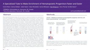 科学海报Fast and Easy Hematopoietic Progenitor Cell Enrichment With SepMate™

 沪公网安备31010102008431号