Z. Liu et al. (nov 2020)
Cell 183 4 1117--1133.e19
Detecting Tumor Antigen-Specific T Cells via Interaction-Dependent Fucosyl-Biotinylation.
Re-activation and clonal expansion of tumor-specific antigen (TSA)-reactive T cells are critical to the success of checkpoint blockade and adoptive transfer of tumor-infiltrating lymphocyte (TIL)-based therapies. There are no reliable markers to specifically identify the repertoire of TSA-reactive T cells due to their heterogeneous composition. We introduce FucoID as a general platform to detect endogenous antigen-specific T cells for studying their biology. Through this interaction-dependent labeling approach,intratumoral TSA-reactive CD4+,CD8+ T cells,and TSA-suppressive CD4+ T cells can be detected and separated from bystander T cells based on their cell-surface enzymatic fucosyl-biotinylation. Compared to bystander TILs,TSA-reactive TILs possess a distinct T cell receptor (TCR) repertoire and unique gene features. Although exhibiting a dysfunctional phenotype,TSA-reactive CD8+ TILs possess substantial capabilities of proliferation and tumor-specific killing. Featuring genetic manipulation-free procedures and a quick turnover cycle,FucoID should have the potential of accelerating the pace of personalized cancer treatment.
View Publication
产品类型:
产品号#:
17858
19853
17858RF
100-0694
19853RF
产品名:
EasySep™人CD14正选试剂盒II
EasySep™小鼠CD8+ T细胞分选试剂盒
RoboSep™ 人CD14正选试剂盒II
EasySep™人CD14正选试剂盒II
RoboSep™ 小鼠CD8+ T细胞分选试剂盒
Park S-W et al. (DEC 2010)
Blood 116 25 5762--72
Efficient differentiation of human pluripotent stem cells into functional CD34+ progenitor cells by combined modulation of the MEK/ERK and BMP4 signaling pathways.
Differentiation of human pluripotent stem cells (hPSCs) into functional cell types is a crucial step in cell therapy. In the present study,we demonstrate that functional CD34(+) progenitor cells can be efficiently produced from human embryonic stem cells (hESCs) and induced pluripotent stem cells (hiPSCs) by combined modulation of 2 signaling pathways. A higher proportion of CD34(+) cells (∼ 20%) could be derived from hPSCs by inhibition of mitogen-activated protein kinase (MAPK) extracellular signal-regulated protein kinase (MEK)/extracellular signal-regulated kinase (ERK) signaling and activation of bone morphogenic protein-4 (BMP4) signaling. hPSC-derived CD34(+) progenitor cells further developed to endothelial and smooth muscle cells with functionality. Moreover,they contributed directly to neovasculogenesis in ischemic mouse hind limbs,thereby resulting in improved blood perfusion and limb salvage. Our results suggest that combined modulation of signaling pathways may be an efficient means of differentiating hPSCs into functional CD34(+) progenitor cells.
View Publication
产品类型:
产品号#:
04434
04444
产品名:
MethoCult™ H4434 Classic
MethoCult™ H4434 Classic
Nguyen HX et al. (AUG 2014)
Journal of Comparative Neurology 522 12 2767--2783
Induction of early neural precursors and derivation of tripotent neural stem cells from human pluripotent stem cells under xeno-free conditions
Human embryonic stem cells (hESC) and induced pluripotent stem cells (hiPSC) can differentiate into many cell types and are important for regenerative medicine; however,further work is needed to reliably differentiate hESC and hiPSC into neural-restricted multipotent derivatives or specialized cell types under conditions that are free from animal products. Toward this goal,we tested the transition of hESC and hiPSC lines onto xeno-free (XF) / feeder-free conditions and evaluated XF substrate preference,pluripotency,and karyotype. Critically,XF transitioned H9 hESC,Shef4 hESC,and iPS6-9 retained pluripotency (Oct-4 and NANOG),proliferation (MKI67 and PCNA),and normal karyotype. Subsequently,XF transitioned hESC and hiPSC were induced with epidermal growth factor (EGF) and basic fibroblast growth factor (bFGF) to generate neuralized spheres containing primitive neural precursors,which could differentiate into astrocytes and neurons,but not oligoprogenitors. Further neuralization of spheres via LIF supplementation and attachment selection on CELLstart substrate generated adherent human neural stem cells (hNSC) with normal karyotype and high proliferation potential under XF conditions. Interestingly,adherent hNSC derived from H9,Shef4,and iPS6-9 differentiated into significant numbers of O4+ oligoprogenitors (∼20-30%) with robust proliferation; however,very few GalC+ cells were observed (∼2-4%),indicative of early oligodendrocytic lineage commitment. Overall,these data demonstrate the transition of multiple hESC and hiPSC lines onto XF substrate and media conditions,and a reproducible neuralization method that generated neural derivatives with multipotent cell fate potential and normal karyotype.
View Publication
产品类型:
产品号#:
05860
05880
05850
05857
05870
05875
85850
85857
85870
85875
产品名:
mTeSR™1
mTeSR™1
Wang H et al. (JAN 2012)
Journal of translational medicine 10 1 167
Oncolytic vaccinia virus GLV-1h68 strain shows enhanced replication in human breast cancer stem-like cells in comparison to breast cancer cells.
BACKGROUND: Recent data suggest that cancer stem cells (CSCs) play an important role in cancer,as these cells possess enhanced tumor-forming capabilities and are responsible for relapses after apparently curative therapies have been undertaken. Hence,novel cancer therapies will be needed to test for both tumor regression and CSC targeting. The use of oncolytic vaccinia virus (VACV) represents an attractive anti-tumor approach and is currently under evaluation in clinical trials. The purpose of this study was to demonstrate whether VACV does kill CSCs that are resistant to irradiation and chemotherapy. METHODS: Cancer stem-like cells were identified and separated from the human breast cancer cell line GI-101A by virtue of increased aldehyde dehydrogenase 1 (ALDH1) activity as assessed by the ALDEFLUOR assay and cancer stem cell-like features such as chemo-resistance,irradiation-resistance and tumor-initiating were confirmed in cell culture and in animal models. VACV treatments were applied to both ALDEFLUOR-positive cells in cell culture and in xenograft tumors derived from these cells. Moreover,we identified and isolated CD44(+)CD24(+)ESA(+) cells from GI-101A upon an epithelial-mesenchymal transition (EMT). These cells were similarly characterized both in cell culture and in animal models. RESULTS: We demonstrated for the first time that the oncolytic VACV GLV-1h68 strain replicated more efficiently in cells with higher ALDH1 activity that possessed stem cell-like features than in cells with lower ALDH1 activity. GLV-1h68 selectively colonized and eventually eradicated xenograft tumors originating from cells with higher ALDH1 activity. Furthermore,GLV-1h68 also showed preferential replication in CD44(+)CD24(+)ESA(+) cells derived from GI-101A upon an EMT induction as well as in xenograft tumors originating from these cells that were more tumorigenic than CD44(+)CD24(-)ESA(+) cells. CONCLUSIONS: Taken together,our findings indicate that GLV-1h68 efficiently replicates and kills cancer stem-like cells. Thus,GLV-1h68 may become a promising agent for eradicating both primary and metastatic tumors,especially tumors harboring cancer stem-like cells that are resistant to chemo and/or radiotherapy and may be responsible for recurrence of tumors.
View Publication
产品类型:
产品号#:
01700
01705
05620
01702
产品名:
ALDEFLUOR™ 试剂盒
ALDEFLUOR™ DEAB试剂, 1.5 mM, 1 mL
MammoCult™ 人源培养基套装
ALDEFLUOR™检测缓冲液
Ostrakhovitch EA et al. (DEC 2012)
Archives of biochemistry and biophysics 528 1 21--31
Directed differentiation of embryonic P19 cells and neural stem cells into neural lineage on conducting PEDOT-PEG and ITO glass substrates.
Differentiation of pluripotent and lineage restricted stem cells such as neural stem cells (NSCs) was studied on conducting substrates of various nature without perturbation of the genome with exogenous genetic material or chemical stimuli. Primary mouse adult neural stem cells (NSCs) and P19 pluripotent embryonal (P19 EC) carcinoma cells were used. Expression levels of neuronal markers β-III-tubulin and neurofilament were evaluated by immunochemistry and flow cytometry. It was shown that the ability of the substrate to induce differentiation directly correlated with its conductivity. Conducting substrates (conducting oxides or doped pi-conjugated organic polymers) with different morphology,structure,and conductivity mechanisms all promoted differentiation of NSC and P19 cells into neuronal lineage to a similar degree without use of additional factors such as poly-L-ornithine coating or retinoic acid,as verified by their morphology and upregulation of the neuronal markers but not astrocyte marker GFAP. However,substrates with low conductance below ca. 10(-4) S cm(-2) did not show this ability. Morphology of differentiating cells was visualized by atomic force microscopy. NSCs cells increased β-III-tubulin expression by 95% and P19 cells by over 30%. Our results suggest that the substrate conductivity is a key factor governing the cell fate. Differentiation of P19 cells into neuronal lineage on conducting substrates was attributed to downregualtion of Akt signaling pathway and increase in expression of dual oxidase 1 (DUOX 1).
View Publication
产品类型:
产品号#:
05700
05701
05702
05703
05704
05715
产品名:
NeuroCult™ 基础培养基(小鼠和大鼠)
NeuroCult™ 扩增添加物(小鼠和大鼠)
NeuroCult™扩增试剂盒(小鼠和大鼠)
NeuroCult™ 分化添加物(小鼠和大鼠)
NeuroCult™ 分化试剂盒(小鼠和大鼠)
NeuroCult™成年中枢神经系统(CNS)组织酶解试剂盒(小鼠和大鼠)
Kim et al. (Oct 2025)
Scientific Reports 15
Attenuation of natural killer cell cytotoxicity by interaction between NKp30 of NK cells and dipeptidase 1 of colon cancer cells
Natural killer (NK) cells play a crucial role in immune surveillance by recognizing and eliminating tumor cells. However,tumors employ various mechanisms to evade NK cell-mediated immunity. NKp30 is a potent activating receptor on NK cells,but its function can be inhibited by specific ligands secreted by cancer cells. Here,we identified dipeptidase 1 (DPEP1) as a novel ligand for NKp30 in KM12C colon cancer cells,using co-immunoprecipitation,confocal microscopy,and flow cytometry. We examined how the DPEP1–NKp30 interaction affects NK cell activity and found that NK cytotoxicity increased in KM12C cells with DPEP1 knockdown but was significantly reduced in HCT116 cells overexpressing DPEP1. We further demonstrated that DPEP1 is secreted via extracellular vesicles and that its interaction with NKp30 suppressed the expression and secretion of perforin 1,granzyme B,CD107a,and interferon-γ in NK92 cells. In a xenograft mouse model treated with NK92 cells,tumors derived from HCT116/DPEP1 cells were significantly larger than those from HCT116/mock cells. Using peripheral blood-derived human NK cells,we confirmed that DPEP1 inhibited both cytotoxicity and granzyme B secretion. These findings suggest that disrupting the DPEP1–NKp30 interaction may enhance NK cell-mediated cytotoxicity and represent a novel therapeutic strategy for cancer immunotherapy. The online version contains supplementary material available at 10.1038/s41598-025-18475-z.
View Publication
Non-integrating episomal plasmid-based reprogramming of human amniotic fluid stem cells into induced pluripotent stem cells in chemically defined conditions.
Amniotic fluid stem cells (AFSC) represent an attractive potential cell source for fetal and pediatric cell-based therapies. However,upgrading them to pluripotency confers refractoriness toward senescence,higher proliferation rate and unlimited differentiation potential. AFSC were observed to rapidly and efficiently reacquire pluripotency which together with their easy recovery makes them an attractive cell source for reprogramming. The reprogramming process as well as the resulting iPSC epigenome could potentially benefit from the unspecialized nature of AFSC. iPSC derived from AFSC also have potential in disease modeling,such as Down syndrome or $\$-thalassemia. Previous experiments involving AFSC reprogramming have largely relied on integrative vector transgene delivery and undefined serum-containing,feeder-dependent culture. Here,we describe non-integrative oriP/EBNA-1 episomal plasmid-based reprogramming of AFSC into iPSC and culture in fully chemically defined xeno-free conditions represented by vitronectin coating and E8 medium,a system that we found uniquely suited for this purpose. The derived AF-iPSC lines uniformly expressed a set of pluripotency markers Oct3/4,Nanog,Sox2,SSEA-1,SSEA-4,TRA-1-60,TRA-1-81 in a pattern typical for human primed PSC. Additionally,the cells formed teratomas,and were deemed pluripotent by PluriTest,a global expression microarray-based in-silico pluripotency assay. However,we found that the PluriTest scores were borderline,indicating a unique pluripotent signature in the defined condition. In the light of potential future clinical translation of iPSC technology,non-integrating reprogramming and chemically defined culture are more acceptable.
View Publication
W. Wang et al. (may 2019)
Nature 569 7755 270--274
CD8+ T cells regulate tumour ferroptosis during cancer immunotherapy.
Cancer immunotherapy restores or enhances the effector function of CD8+ T cells in the tumour microenvironment1,2. CD8+ T cells activated by cancer immunotherapy clear tumours mainly by inducing cell death through perforin-granzyme and Fas-Fas ligand pathways3,4. Ferroptosis is a form of cell death that differs from apoptosis and results from iron-dependent accumulation of lipid peroxide5,6. Although it has been investigated in vitro7,8,there is emerging evidence that ferroptosis might be implicated in a variety of pathological scenarios9,10. It is unclear whether,and how,ferroptosis is involved in T cell immunity and cancer immunotherapy. Here we show that immunotherapy-activated CD8+ T cells enhance ferroptosis-specific lipid peroxidation in tumour cells,and that increased ferroptosis contributes to the anti-tumour efficacy of immunotherapy. Mechanistically,interferon gamma (IFNgamma) released from CD8+ T cells downregulates the expression of SLC3A2 and SLC7A11,two subunits of the glutamate-cystine antiporter system xc-,impairs the uptake of cystine by tumour cells,and as a consequence,promotes tumour cell lipid peroxidation and ferroptosis. In mouse models,depletion of cystine or cysteine by cyst(e)inase (an engineered enzyme that degrades both cystine and cysteine) in combination with checkpoint blockade synergistically enhanced T cell-mediated anti-tumour immunity and induced ferroptosis in tumour cells. Expression of system xc- was negatively associated,in cancer patients,with CD8+ T cell signature,IFNgamma expression,and patient outcome. Analyses of human transcriptomes before and during nivolumab therapy revealed that clinical benefits correlate with reduced expression of SLC3A2 and increased IFNgamma and CD8. Thus,T cell-promoted tumour ferroptosis is an anti-tumour mechanism,and targeting this pathway in combination with checkpoint blockade is a potential therapeutic approach.
View Publication
产品类型:
产品号#:
17953
17953RF
19853
19853RF
100-0710
产品名:
EasySep™人CD8+ T细胞分选试剂盒
RoboSep™ 人CD8+ T细胞分选试剂盒
EasySep™小鼠CD8+ T细胞分选试剂盒
RoboSep™ 小鼠CD8+ T细胞分选试剂盒
EasySep™人CD8+ T细胞分选试剂盒
Cai J et al. (JAN 2004)
Journal of neurochemistry 88 1 212--26
Membrane properties of rat embryonic multipotent neural stem cells.
We have characterized several potential stem cell markers and defined the membrane properties of rat fetal (E10.5) neural stem cells (NSC) by immunocytochemistry,electrophysiology and microarray analysis. Immunocytochemical analysis demonstrates specificity of expression of Sox1,ABCG2/Bcrp1,and shows that nucleostemin labels both progenitor and stem cell populations. NSCs,like hematopoietic stem cells,express high levels of aldehyde dehydrogenase (ALDH) as assessed by Aldefluor labeling. Microarray analysis of 96 transporters and channels showed that Glucose transporter 1 (Glut1/Slc2a1) expression is unique to fetal NSCs or other differentiated cells. Electrophysiological examination showed that fetal NSCs respond to acetylcholine and its agonists,such as nicotine and muscarine. NSCs express low levels of tetrodotoxin (TTX) sensitive and insensitive sodium channels and calcium channels while expressing at least three kinds of potassium channels. We find that gap junction communication is mediated by connexin (Cx)43 and Cx45,and is essential for NSC survival and proliferation. Overall,our results show that fetal NSCs exhibit a unique signature that can be used to determine their location and assess their ability to respond to their environment.
View Publication

 EasySep™小鼠TIL(CD45)正选试剂盒


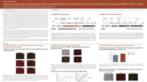 科学海报Generation, Maintenance and Cryopreservation of Neural Progenitor Cells Derived from Human Pluripotent Stem Cells Using the STEMdiff™ Neural System
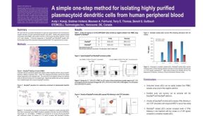 科学海报Isolation of Plasmacytoid Dendritic Cells from Human Peripheral Blood
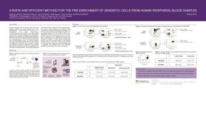 科学海报Pre-Enrichment of Dendritic Cells from Human Peripheral Blood Samples
 沪公网安备31010102008431号