Characterization of hematopoietic progenitor mobilization in protease-deficient mice.
Recent evidence suggests that protease release by neutrophils in the bone marrow may contribute to hematopoietic progenitor cell (HPC) mobilization. Matrix metalloproteinase-9 (MMP-9),neutrophil elastase (NE),and cathepsin G (CG) accumulate in the bone marrow during granulocyte colony-stimulating factor (G-CSF) treatment,where they are thought to degrade key substrates including vascular cell adhesion molecule-1 (VCAM-1) and CXCL12. To test this hypothesis,HPC mobilization was characterized in transgenic mice deficient in one or more hematopoietic proteases. Surprisingly,HPC mobilization by G-CSF was normal in MMP-9-deficient mice,NE x CG-deficient mice,or mice lacking dipeptidyl peptidase I,an enzyme required for the functional activation of many hematopoietic serine proteases. Moreover,combined inhibition of neutrophil serine proteases and metalloproteinases had no significant effect on HPC mobilization. VCAM-1 expression on bone marrow stromal cells decreased during G-CSF treatment of wild-type mice but not NE x CG-deficient mice,indicating that VCAM-1 cleavage is not required for efficient HPC mobilization. G-CSF induced a significant decrease in CXCL12 alpha protein expression in the bone marrow of Ne x CG-deficient mice,indicating that these proteases are not required to down-regulate CXCL12 expression. Collectively,these data suggest a complex model in which both protease-dependent and -independent pathways may contribute to HPC mobilization.
View Publication
Macrophages enhance contractile force in iPSC-derived human engineered cardiac tissue
SUMMARY Resident cardiac macrophages are critical mediators of cardiac function. Despite their known importance to cardiac electrophysiology and tissue maintenance,there are currently no stem-cell-derived models of human engineered cardiac tissues (hECTs) that include resident macrophages. In this study,we made an induced pluripotent stem cell (iPSC)-derived hECT model with a resident population of macrophages (iM0) to better recapitulate the native myocardium and characterized their impact on tissue function. Macrophage retention within the hECTs was confirmed via immunofluorescence after 28 days of cultivation. The inclusion of iM0s significantly impacted hECT function,increasing contractile force production. A potential mechanism underlying these changes was revealed by the interrogation of calcium signaling,which demonstrated the modulation of ?-adrenergic signaling in +iM0 hECTs. Collectively,these findings demonstrate that macrophages significantly enhance cardiac function in iPSC-derived hECT models,emphasizing the need to further explore their contributions not only in healthy hECT models but also in the contexts of disease and injury. In brief Lock and Graney et al. develop a human engineered cardiac tissue with an incorporated iPSC-derived macrophage population to better mimic the complex cell landscape of the native myocardium. Macrophage inclusion leads to increased contractile function of the tissue,which is attributed to macrophage stimulation of the cardiomyocyte ?-adrenergic signaling pathway. Graphical Abstract
View Publication
产品类型:
产品号#:
05310
100-0276
100-1130
产品名:
STEMdiff™ 造血试剂盒
mTeSR™ Plus
mTeSR™ Plus
Sandrin V et al. (AUG 2002)
Blood 100 3 823--32
Lentiviral vectors pseudotyped with a modified RD114 envelope glycoprotein show increased stability in sera and augmented transduction of primary lymphocytes and CD34+ cells derived from human and nonhuman primates.
Generating lentiviral vectors pseudotyped with different viral glycoproteins (GPs) may modulate the physicochemical properties of the vectors,their interaction with the host immune system,and their host range. We have investigated the capacity of a panel of GPs of both retroviral (amphotropic murine leukemia virus [MLV-A]; gibbon ape leukemia virus [GALV]; RD114,feline endogenous virus) and nonretroviral (fowl plague virus [FPV]; Ebola virus [EboV]; vesicular stomatitis virus [VSV]; lymphocytic choriomeningitis virus [LCMV]) origins to pseudotype lentiviral vectors derived from simian immunodeficiency virus (SIVmac251). SIV vectors were efficiently pseudotyped with the FPV hemagglutinin,VSV-G,LCMV,and MLV-A GPs. In contrast,the GALV and RD114 GPs conferred much lower infectivity to the vectors. Capitalizing on the conservation of some structural features in the transmembrane domains and cytoplasmic tails of the incorporation-competent MLV-A GP and in RD114 and GALV GPs,we generated chimeric GPs encoding the extracellular and transmembrane domains of GALV or RD114 GPs fused to the cytoplasmic tail (designated TR) of MLV-A GP. Importantly,SIV-derived vectors pseudotyped with these GALV/TR and RD114/TR GP chimeras had significantly higher titers than vectors coated with the parental GPs. Additionally,RD114/TR-pseudotyped vectors were efficiently concentrated and were resistant to inactivation induced by the complement of both human and macaque sera,indicating that modified RD114 GP-pseudotyped lentiviral vectors may be of particular interest for in vivo gene transfer applications. Furthermore,as compared to vectors pseudotyped with other retroviral GPs or with VSV-G,RD114/TR-pseudotyped vectors showed augmented transduction of human and macaque primary blood lymphocytes and CD34+ cells.
View Publication
产品类型:
产品号#:
02690
09600
09650
产品名:
StemSpan™ CC100
StemSpan™ SFEM
StemSpan™ SFEM
Abeysinghe HCS et al. (SEP 2015)
Stem cell research & therapy 6 1 186
Pre-differentiation of human neural stem cells into GABAergic neurons prior to transplant results in greater repopulation of the damaged brain and accelerates functional recovery after transient ischemic stroke.
INTRODUCTION Despite attempts to prevent brain injury during the hyperacute phase of stroke,most sufferers end up with significant neuronal loss and functional deficits. The use of cell-based therapies to recover the injured brain offers new hope. In the current study,we employed human neural stem cells (hNSCs) isolated from subventricular zone (SVZ),and directed their differentiation into GABAergic neurons followed by transplantation to ischemic brain. METHODS Pre-differentiated GABAergic neurons,undifferentiated SVZ-hNSCs or media alone were stereotaxically transplanted into the rat brain (n=7/group) 7 days after endothelin-1 induced stroke. Neurological outcome was assessed by neurological deficit scores and the cylinder test. Transplanted cell survival,cellular phenotype and maturation were assessed using immunohistochemistry and confocal microscopy. RESULTS Behavioral assessments revealed accelerated improvements in motor function 7 days post-transplant in rats treated with pre-differentiated GABAergic cells in comparison to media alone and undifferentiated hNSC treated groups. Histopathology 28 days-post transplant indicated that pre-differentiated cells maintained their GABAergic neuronal phenotype,showed evidence of synaptogenesis and up-regulated expression of both GABA and calcium signaling proteins associated with neurotransmission. Rats treated with pre-differentiated cells also showed increased neurogenic activity within the SVZ at 28 days,suggesting an additional trophic role of these GABAergic cells. In contrast,undifferentiated SVZ-hNSCs predominantly differentiated into GFAP-positive astrocytes and appeared to be incorporated into the glial scar. CONCLUSION Our study is the first to show enhanced exogenous repopulation of a neuronal phenotype after stroke using techniques aimed at GABAergic cell induction prior to delivery that resulted in accelerated and improved functional recovery.
View Publication
Lin S et al. (JAN 2010)
Journal of visualized experiments : JoVE 39 11330
Video bioinformatics analysis of human embryonic stem cell colony growth.
Because video data are complex and are comprised of many images,mining information from video material is difficult to do without the aid of computer software. Video bioinformatics is a powerful quantitative approach for extracting spatio-temporal data from video images using computer software to perform dating mining and analysis. In this article,we introduce a video bioinformatics method for quantifying the growth of human embryonic stem cells (hESC) by analyzing time-lapse videos collected in a Nikon BioStation CT incubator equipped with a camera for video imaging. In our experiments,hESC colonies that were attached to Matrigel were filmed for 48 hours in the BioStation CT. To determine the rate of growth of these colonies,recipes were developed using CL-Quant software which enables users to extract various types of data from video images. To accurately evaluate colony growth,three recipes were created. The first segmented the image into the colony and background,the second enhanced the image to define colonies throughout the video sequence accurately,and the third measured the number of pixels in the colony over time. The three recipes were run in sequence on video data collected in a BioStation CT to analyze the rate of growth of individual hESC colonies over 48 hours. To verify the truthfulness of the CL-Quant recipes,the same data were analyzed manually using Adobe Photoshop software. When the data obtained using the CL-Quant recipes and Photoshop were compared,results were virtually identical,indicating the CL-Quant recipes were truthful. The method described here could be applied to any video data to measure growth rates of hESC or other cells that grow in colonies. In addition,other video bioinformatics recipes can be developed in the future for other cell processes such as migration,apoptosis,and cell adhesion.
View Publication
产品类型:
产品号#:
05850
05857
05870
05875
85850
85857
85870
85875
产品名:
mTeSR™1
mTeSR™1
S. E. Wamaitha et al. ( 2020)
Nature communications 11 1 764
IGF1-mediated human embryonic stem cell self-renewal recapitulates the embryonic niche.
Our understanding of the signalling pathways regulating early human development is limited,despite their fundamental biological importance. Here,we mine transcriptomics datasets to investigate signalling in the human embryo and identify expression for the insulin and insulin growth factor 1 (IGF1) receptors,along with IGF1 ligand. Consequently,we generate a minimal chemically-defined culture medium in which IGF1 together with Activin maintain self-renewal in the absence of fibroblast growth factor (FGF) signalling. Under these conditions,we derive several pluripotent stem cell lines that express pluripotency-associated genes,retain high viability and a normal karyotype,and can be genetically modified or differentiated into multiple cell lineages. We also identify active phosphoinositide 3-kinase (PI3K)/AKT/mTOR signalling in early human embryos,and in both primed and na{\{i}}ve pluripotent culture conditions. This demonstrates that signalling insights from human blastocysts can be used to define culture conditions that more closely recapitulate the embryonic niche."
View Publication
产品类型:
产品号#:
05010
85850
85857
产品名:
STEMdiff™ 心室肌细胞分化试剂盒
mTeSR™1
mTeSR™1
Y. Zhang et al. (aug 2019)
Nature communications 10 1 3667
Receptor type protein tyrosine phosphatase-sigma (PTPsigma) is primarily expressed by adult neurons and regulates neural regeneration. We recently discovered that PTPsigma is also expressed by hematopoietic stem cells (HSCs). Here,we describe small molecule inhibitors of PTPsigma that promote HSC regeneration in vivo. Systemic administration of the PTPsigma inhibitor,DJ001,or its analog,to irradiated mice promotes HSC regeneration,accelerates hematologic recovery,and improves survival. Similarly,DJ001 administration accelerates hematologic recovery in mice treated with 5-fluorouracil chemotherapy. DJ001 displays high specificity for PTPsigma and antagonizes PTPsigma via unique non-competitive,allosteric binding. Mechanistically,DJ001 suppresses radiation-induced HSC apoptosis via activation of the RhoGTPase,RAC1,and induction of BCL-XL. Furthermore,treatment of irradiated human HSCs with DJ001 promotes the regeneration of human HSCs capable of multilineage in vivo repopulation. These studies demonstrate the therapeutic potential of selective,small-molecule PTPsigma inhibitors for human hematopoietic regeneration.
View Publication


 EasySep™小鼠TIL(CD45)正选试剂盒




 挂图Identification of Colonies Derived from Mouse Hematopoietic Progenitors Representative colony images and tips for identifying progenitor subtypes in CFU assays
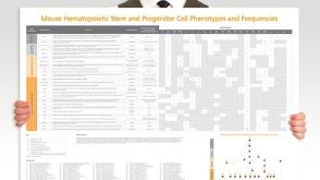 挂图Mouse Hematopoietic Stem and Progenitor Cell Phenotyping Overview of mouse HSPC subset surface markers and frequencies发布日期: 08/10/2018
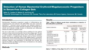 科学海报Detection of Human Bipotential Erythroid-Megakaryocytic Progenitors in Serum-Free Collagen Gels

 沪公网安备31010102008431号