RNA Sequencing of H3N2 Influenza Virus-Infected Human Nasal Epithelial Cells from Multiple Subjects Reveals Molecular Pathways Associated with Tissue Injury and Complications.
The human nasal epithelium is the primary site of exposure to influenza virus,the initiator of host responses to influenza and the resultant pathologies. Influenza virus may cause serious respiratory infection resulting in major complications,as well as severe impairment of the airways. Here,we elucidated the global transcriptomic changes during H3N2 infection of human nasal epithelial cells from multiple individuals. Using RNA sequencing,we characterized the differentially-expressed genes and pathways associated with changes occurring at the nasal epithelium following infection. We used in vitro differentiated human nasal epithelial cell culture model derived from seven different donors who had no concurrent history of viral infections. Statistical analysis highlighted strong transcriptomic signatures significantly associated with 24 and 48 h after infection,but not at the earlier 8-h time point. In particular,we found that the influenza infection induced in the nasal epithelium early and altered responses in interferon gamma signaling,B-cell signaling,apoptosis,necrosis,smooth muscle proliferation,and metabolic alterations. These molecular events initiated at the infected nasal epithelium may potentially adversely impact the airway,and thus the genes we identified could serve as potential diagnostic biomarkers or therapeutic targets for influenza infection and associated disease management.
View Publication
产品类型:
产品号#:
05001
05022
05021
产品名:
PneumaCult™-ALI 培养基
PneumaCult™-ALI 培养基含6.5 mm Transwell®插件
PneumaCult™-ALI 培养基含12 mm Transwell®插件
Du A et al. (MAY 2012)
Developmental Biology 365 1 175--188
Arx is required for normal enteroendocrine cell development in mice and humans
Enteroendocrine cells of the gastrointestinal (GI) tract play a central role in metabolism,digestion,satiety and lipid absorption,yet their development remains poorly understood. Here we show that Arx,a homeodomain-containing transcription factor,is required for the normal development of mouse and human enteroendocrine cells. Arx expression is detected in a subset of Neurogenin3 (Ngn3)-positive endocrine progenitors and is also found in a subset of hormone-producing cells. In mice,removal of Arx from the developing endoderm results in a decrease of enteroendocrine cell types including gastrin-,glucagon/GLP-1-,CCK-,secretin-producing cell populations and an increase of somatostatin-expressing cells. This phenotype is also observed in mice with endocrine-progenitor-specific Arx ablation suggesting that Arx is required in the progenitor for enteroendocrine cell development. In addition,depletion of human ARX in developing human intestinal tissue results in a profound deficit in expression of the enteroendocrine cell markers CCK,secretin and glucagon while expression of a pan-intestinal epithelial marker,CDX2,and other non-endocrine markers remained unchanged. Taken together,our findings uncover a novel and conserved role of Arx in mammalian endocrine cell development and provide a potential cause for the chronic diarrhea seen in both humans and mice carrying Arx mutations.
View Publication
产品类型:
产品号#:
05850
05857
05870
05875
85850
85857
85870
85875
产品名:
mTeSR™1
mTeSR™1
Yoon T-MM et al. (SEP 2010)
Stem Cell Reviews and Reports 6 3 425--437
Human embryonic stem cells (hESCs) cultured under distinctive feeder-free culture conditions display global gene expression patterns similar to hESCs from feeder-dependent culture conditions.
Human embryonic stem cell (hESC)-based assay systems and genetically modified hESCs are very useful tools for screening drugs that regulate stemness and differentiation and for studying the molecular mechanisms involved in hESC fate determination. For these types of studies,feeder cell-dependent cultures of hESCs are often problematic because the physiology of the feeder cells is perturbed by the drug treatments or genetic modifications,which potentially obscures research outcomes. In this study,we evaluated three commonly used feeder-free culture conditions to determine whether they supported the undifferentiated growth of hESCs and to determine whether the hESCs grown in these conditions displayed gene expression patterns that were similar to the expression patterns of feeder cell-dependent hESCs. Our results demonstrate that hESCs grown in the three feeder-free conditions expressed undifferentiation marker genes as strongly as hESCs that were grown in the feeder-dependent cultures. Furthermore,genome-wide gene expression profiles indicated that the gene expression patterns of hESCs that were grown under feeder-free or feeder-dependent culture conditions were highly similar. These results indicate that the feeder-free culture conditions support the undifferentiated growth of hESCs as effectively as the feeder-dependent culture conditions. Therefore,feeder-free culture conditions are potentially suitable for drug screening and for the genetic manipulation of hESCs in basic research.
View Publication
产品类型:
产品号#:
05850
05857
05870
05875
85850
85857
85870
85875
产品名:
mTeSR™1
mTeSR™1
Felgentreff K et al. (JUN 2014)
Proceedings of the National Academy of Sciences of the United States of America 111 24 8889--94
Differential role of nonhomologous end joining factors in the generation, DNA damage response, and myeloid differentiation of human induced pluripotent stem cells.
Nonhomologous end-joining (NHEJ) is a key pathway for efficient repair of DNA double-strand breaks (DSBs) and V(D)J recombination. NHEJ defects in humans cause immunodeficiency and increased cellular sensitivity to ionizing irradiation (IR) and are variably associated with growth retardation,microcephaly,and neurodevelopmental delay. Repair of DNA DSBs is important for reprogramming of somatic cells into induced pluripotent stem cells (iPSCs). To compare the specific contribution of DNA ligase 4 (LIG4),Artemis,and DNA-protein kinase catalytic subunit (PKcs) in this process and to gain insights into phenotypic variability associated with these disorders,we reprogrammed patient-derived fibroblast cell lines with NHEJ defects. Deficiencies of LIG4 and of DNA-PK catalytic activity,but not Artemis deficiency,were associated with markedly reduced reprogramming efficiency,which could be partially rescued by genetic complementation. Moreover,we identified increased genomic instability in LIG4-deficient iPSCs. Cell cycle synchronization revealed a severe defect of DNA repair and a G0/G1 cell cycle arrest,particularly in LIG4- and DNA-PK catalytically deficient iPSCs. Impaired myeloid differentiation was observed in LIG4-,but not Artemis- or DNA-PK-mutated iPSCs. These results indicate a critical importance of the NHEJ pathway for somatic cell reprogramming,with a major role for LIG4 and DNA-PKcs and a minor,if any,for Artemis.
View Publication
产品类型:
产品号#:
05850
05857
05870
05875
07920
85850
85857
85870
85875
07922
产品名:
ACCUTASE™
mTeSR™1
mTeSR™1
ACCUTASE™
Matsuura K et al. (MAR 2015)
Tissue engineering. Part C,Methods 21 3 330--338
Elimination of remaining undifferentiated induced pluripotent stem cells in the process of human cardiac cell sheet fabrication using a methionine-free culture condition.
Cardiac tissue engineering is a promising method for regenerative medicine. Although we have developed human cardiac cell sheets by integration of cell sheet-based tissue engineering and scalable bioreactor culture,the risk of contamination by induced pluripotent stem (iPS) cells in cardiac cell sheets remains unresolved. In the present study,we established a novel culture method to fabricate human cardiac cell sheets with a decreased risk of iPS cell contamination while maintaining viabilities of iPS cell-derived cells,including cardiomyocytes and fibroblasts,using a methionine-free culture condition. When cultured in the methionine-free condition,human iPS cells did not survive without feeder cells and could not proliferate or form colonies on feeder cells or in coculture with cells for cardiac cell sheet fabrication. When iPS cell-derived cells after the cardiac differentiation were transiently cultured in the methionine-free condition,gene expression of OCT3/4 and NANOG was downregulated significantly compared with that in the standard culture condition. Furthermore,in fabricated cardiac cell sheets,spontaneous and synchronous beating was observed in the whole area while maintaining or upregulating the expression of various cardiac and extracellular matrix genes. These findings suggest that human iPS cells are methionine dependent and a methionine-free culture condition for cardiac cell sheet fabrication might reduce the risk of iPS cell contamination.
View Publication
产品类型:
产品号#:
05850
05857
05870
05875
85850
85857
85870
85875
产品名:
mTeSR™1
mTeSR™1
Kamata M et al. (NOV 2010)
Human gene therapy 21 11 1555--67
Generation of human induced pluripotent stem cells bearing an anti-HIV transgene by a lentiviral vector carrying an internal murine leukemia virus promoter.
The recent development of induced pluripotent stem cells (iPSCs) by ectopic expression of defined reprogramming factors offers enormous therapeutic opportunity. To deliver these factors,murine leukemia virus (MLV)-based vectors have been broadly used in the setting of hematopoietic stem cell transplantation. However,MLV vectors have been implicated in malignancy induced by insertional mutagenesis,whereas lentiviral vectors have not. Furthermore,the infectivity of MLV vectors is limited to dividing cells,whereas lentiviral vectors can also transduce nondividing cells. One important characteristic of MLV vectors is a self-silencing property of the promoter element in pluripotent stem cells,allowing temporal transgene expression in a nonpluripotent state before iPSC derivation. Here we test iPSC generation using a novel chimeric vector carrying a mutant MLV promoter internal to a lentiviral vector backbone,thereby containing the useful properties of both types of vectors. Transgene expression of this chimeric vector was highly efficient compared with that of MLV vectors and was silenced specifically in human embryonic stem cells. Human fetal fibroblasts transduced with the vector encoding each factor were efficiently reprogrammed into a pluripotent state,and these iPSCs had potential to differentiate into a variety of cell types. To explore the possibility of iPSCs for gene therapy,we established iPSC clones expressing a short hairpin RNA (shRNA) targeting chemokine receptor 5 (CCR5),the main coreceptor for HIV-1. Using a reporter construct for CCR5 expression,we confirmed that CCR5 shRNA was expressed and specifically knocked down the reporter expression in iPSCs. These data indicate that our chimeric lentiviral vector is a valuable tool for generation of iPSCs and the combination with vectors encoding transgenes allows for rapid establishment of desired genetically engineered iPSC lines.
View Publication
产品类型:
产品号#:
05850
05857
05870
05875
85850
85857
85870
85875
27845
27945
27840
27865
27940
27965
产品名:
mTeSR™1
mTeSR™1
Jin F et al. (JUL 2005)
Cancer research 65 14 6354--63
Activation of nuclear factor-kappaB contributes to induction of death receptors and apoptosis by the synthetic retinoid CD437 in DU145 human prostate cancer cells.
Activation of the transcription factor,nuclear factor-kappaB (NF-kappaB),results in up-regulation of not only antiapoptotic genes but also proapoptotic genes,including death receptor 4 (DR4) and death receptor 5 (DR5). Therefore,NF-kappaB activation either suppresses or promotes apoptosis depending on the type of stimulus or cell context. We showed previously that the synthetic retinoid,6-[3-(1-adamantyl)-4-hydroxyphenyl]-2-naphthalene carboxylic acid (CD437),effectively induces apoptosis particularly in androgen-independent prostate carcinoma cells. This effect was associated with the ability of CD437 to induce the expression of DR4 and DR5. In the present study,we examined the hypothesis that NF-kappaB activation plays a role in CD437-induced death receptor expression and apoptosis. Treatment of DU145 cells with CD437 resulted in a rapid decrease (textgreater or = 3 hours) of IkappaBalpha,which was accompanied by increased translocation of the NF-kappaB subunit p65 from the cytoplasm to the nucleus and increased NF-kappaB DNA-binding activity (textgreater or = 4 hours). The NF-kappaB inhibitor,helenalin,inhibited CD437-induced IkappaBalpha reduction and p65 nuclear translocation. Accordingly,it also abrogated CD437-induced up-regulation of DR4,activation of caspase-8 and caspase-3,and increased DNA fragmentation. Overexpression of an IkappaBalpha dominant-negative mutant blocked not only CD437-induced p65 nuclear translocation but also DR4 up-regulation,caspase activation,and DNA fragmentation. CD437 was unable to decrease IkappaBalpha protein levels and up-regulate DR4 expression in CD437-resistant DU145 cells. Moreover,knockdown of Fas-associated death domain,caspase-8,and DR4,respectively,suppressed CD437-induced apoptosis. Collectively,these results indicate that CD437 activates NF-kappaB via decreasing IkappaBalpha protein and thereby induces DR4 expression and subsequent apoptosis in DU145 cells.
View Publication
L. Wang et al. (may 2020)
Science advances 6 21 eaba6357
In situ repair abilities of human umbilical cord-derived mesenchymal stem cells and autocrosslinked hyaluronic acid gel complex in rhesus monkeys with intrauterine adhesion.
Increasing occurrence of moderate to severe intrauterine adhesion (IUA) is seriously affecting the quality of human life. The aim of the study was to establish IUA models in nonhuman primates and to explore the dual repair effects of human umbilical cord-derived mesenchymal stem cells (huMSCs) loaded on autocrosslinked hyaluronic acid gel (HA-GEL) on endometrial damage and adhesion. Here,we recorded the menstrual cycle data in detail with uterine cavities observed and endometrial tissues detected after intervention,and the thicker endometria,decreased amount of fibrotic formation,increased number of endometrium glands,etc.,suggested that both HA-GEL and huMSC/HA-GEL complexes could partially repair IUA caused by mechanical injury,but huMSC/HA-GEL complex transplantation had notable dual repair effects: a reliable antiadhesion property and the promotion of endometrial regeneration.
View Publication
Y. W. Chang et al. (Feb 2026)
Stem Cell Reports 21 3
Reconstitution of the cellular niche requirements for primordial germ cell-like cell progression in humans
Human primordial germ cell-like cells (hPGCLCs) can be specified from human-induced pluripotent stem cells (hiPSCs),offering a valuable model for human germ cell development. However,further maturation steps of hPGCLCs rely on mouse feeders,or co-culture with mouse gonadal somatic cells. Exposure of hPGCLCs to human cellular niche has not been attempted. Here,we co-cultured female hPGCLCs in two distinct niche compartments. In reconstituted fetal ovary (rOv) culture,human fetal germ cells proliferated and initiated meiosis,while hPGCLCs upregulated gonadal germ cell markers such as DDX4. Additionally,hPGCLCs were supported and matured into migratory hPGCLCs in 3D co-culture with amnion-like cells (AMLCs). Compared to rOv,hPGCLCs in PGCLC/AMLC aggregates were less prone to dedifferentiate. In both niches,stem cell factor (SCF) was crucial for the survival of hPGCLCs. Together,this work underscores that a shift in niche is required for the further germ cell development of hPGCLCs. Graphical abstract Highlights•Reconstituted human fetal ovaries (rOvs) support meiosis entry of fetal germ cells•The rOvs support hPGCLCs to upregulate gonadal germ cell markers•hPGCLCs show less dedifferentiation in amnion-like cell aggregates compared to rOvs•SCF is not required for survival of fetal germ cells,but crucial for hPGCLCs Chuva de Sousa Lopes and colleagues used in vitro reconstituted human fetal ovaries (rOvs) as cellular niche to mature human primordial germ cell-like cells (hPGCLCs). hPGCLCs in rOvs matured but were prone to dedifferentiate. By contrast,hPGCLCs cultured with amnion-like cells were maintained without dedifferentiation and acquired migratory fate. In both systems,SCF was crucial for the survival of hPGCLCs.
View Publication
产品类型:
产品号#:
100-0276
100-0483
100-0484
100-1130
34811
34815
34821
34825
34850
34860
产品名:
mTeSR™ Plus
Hausser Scientificᵀᴹ 明线血球计数板
ReLeSR™
mTeSR™ Plus
AggreWell™ 800 24孔板,1个
AggreWell™ 800 24孔板,5个
AggreWell™ 800 6孔板,1个
AggreWell™ 800 6孔板,5个
AggreWell™ 800 24孔板启动套装
AggreWell™ 800 6孔板启动套装
Davenport C et al. (JUN 2016)
Stem Cells
The Anterior-Posterior Patterning of Definitive Endoderm Generated from Human Embryonic Stem Cells Depends on the Differential Signaling of Retinoic Acid, Wnt- and BMP-Signaling.
As known from model organisms,such as frog,fish,mouse and chicken,the anterior-posterior patterning of the definitive endoderm (DE) into distinct domains is controlled by a variety of signaling interactions between the DE and its surrounding mesoderm. This includes Wnt/FGFs and BMPs in the posterior half and all-trans-retinoic acid,TGF-$$-ligands,Wnt- and BMP-inhibitors in the anterior half of the DE sheet. However,it is currently unclear how these embryonic tissue interactions can be translated into a defined differentiation protocol for human embryonic stem cells. Activin A has been proposed to direct DE into a SOX2-positive foregut-like cell type. Due to the pleiotropic nature of SOX2 in pluripotency and developing cells of the foregut we purified DE-cells by magnetic cell sorting and tested the effects of anteriorizing and posteriorizing factors on pure endoderm. We show in contrast to previous studies that the generation of the foregut marked by SOX2/FOXA2 double-positive cells does not depend on activin A/TGF-$$-signaling but is mediated by the inhibition of Wnt- and BMP-signaling. Retinoic acid can posteriorize and at the same time dorsalize the foregut towards a PDX1-positive pancreatic duodenal cell type whereas active Wnt/beta-catenin signaling synergistically with FGF-2,BMP-4 and RA induces the formation of CDX2-positive posterior endoderm. Thus,these results provide new insights into the mechanisms behind cell specification of human DE derived from pluripotent stem cells. This article is protected by copyright. All rights reserved.
View Publication


 EasySep™小鼠TIL(CD45)正选试剂盒





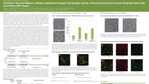 科学海报BrainPhys™ Neuronal Medium: A Medium Optimized to Support the Synaptic Activity of Neurons Derived from Human Pluripotent Stem Cells and Primary CNS Tissues

 沪公网安备31010102008431号