
 EasySep™小鼠TIL(CD45)正选试剂盒


搜索结果: 'methocult media formulations for human hematopoietic cells serum containing'
-

产品类型:
产品号#:
05850
05857
05870
05875
85850
85857
85870
85875
产品名:
mTeSR™1
mTeSR™1
-
产品类型:
产品号#:
05850
05857
05870
05875
85850
85857
85870
85875
产品名:
mTeSR™1
mTeSR™1
-
产品类型:
产品号#:
05850
05857
05870
05875
85850
85857
85870
85875
产品名:
mTeSR™1
mTeSR™1
-
产品类型:
产品号#:
05850
05857
05870
05875
85850
85857
85870
85875
产品名:
mTeSR™1
mTeSR™1
-
产品类型:
产品号#:
72052
72054
100-1042
产品名:
CHIR99021
CHIR99021
CHIR99021
-
产品类型:
产品号#:
05850
05857
05870
05875
07920
85850
85857
85870
85875
07922
产品名:
ACCUTASE™
mTeSR™1
mTeSR™1
ACCUTASE™
-
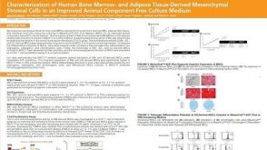 科学海报Characterization of Human Bone Marrow- and Adipose Tissue-Derived Mesenchymal Stromal Cells in an Improved Animal Component-Free Culture Medium
产品类型:
Conference:
ISSCR 2019
产品号#:
产品名:
发布日期: 11/12/2019 -
产品类型:
产品号#:
05790
产品名:
BrainPhys™神经元培养基
-
产品类型:
产品号#:
17954
20144
17954RF
100-0971
产品名:
EasySep™人B细胞分选试剂盒
EasySep™缓冲液
RoboSep™ 人B细胞分选试剂盒
EasySep™人B细胞分离试剂盒
-
产品类型:
产品号#:
85415
85420
产品名:
SepMate™-15 (IVD)
SepMate™-15 (IVD)
-
产品类型:
产品号#:
18000
产品名:
EasySep™磁极
-
产品类型:
产品号#:
04230
05850
05857
05870
05875
07923
60062
60062AD
60062AD.1
60062BT
60062FI
60062FI.1
60062PE
60062PE.1
85850
85857
85870
85875
05270
05275
产品名:
MethoCult™ H4230
Dispase (1 U/mL)
抗人SSEA-4抗体,克隆号MC-813-70,生物素
抗人SSEA-4抗体,克隆号MC-813-70,FITC
抗人SSEA-4抗体, 克隆号MC-813-70,FITC
抗人SSEA-4抗体,克隆号MC-813-70,PE
抗人SSEA-4抗体,克隆号MC-813-70,PE
mTeSR™1
mTeSR™1
STEMdiff™ APEL™2 培养基
STEMdiff™ APEL™2 培养基

 沪公网安备31010102008431号