Hicar MD et al. (JUL 2010)
Journal of acquired immune deficiency syndromes (1999) 54 3 223--35
Pseudovirion particles bearing native HIV envelope trimers facilitate a novel method for generating human neutralizing monoclonal antibodies against HIV.
Monomeric HIV envelope vaccines fail to elicit broadly neutralizing antibodies or to protect against infection. Neutralizing antibodies against HIV bind to native functionally active Env trimers on the virion surface. Gag-Env pseudovirions recapitulate the native trimer and could serve as an effective epitope presentation platform for study of the neutralizing antibody response in HIV-infected individuals. To address if pseudovirions can recapitulate native HIV virion epitope structures,we carefully characterized these particles,concentrating on the antigenic structure of the coreceptor binding site. By blue native gel shift assays,Gag-Env pseudovirions were shown to contain native trimers that were competent for binding to neutralizing monoclonal antibodies. In enzyme-linked immunosorbent assay,pseudovirions exhibited increased binding of known CD4-induced antibodies after addition of CD4. Using flow cytometric analysis,fluorescently labeled pseudovirions specifically identified a subset of antigen-specific B cells in HIV-infected subjects. Interestingly,the sequence of one of these novel human antibodies,identified during cloning of single HIV-specific B cells and designated 2C6,exhibited homology to mAb 47e,a known anti-CD4-induced coreceptor binding site antibody. The secreted monoclonal antibody 2C6 did not bind monomeric gp120,but specifically bound envelope on pseudovirions. A recombinant form of the antibody 2C6 acted as a CD4-induced epitope-specific antibody in neutralization assays,yet did not bind monomeric gp120. These findings imply specificity against a quaternary epitope presented on the pseudovirion envelope spike. These data demonstrate that Gag-Env pseudovirions recapitulate CD4 and coreceptor binding pocket antigenic structures and can facilitate identification of B-cell clones that secrete neutralizing antibodies.
View Publication
产品类型:
产品号#:
18054
18054RF
产品名:
Chun T-W et al. (NOV 2010)
AIDS (London,England) 24 18 2803--8
Rebound of plasma viremia following cessation of antiretroviral therapy despite profoundly low levels of HIV reservoir: implications for eradication.
OBJECTIVES: Sustained suppression of plasma viremia in HIV-infected individuals is attainable with antiretroviral therapy (ART); however,eradication of virus that would allow discontinuation of ART has been hampered by the persistence of HIV reservoirs. It is of great interest to identify individuals who had received ART for prolonged periods of time with extremely low or undetectable HIV reservoirs and monitor plasma viremia following discontinuation of therapy. METHODS: We measured the size of HIV reservoirs in CD4(+) T cells of individuals on long-term ART and monitored plasma viremia following cessation of ART in one individual with an exceptionally low viral burden after a decade of therapy. RESULTS: We demonstrated undetectable levels of HIV DNA in the blood of eight of 45 infected individuals on long-term ART. Among those eight individuals,the frequency of cells carrying infectious virus was significantly lower in those who initiated ART during the early versus the chronic phase of infection. One individual with undetectable HIV DNA in both blood and tissue and a profoundly low level of infectious virus experienced plasma viral rebound 50 days following discontinuation of ART. CONCLUSIONS: Our data suggest that a significant reduction in the size of viral reservoirs may be achievable in selected individuals who initiate standard ART early in infection. However,given re-emergence of plasma viremia in an individual with an extraordinarily low viral burden,therapeutic strategies aimed at specifically targeting these extremely rare HIV-infected cells with novel interventions may be necessary in order to achieve eradication of virus.
View Publication
Wilson KD et al. (JUN 2009)
Stem cells and development 18 5 749--58
MicroRNA profiling of human-induced pluripotent stem cells.
MicroRNAs (miRNAs) are a newly discovered endogenous class of small noncoding RNAs that play important posttranscriptional regulatory roles by targeting mRNAs for cleavage or translational repression. Accumulating evidence now supports the importance of miRNAs for human embryonic stem cell (hESC) self-renewal,pluripotency,and differentiation. However,with respect to induced pluripotent stem cells (iPSC),in which embryonic-like cells are reprogrammed from adult cells using defined factors,the role of miRNAs during reprogramming has not been well-characterized. Determining the miRNAs that are associated with reprogramming should yield significant insight into the specific miRNA expression patterns that are required for pluripotency. To address this lack of knowledge,we use miRNA microarrays to compare the microRNA-omes" of human iPSCs�
View Publication
产品类型:
产品号#:
05850
05857
05870
05875
85850
85857
85870
85875
产品名:
mTeSR™1
mTeSR™1
Lufino MMP et al. (JAN 2011)
Methods in molecular biology (Clifton,N.J.) 767 369--87
Episomal transgene expression in pluripotent stem cells.
Herpes simplex type 1 (HSV-1) amplicon vectors possess a number of features that make them excellent vectors for the delivery of transgenes into stem cells. HSV-1 amplicon vectors are capable of efficiently transducing both dividing and nondividing cells and since the virus is quite large,152 kb,it is of sufficient size to allow for incorporation of entire genomic DNA loci with native promoters. HSV-1 amplicon vectors can also be used to incorporate and deliver to cells a variety of sequences that allow extrachromosomal retention. These elements offer advantages over integrating vectors as they avoid transgene silencing and insertional mutagenesis. The construction of amplicon vectors carrying extrachromosomal retention elements,their packaging into HSV-1 viral particles,and the use of HSV-1 amplicons for stem cell transduction will be described.
View Publication
产品类型:
产品号#:
05850
05857
05870
05875
85850
85857
85870
85875
产品名:
mTeSR™1
mTeSR™1
Ware CB et al. (MAR 2014)
Proceedings of the National Academy of Sciences of the United States of America 111 12 4484--9
Derivation of naive human embryonic stem cells.
The naïve pluripotent state has been shown in mice to lead to broad and more robust developmental potential relative to primed mouse epiblast cells. The human naïve ES cell state has eluded derivation without the use of transgenes,and forced expression of OCT4,KLF4,and KLF2 allows maintenance of human cells in a naïve state [Hanna J,et al. (2010) Proc Natl Acad Sci USA 107(20):9222-9227]. We describe two routes to generate nontransgenic naïve human ES cells (hESCs). The first is by reverse toggling of preexisting primed hESC lines by preculture in the histone deacetylase inhibitors butyrate and suberoylanilide hydroxamic acid,followed by culture in MEK/ERK and GSK3 inhibitors (2i) with FGF2. The second route is by direct derivation from a human embryo in 2i with FGF2. We show that human naïve cells meet mouse criteria for the naïve state by growth characteristics,antibody labeling profile,gene expression,X-inactivation profile,mitochondrial morphology,microRNA profile and development in the context of teratomas. hESCs can exist in a naïve state without the need for transgenes. Direct derivation is an elusive,but attainable,process,leading to cells at the earliest stage of in vitro pluripotency described for humans. Reverse toggling of primed cells to naïve is efficient and reproducible.
View Publication
产品类型:
产品号#:
05860
05880
产品名:
Szkolnicka D et al. ( 2014)
Current protocols in stem cell biology 30 1G.5.1--------12
Deriving functional hepatocytes from pluripotent stem cells.
Despite major progress in the management of human liver disease,the only cure for a critically failing organ is liver transplantation. While a highly successful approach,the use of cadaveric organs as a routine treatment option is severely limited by organ availability. Therefore,the use of cell-based therapies has been explored to provide support for the failing liver. In addition to developing new treatments,there is also an imperative to develop better human models 'in a dish'. Such approaches will undoubtedly lead to a better understanding of the disease process,offering new treatment or preventative strategies. With both approaches in mind,we have developed robust hepatocyte differentiation methodologies for use with pluripotent stem cells. Importantly,our procedure is highly efficient (∼ 90%) and delivers active,drug-inducible,and predictive human hepatocyte populations.
View Publication
产品类型:
产品号#:
05850
05857
05870
05875
07174
85850
85857
85870
85875
100-0485
100-1077
产品名:
mTeSR™1
mTeSR™1
温和细胞解离试剂
ReLeSR™
J. M. Crook and E. Tomaskovic-Crook ( 2017)
Methods in molecular biology (Clifton,N.J.) 1590 199--206
Culturing and Cryobanking Human Neural Stem Cells.
The discovery and study of human neural stem cells has advanced our understanding of human neurogenesis,and the development of novel therapeutics based on neural cell replacement. Here,we describe methods to culture and cryopreserve human neural stem cells (hNSCs) for expansion and banking. Importantly,the protocols ensure that the multipotency of hNSCs is preserved to enable differentiation to neurons and supporting neuroglia.
View Publication
Steward CG et al. (FEB 2005)
Biology of blood and marrow transplantation : journal of the American Society for Blood and Marrow Transplantation 11 2 115--21
High peripheral blood progenitor cell counts enable autologous backup before stem cell transplantation for malignant infantile osteopetrosis.
Autosomal recessive osteopetrosis (OP) is a rare,lethal disorder in which osteoclasts are absent or nonfunctional,resulting in a bone marrow cavity insufficient to support hematopoiesis. Because osteoclasts are derived from hematopoietic precursors,allogeneic hematopoietic cell transplantation can cure the bony manifestations of the disorder. However,high rates of graft failure have been observed in this population. It is not possible to harvest bone marrow from these patients for reinfusion should graft failure be observed. We report that 8 of 10 patients with OP had high numbers of circulating CD34(+) cells (3% +/- 0.9%). This increased proportion of peripheral CD34(+) cells made it possible to harvest 2 x 10(6) CD34(+) cells per kilogram with a total volume of blood ranging from 8.3 to 83.7 mL (1.3-11.6 mL/kg). In addition,colony-forming assays documented significantly more colony-forming unit-granulocyte-macrophage and burst-forming unit-erythroid in the blood of osteopetrotic patients compared with controls; the numbers of colony-forming units approximated those found in control marrow. We conclude that OP patients with high levels of circulating CD34(+) are candidates for peripheral blood autologous harvest by limited exchange transfusion. These cells are then available for reinfusion should graft failure be observed in patients for whom retransplantation is impractical.
View Publication
产品类型:
产品号#:
05401
05402
05411
产品名:
MesenCult™ MSC 基础培养基(人)
MesenCult™ MSC刺激添加物(人)
MesenCult™ 增殖试剂盒(人)
Schwartz C et al. (JUN 2015)
Blood 125 25 3896--904
Eosinophil-specific deletion of IκBα in mice reveals a critical role of NF-κB-induced Bcl-xL for inhibition of apoptosis.
Eosinophils are associated with type 2 immune responses to allergens and helminths. They release various proinflammatory mediators and toxic proteins on activation and are therefore considered proinflammatory effector cells. Eosinophilia is promoted by the cytokines interleukin (IL)-3,IL-5,and granulocyte macrophage-colony-stimulating factor (GM-CSF) and can result from enhanced de novo production or reduced apoptosis. In this study,we show that only IL-5 induces differentiation of eosinophils from bone marrow precursors,whereas IL-5,GM-CSF,and to a lesser extent IL-3 promote survival of mature eosinophils. The receptors for these cytokines use the common β chain,which serves as the main signaling unit linked to signal transducer and activator of transcription 5,p38 mitogen-activated protein kinase,and nuclear factor (NF)-κB pathways. Inhibition of NF-κB induced apoptosis of in vitro cultured eosinophils. Selective deletion of IκBα in vivo resulted in enhanced expression of Bcl-xL and reduced apoptosis during helminth infection. Retroviral overexpression of Bcl-xL promoted survival,whereas pharmacologic inhibition of Bcl-xL in murine or human eosinophils induced rapid apoptosis. These results suggest that therapeutic strategies targeting Bcl-xL in eosinophils could improve health conditions in allergic inflammatory diseases.
View Publication

 EasySep™小鼠TIL(CD45)正选试剂盒


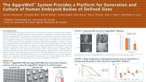 科学海报The Aggrewell™ System Provides a Platform for Generation and Culture of Human Embryoid Bodies of Defined Sizes
 沪公网安备31010102008431号