Deep sequencing reveals low incidence of endogenous LINE-1 retrotransposition in human induced pluripotent stem cells
Long interspersed element-1 (LINE-1 or L1) retrotransposition induces insertional mutations that can result in diseases. It was recently shown that the copy number of L1 and other retroelements is stable in induced pluripotent stem cells (iPSCs). However,by using an engineered reporter construct over-expressing L1,another study suggests that reprogramming activates L1 mobility in iPSCs. Given the potential of human iPSCs in therapeutic applications,it is important to clarify whether these cells harbor somatic insertions resulting from endogenous L1 retrotransposition. Here,we verified L1 expression during and after reprogramming as well as potential somatic insertions driven by the most active human endogenous L1 subfamily (L1Hs). Our results indicate that L1 over-expression is initiated during the reprogramming process and is subsequently sustained in isolated clones. To detect potential somatic insertions in iPSCs caused by L1Hs retotransposition,we used a novel sequencing strategy. As opposed to conventional sequencing direction,we sequenced from the 3' end of L1Hs to the genomic DNA,thus enabling the direct detection of the polyA tail signature of retrotransposition for verification of true insertions. Deep coverage sequencing thus allowed us to detect seven potential somatic insertions with low read counts from two iPSC clones. Negative PCR amplification in parental cells,presence of a polyA tail and absence from seven L1 germline insertion databases highly suggested true somatic insertions in iPSCs. Furthermore,these insertions could not be detected in iPSCs by PCR,likely due to low abundance. We conclude that L1Hs retrotransposes at low levels in iPSCs and therefore warrants careful analyses for genotoxic effects.
View Publication
产品类型:
产品号#:
05850
05857
05870
05875
85850
85857
85870
85875
产品名:
mTeSR™1
mTeSR™1
Darabi R and Perlingeiro RCR ( 2016)
1357 423--439
Derivation of Skeletal Myogenic Precursors from Human Pluripotent Stem Cells Using Conditional Expression of PAX7.
Cell-based therapies are considered as one of the most promising approaches for the treatment of degenerating pathologies including muscle disorders and dystrophies. Advances in the approach of reprogramming somatic cells into induced pluripotent stem (iPS) cells allow for the possibility of using the patient's own pluripotent cells to generate specific tissues for autologous transplantation. In addition,patient-specific tissue derivatives have been shown to represent valuable material for disease modeling and drug discovery. Nevertheless,directed differentiation of pluripotent stem cells into a specific lineage is not a trivial task especially in the case of skeletal myogenesis,which is generally poorly recapitulated during the in vitro differentiation of pluripotent stem cells.Here,we describe a practical and efficient method for the derivation of skeletal myogenic precursors from differentiating human pluripotent stem cells using controlled expression of PAX7. Flow cytometry (FACS) purified myogenic precursors can be expanded exponentially and differentiated in vitro into myotubes,enabling researchers to use these cells for disease modeling as well as therapeutic purposes.
View Publication
产品类型:
产品号#:
05850
05857
05870
05875
85850
85857
85870
85875
产品名:
mTeSR™1
mTeSR™1
Wrighton PJ et al. (DEC 2014)
Proceedings of the National Academy of Sciences of the United States of America 111 51 18126--18131
Signals from the surface modulate differentiation of human pluripotent stem cells through glycosaminoglycans and integrins.
The fate decisions of human pluripotent stem (hPS) cells are governed by soluble and insoluble signals from the microenvironment. Many hPS cell differentiation protocols use Matrigel,a complex and undefined substrate that engages multiple adhesion and signaling receptors. Using defined surfaces programmed to engage specific cell-surface ligands (i.e.,glycosaminoglycans and integrins),the contribution of specific matrix signals can be dissected. For ectoderm and motor neuron differentiation,peptide-modified surfaces that can engage both glycosaminoglycans and integrins are effective. In contrast,surfaces that interact selectively with glycosaminoglycans are superior to Matrigel in promoting hPS cell differentiation to definitive endoderm and mesoderm. The modular surfaces were used to elucidate the signaling pathways underlying these differences. Matrigel promotes integrin signaling,which in turn inhibits mesendoderm differentiation. The data indicate that integrin-activating surfaces stimulate Akt signaling via integrin-linked kinase (ILK),which is antagonistic to endoderm differentiation. The ability to attribute cellular responses to specific interactions between the cell and the substrate offers new opportunities for revealing and controlling the pathways governing cell fate.
View Publication
产品类型:
产品号#:
05850
05857
05870
05875
85850
85857
85870
85875
产品名:
mTeSR™1
mTeSR™1
Fuerstenau-Sharp M et al. (MAY 2015)
PloS one 10 5 e0126596
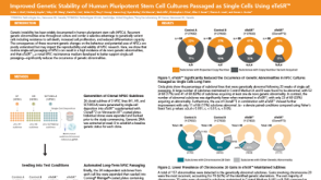
Generation of highly purified human cardiomyocytes from peripheral blood mononuclear cell-derived induced pluripotent stem cells.
Induced pluripotent stem (iPS) cells have an enormous potential for physiological studies. A novel protocol was developed combining the derivation of iPS from peripheral blood with an optimized directed differentiation to cardiomyocytes and a subsequent metabolic selection. The human iPS cells were retrovirally dedifferentiated from activated T cells. The subsequent optimized directed differentiation protocol yielded 30-45% cardiomyocytes at day 16 of differentiation. The derived cardiomyocytes expressed appropriate structural markers like cardiac troponin T,$\$-actinin and myosin light chain 2 (MLC2V). In a subsequent metabolic selection with lactate,the cardiomyocytes content could be increased to more than 90%. Loss of cardiomyocytes during metabolic selection were less than 50%,whereas alternative surface antibody-based selection procedures resulted in loss of up to 80% of cardiomyocytes. Electrophysiological characterization confirmed the typical cardiac features and the presence of ventricular,atrial and nodal-like action potentials within the derived cardiomyocyte population. Our combined and optimized protocol is highly robust and applicable for scalable cardiac differentiation. It provides a simple and cost-efficient method without expensive equipment for generating large numbers of highly purified,functional cardiomyocytes. It will further enhance the applicability of iPS cell-derived cardiomyocytes for disease modeling,drug discovery,and regenerative medicine.
View Publication
产品类型:
产品号#:
05850
05857
05870
05875
85850
85857
85870
85875
产品名:
mTeSR™1
mTeSR™1
Miki T et al. (MAY 2011)
Tissue engineering. Part C,Methods 17 5 557--68
Hepatic differentiation of human embryonic stem cells is promoted by three-dimensional dynamic perfusion culture conditions.
The developmental potential of human embryonic stem cells (hESCs) holds great promise to provide a source of human hepatocytes for use in drug discovery,toxicology,hepatitis research,and extracorporeal bioartificial liver support. There are,however,limitations to induce fully functional hepatocytes on conventional two-dimensional (2D) static culture. It had been shown that dynamic three-dimensional (3D) perfusion culture is superior to induce maturation in fetal hepatocytes and prolong hepatic functions of primary adult hepatocytes. We investigated the potential of using a four-compartment 3D perfusion culture to induce hepatic differentiation in hESC. Undifferentiated hESC were inoculated into hollow fiber-based 3D perfusion bioreactors with integral oxygenation. Hepatic differentiation was induced with a multistep growth factor cocktail protocol. Parallel controls were operated under equal perfusion conditions without the growth factor supplementations to allow for spontaneous differentiation,as well as in conventional 2D static conditions using growth factors. Metabolism,hepatocyte-specific gene expression,protein expression,and hepatic function were evaluated after 20 days. Significantly upregulated hepatic gene expression was observed in the hepatic differentiation 3D culture group. Ammonia metabolism activity and albumin production was observed in the 3D directed differentiation culture. Drug-induced cytochrome P450 gene expression was increased with rifampicin induction. Using flow cytometry analysis the mature hepatocyte marker asialoglycoprotein receptor was found on up to 30% of the cells in the 3D system with directed hepatic differentiation. Histological and immunohistochemical analysis revealed structural formation of hepatic and biliary marker-positive cells. In contrast to 2D culture,the 3D perfusion culture induced more functional maturation in hESC-derived hepatic cells. 3D perfusion bioreactor technologies may be useful for further studies on generating hESC-derived hepatic cells.
View Publication
产品类型:
产品号#:
05850
05857
05870
05875
85850
85857
85870
85875
产品名:
mTeSR™1
mTeSR™1
Kallas A et al. (APR 2011)
PLoS ONE 6 4 e19114
Nocodazole treatment decreases expression of pluripotency markers nanog and Oct4 in human embryonic stem cells
Nocodazole is a known destabiliser of microtubule dynamics and arrests cell-cycle at the G2/M phase. In the context of the human embryonic stem cell (hESC) it is important to understand how this arrest influences the pluripotency of cells. Here we report for the first time the changes in the expression of transcription markers Nanog and Oct4 as well as SSEA-3 and SSEA-4 in human embryonic cells after their treatment with nocodazole. Multivariate permeabilised-cell flow cytometry was applied for characterising the expression of Nanog and Oct4 during different cell cycle phases. Among untreated hESC we detected Nanog-expressing cells,which also expressed Oct4,SSEA-3 and SSEA-4. We also found another population expressing SSEA-4,but without Nanog,Oct4 and SSEA-3 expression. Nocodazole treatment resulted in a decrease of cell population positive for all four markers Nanog,Oct4,SSEA-3,SSEA-4. Nocodazole-mediated cell-cycle arrest was accompanied by higher rate of apoptosis and upregulation of p53. Twenty-four hours after the release from nocodazole block,the cell cycle of hESC normalised,but no increase in the expression of transcription markers Nanog and Oct4 was detected. In addition,the presence of ROCK-2 inhibitor Y-27632 in the medium had no effect on increasing the expression of pluripotency markers Nanog and Oct4 or decreasing apoptosis or the level of p53. The expression of SSEA-3 and SSEA-4 increased in Nanog-positive cells after wash-out of nocodazole in the presence and in the absence of Y-27632. Our data show that in hESC nocodazole reversible blocks cell cycle,which is accompanied by irreversible loss of expression of pluripotency markers Nanog and Oct4.
View Publication
产品类型:
产品号#:
05850
05857
05870
05875
85850
85857
85870
85875
产品名:
mTeSR™1
mTeSR™1
Cox JL et al. (AUG 2011)
Journal of Cell Science 124 Pt 15 2654--65
Banf1 is required to maintain the self-renewal of both mouse and human embryonic stem cells.
Self-renewal is a complex biological process necessary for maintaining the pluripotency of embryonic stem cells (ESCs). Recent studies have used global proteomic techniques to identify proteins that associate with the master regulators Oct4,Nanog and Sox2 in ESCs or in ESCs during the early stages of differentiation. Through an unbiased proteomic screen,Banf1 was identified as a Sox2-associated protein. Banf1 has been shown to be essential for worm and fly development but,until now,its role in mammalian development and ESCs has not been explored. In this study,we examined the effect of knocking down Banf1 on ESCs. We demonstrate that the knockdown of Banf1 promotes the differentiation of mouse ESCs and decreases the survival of both mouse and human ESCs. For mouse ESCs,we demonstrate that knocking down Banf1 promotes their differentiation into cells that exhibit markers primarily associated with mesoderm and trophectoderm. Interestingly,knockdown of Banf1 disrupts the survival of human ESCs without significantly reducing the expression levels of the master regulators Sox2,Oct4 and Nanog or inducing the expression of markers of differentiation. Furthermore,we determined that the knockdown of Banf1 alters the cell cycle distribution of both human and mouse ESCs by causing an uncharacteristic increase in the proportion of cells in the G2-M phase of the cell cycle.
View Publication
产品类型:
产品号#:
05850
05857
05870
05875
85850
85857
85870
85875
产品名:
mTeSR™1
mTeSR™1
Tan Y et al. (JAN 2012)
Journal of biomechanics 45 1 123--8
Probing the mechanobiological properties of human embryonic stem cells in cardiac differentiation by optical tweezers.
Human embryonic stem cells (hESC) and hESC-derived cardiomyocytes (hESC-CM) hold great promise for the treatment of cardiovascular diseases. However the mechanobiological properties of hESC and hESC-CM remains elusive. In this paper,we examined the dynamic and static micromechanical properties of hESC and hESC-CM,by manipulating via optical tweezers at the single-cell level. Theoretical approaches were developed to model the dynamic and static mechanical responses of cells during optical stretching. Our experiments showed that the mechanical stiffness of differentiated hESC-CM increased after cardiac differentiation. Such stiffening could associate with increasingly organized myofibrillar assembly that underlines the functional characteristics of hESC-CM. In summary,our findings lay the ground work for using hESC-CMs as models to study mechanical and contractile defects in heart diseases.
View Publication
产品类型:
产品号#:
05850
05857
05870
05875
85850
85857
85870
85875
产品名:
mTeSR™1
mTeSR™1
Moschidou D et al. (OCT 2012)
Molecular therapy : the journal of the American Society of Gene Therapy 20 10 1953--67
Valproic acid confers functional pluripotency to human amniotic fluid stem cells in a transgene-free approach.
Induced pluripotent stem cells (iPSCs) with potential for therapeutic applications can be derived from somatic cells via ectopic expression of a set of limited and defined transcription factors. However,due to risks of random integration of the reprogramming transgenes into the host genome,the low efficiency of the process,and the potential risk of virally induced tumorigenicity,alternative methods have been developed to generate pluripotent cells using nonintegrating systems,albeit with limited success. Here,we show that c-KIT+ human first-trimester amniotic fluid stem cells (AFSCs) can be fully reprogrammed to pluripotency without ectopic factors,by culture on Matrigel in human embryonic stem cell (hESC) medium supplemented with the histone deacetylase inhibitor (HDACi) valproic acid (VPA). The cells share 82% transcriptome identity with hESCs and are capable of forming embryoid bodies (EBs) in vitro and teratomas in vivo. After long-term expansion,they maintain genetic stability,protein level expression of key pluripotency factors,high cell-division kinetics,telomerase activity,repression of X-inactivation,and capacity to differentiate into lineages of the three germ layers,such as definitive endoderm,hepatocytes,bone,fat,cartilage,neurons,and oligodendrocytes. We conclude that AFSC can be utilized for cell banking of patient-specific pluripotent cells for potential applications in allogeneic cellular replacement therapies,pharmaceutical screening,and disease modeling.
View Publication
产品类型:
产品号#:
05850
05857
05870
05875
85850
85857
85870
85875
产品名:
mTeSR™1
mTeSR™1
Deng Y et al. (NOV 2013)
Acta Biomaterialia 9 11 8840--8850
Long-term self-renewal of human pluripotent stem cells on peptide-decorated poly(OEGMA-co-HEMA) brushes under fully defined conditions
Realization of the full potential of human induced pluripotent stem cells (hiPSC) in clinical applications requires the development of well-defined culture conditions for their long-term growth and directed differentiation. This paper describes a novel fully defined synthetic peptide-decorated substrate that supports self-renewal of hiPSC in commercially available xeno-free,chemically defined medium. The Au surface was deposited by a poly(OEGMA-co-HEMA) film,using the surface-initiated polymerization method (SIP) with the further step of carboxylation. The hiPSC generated from umbilical cord mesenchymal cells were successfully cultured for 10 passages on the peptide-tethered poly(OEGMA-co-HEMA) brushes for the first time. Cells maintained their characteristic morphology,proliferation and expressed high levels of markers of pluripotency,similar to the cells cultured on Matrigel???. Moreover,the cell adhesion could be tuned by the pattern and peptide concentration on the substrate. This well-defined,xeno-free and safe substrate,which supports long-term proliferation and self-renewal of hiPSC,will not only help to accelerate the translational perspectives of hiPSC,but also provide a platform to elucidate the underlying molecular mechanisms that regulate stem cell proliferation and differentiation via SIP technology. ?? 2013 Acta Materialia Inc. Published by Elsevier Ltd. All rights reserved.
View Publication
产品类型:
产品号#:
05850
05857
05870
05875
85850
85857
85870
85875
产品名:
mTeSR™1
mTeSR™1
S. D. Maldonado et al. (aug 2022)
Journal of immunology (Baltimore,Md. : 1950) 209 4 675--683
Human Plasmacytoid Dendritic Cells Express C-Type Lectin Receptors and Attach and Respond to Aspergillus fumigatus.
Plasmacytoid dendritic cells (pDCs) have been implicated as having a role in antifungal immunity,but mechanisms of their interaction with fungi and the resulting cellular responses are not well understood. In this study,we identify the direct and indirect biological response of human pDCs to the fungal pathogen Aspergillus fumigatus and characterize the expression and regulation of antifungal receptors on the pDC surface. Results indicate pDCs do not phagocytose Aspergillus conidia,but instead bind hyphal surfaces and undergo activation and maturation via the upregulation of costimulatory and maturation markers. Measuring the expression of C-type lectin receptors dectin-1,dectin-2,dectin-3,and mannose receptor on human pDCs revealed intermediate expression of each receptor compared with monocytes. The specific dectin-1 agonist curdlan induced pDC activation and maturation in a cell-intrinsic and cell-extrinsic manner. The indirect activation of pDCs by curdlan was much stronger than direct stimulation and was mediated through cytokine production by other PBMCs. Overall,our data indicate pDCs express various C-type lectin receptors,recognize and respond to Aspergillus hyphal Ag,and serve as immune enhancers or modulators in the overarching fungal immune response.
View Publication

 EasySep™小鼠TIL(CD45)正选试剂盒


 沪公网安备31010102008431号