Lindgren AG et al. (JAN 2015)
Cell regeneration (London,England) 4 1 1
ETV2 expression increases the efficiency of primitive endothelial cell derivation from human embryonic stem cells.
BACKGROUND: Endothelial cells line the luminal surface of blood vessels and form a barrier between the blood and other tissues of the body. Ets variant 2 (ETV2) is transiently expressed in both zebrafish and mice and is necessary and sufficient for vascular endothelial cell specification. Overexpression of this gene in early zebrafish and mouse embryos results in ectopic appearance of endothelial cells. Ectopic expression of ETV2 in later development results in only a subset of cells responding to the signal.backslashnbackslashnFINDINGS: We have examined the expression pattern of ETV2 in differentiating human embryonic stem cells (ESCs) to determine when the peak of ETV2 expression occurs. We show that overexpression of ETV2 in differentiating human ESC is able to increase the number of endothelial cells generated when administered during or after the endogenous peak of gene expression.backslashnbackslashnCONCLUSIONS: Addition of exogenous ETV2 to human ESCs significantly increased the number of cells expressing angioblast genes without arterial or venous specification. This may be a viable solution to generate in vitro endothelial cells for use in research and in the clinic.
View Publication
产品类型:
产品号#:
05850
05857
05870
05875
85850
85857
85870
85875
产品名:
mTeSR™1
mTeSR™1
Hu W et al. (AUG 2015)
Cell stem cell 17 2 204--12
Direct Conversion of Normal and Alzheimer's Disease Human Fibroblasts into Neuronal Cells by Small Molecules.
Neuronal conversion from human fibroblasts can be induced by lineage-specific transcription factors; however,the introduction of ectopic genes limits the therapeutic applications of such induced neurons (iNs). Here,we report that human fibroblasts can be directly converted into neuronal cells by a chemical cocktail of seven small molecules,bypassing a neural progenitor stage. These human chemical-induced neuronal cells (hciNs) resembled hiPSC-derived neurons and human iNs (hiNs) with respect to morphology,gene expression profiles,and electrophysiological properties. This approach was further applied to generate hciNs from familial Alzheimer's disease patients. Taken together,our transgene-free and chemical-only approach for direct reprogramming of human fibroblasts into neurons provides an alternative strategy for modeling neurological diseases and for regenerative medicine.
View Publication
产品类型:
产品号#:
72052
72054
72112
72114
72292
72302
72304
72307
72308
72392
72394
72462
72642
73792
73794
100-1042
100-0249
100-1044
产品名:
CHIR99021
CHIR99021
Forskolin
Forskolin
丙戊酸(钠盐)
Y-27632(二盐酸盐)
Y-27632(二盐酸盐)
Y-27632(二盐酸盐)
Y-27632(二盐酸盐)
RepSox(盐酸盐)
RepSox(盐酸盐)
Gö6983
SP600125
RepSox
RepSox
CHIR99021
Forskolin
Y-27632(二盐酸盐)
Vauchez K et al. (NOV 2009)
Molecular therapy : the journal of the American Society of Gene Therapy 17 11 1948--58
Aldehyde dehydrogenase activity identifies a population of human skeletal muscle cells with high myogenic capacities.
Aldehyde dehydrogenase 1A1 (ALDH) activity is one hallmark of human bone marrow (BM),umbilical cord blood (UCB),and peripheral blood (PB) primitive progenitors presenting high reconstitution capacities in vivo. In this study,we have identified ALDH(+) cells within human skeletal muscles,and have analyzed their phenotypical and functional characteristics. Immunohistofluorescence analysis of human muscle tissue sections revealed rare endomysial cells. Flow cytometry analysis using the fluorescent substrate of ALDH,Aldefluor,identified brightly stained (ALDH(br)) cells with low side scatter (SSC(lo)),in enzymatically dissociated muscle biopsies,thereafter abbreviated as SMALD(+) (for skeletal muscle ALDH(+)) cells. Phenotypical analysis discriminated two sub-populations according to CD34 expression: SMALD(+)/CD34(-) and SMALD(+)/CD34(+) cells. These sub-populations did not initially express endothelial (CD31),hematopoietic (CD45),and myogenic (CD56) markers. Upon sorting,however,whereas SMALD(+)/CD34(+) cells developed in vitro as a heterogeneous population of CD56(-) cells able to differentiate in adipoblasts,the SMALD(+)/CD34(-) fraction developed in vitro as a highly enriched population of CD56(+) myoblasts able to form myotubes. Moreover,only the SMALD(+)/CD34(-) population maintained a strong myogenic potential in vivo upon intramuscular transplantation. Our results suggest that ALDH activity is a novel marker for a population of new human skeletal muscle progenitors presenting a potential for cell biology and cell therapy.
View Publication
Sharma A et al. (JUN 2013)
Journal of Biological Chemistry 288 25 18439--18447
The role of SIRT6 protein in aging and reprogramming of human induced pluripotent stem cells
Aging is known to be the single most important risk factor for multiple diseases. Sirtuin 6,or SIRT6,has recently been identified as a critical regulator of transcription,genome stability,telomere integrity,DNA repair,and metabolic homeostasis. A knockout mouse model of SIRT6 has displayed dramatic phenotypes of accelerated aging. In keeping with its role in aging,we demonstrated that human dermal fibroblasts (HDFs) from older human subjects were more resistant to reprogramming by classic Yamanaka factors than those from younger human subjects,but the addition of SIRT6 during reprogramming improved such efficiency in older HDFs substantially. Despite the importance of SIRT6,little is known about the molecular mechanism of its regulation. We show,for the first,time posttranscriptional regulation of SIRT6 by miR-766 and inverse correlation in the expression of this microRNA in HDFs from different age groups. Our results suggest that SIRT6 regulates miR-766 transcription via a feedback regulatory loop,which has implications for the modulation of SIRT6 expression in reprogramming of aging cells.
View Publication
产品类型:
产品号#:
05850
05857
05870
05875
85850
85857
85870
85875
产品名:
mTeSR™1
mTeSR™1
Bao X et al. ( 2016)
Methods in molecular biology (Clifton,N.J.) 1481 183--196
Directed Endothelial Progenitor Differentiation from Human Pluripotent Stem Cells Via Wnt Activation Under Defined Conditions.
Efficient derivation of endothelial cells and their progenitors from human pluripotent stem cells (hPSCs) can facilitate studies of human vascular development,disease modeling,drug discovery,and cell-based therapy. Here we provide a detailed protocol for directing hPSCs to functional endothelial cells and their progenitors in a completely defined,growth factor- and serum-free system by temporal modulation of Wnt/$$-catenin signaling via small molecules. We demonstrate a 10-day,two-stage process that recapitulates endothelial cell development,in which hPSCs first differentiate to endothelial progenitors that then generate functional endothelial cells and smooth muscle cells. Methods to characterize endothelial cell identity and function are also described.
View Publication
产品类型:
产品号#:
05850
05857
05870
05875
05940
85850
85857
85870
85875
产品名:
mTeSR™1
mTeSR™1
Chen C et al. (NOV 2016)
JCI insight 1 19 e88632
Humanized neuronal chimeric mouse brain generated by neonatally engrafted human iPSC-derived primitive neural progenitor cells.
The creation of a humanized chimeric mouse nervous system permits the study of human neural development and disease pathogenesis using human cells in vivo. Humanized glial chimeric mice with the brain and spinal cord being colonized by human glial cells have been successfully generated. However,generation of humanized chimeric mouse brains repopulated by human neurons to possess a high degree of chimerism have not been well studied. Here we created humanized neuronal chimeric mouse brains by neonatally engrafting the distinct and highly neurogenic human induced pluripotent stem cell (hiPSC)-derived rosette-type primitive neural progenitors. These neural progenitors predominantly differentiate to neurons,which disperse widely throughout the mouse brain with infiltration of the cerebral cortex and hippocampus at 6 and 13 months after transplantation. Building upon the hiPSC technology,we propose that this potentially unique humanized neuronal chimeric mouse model will provide profound opportunities to define the structure,function,and plasticity of neural networks containing human neurons derived from a broad variety of neurological disorders.
View Publication
(Sep 2024)
International Journal of Molecular Sciences 25 19
Mesenchymal Stem Cells Derived from Human Urine-Derived iPSCs Exhibit Low Immunogenicity and Reduced Immunomodulatory Profile
Human-induced pluripotent stem cell (iPSC)-derived mesenchymal stem cells (iMSCs) represent a promising and renewable cell source for therapeutic applications. A systematic evaluation of the immunological properties and engraftment potential of iMSCs generated from urine-derived iPSCs is lacking,which has impeded their broader application. In this study,we differentiated urine-derived iPSCs into iMSCs and assessed their fundamental MSC characteristics,immunogenicity,immunomodulatory capacity and in vivo engraftment. Compared to umbilical cord-derived MSCs (UCMSCs),iMSCs demonstrated an enhanced proliferative capacity,a higher level of regenerative gene expression,and lower immunogenicity,strengthening resistance to apoptosis induced by allogeneic peripheral blood mononuclear cells (PBMCs) and the NK-92 cell line. In addition,iMSCs exhibited a diminished ability to inhibit T cell proliferation and activation compared with UCMSCs. Transcriptomic analyses further revealed the decreased expression of immune regulatory factors in iMSCs. After transfusion into mouse models,iMSCs engrafted in the lungs,liver,and spleen and exhibited the ability to migrate to tumor tissues. Our results indicated that iMSCs generated from urine-derived iPSCs have a significant replicative capacity,low immunogenicity and unique immunomodulatory properties,and hence offer obvious advantages in immune privilege and allogenic therapeutic application prospects.
View Publication
产品类型:
产品号#:
05240
85850
85857
产品名:
STEMdiff™ 间充质祖细胞试剂盒
mTeSR™1
mTeSR™1
(Apr 2024)
Nature Communications 15
Single-cell analyses reveal transient retinal progenitor cells in the ciliary margin of developing human retina
The emergence of retinal progenitor cells and differentiation to various retinal cell types represent fundamental processes during retinal development. Herein,we provide a comprehensive single cell characterisation of transcriptional and chromatin accessibility changes that underline retinal progenitor cell specification and differentiation over the course of human retinal development up to midgestation. Our lineage trajectory data demonstrate the presence of early retinal progenitors,which transit to late,and further to transient neurogenic progenitors,that give rise to all the retinal neurons. Combining single cell RNA-Seq with spatial transcriptomics of early eye samples,we demonstrate the transient presence of early retinal progenitors in the ciliary margin zone with decreasing occurrence from 8 post-conception week of human development. In retinal progenitor cells,we identified a significant enrichment for transcriptional enhanced associate domain transcription factor binding motifs,which when inhibited led to loss of cycling progenitors and retinal identity in pluripotent stem cell derived organoids. Formation of the retina during development involves the coordinated action of retinal progenitor cells and their differentiated cell types,which is key for producing a functioning eye. Here the authors provide a detailed atlas of human retinal development,combining scRNA-seq and spatial transcriptomics,and identify key genetic factors that mediate retinal progenitor cell proliferation and differentiation.
View Publication
产品类型:
产品号#:
85850
85857
产品名:
mTeSR™1
mTeSR™1
S. L. Calzi et al. (Aug 2025)
Cells 14 17
Targeting Diabetic Retinopathy with Human iPSC-Derived Vascular Reparative Cells in a Type 2 Diabetes Model
Purpose: To investigate the therapeutic potential of inducible pluripotent stem cell (hiPSC)-based vascular repair,we evaluated two vascular reparative cell populations,CD34+ cells derived from hiPSC (hiPSC-CD34+) and endothelial colony forming cells (ECFCs) derived from hiPSC (iPS-ECFCs),alone and in combination,in a type 2 diabetic (db/db) mouse model of DR. Methods: hiPSC-CD34+ cells (1 × 10⁴) or iPSC- ECFCs (1 × 10⁵) alone or in combination (1.1 × 10⁵) were injected into the vitreous of immunosuppressed db/db mice with six months of established diabetes. One month post-injection,mice underwent electroretinography (ERG) and optical coherence tomography (OCT) to evaluate functional and structural retinal recovery with iPSC administration. Immunohistochemistry (IHC) was used to assess recruitment and incorporation of cells into the retinal vasculature. Retinas from the experimental groups were analyzed using Functional Proteomics via Reverse Phase Protein Array (RPPA). Results: Functional assessment via ERG demonstrated significant improvements in retinal response in the diabetic cohorts treated with either hiPSC-derived CD34+ cells or hiPSC-ECFCs. Retinal thickness,assessed by OCT,was restored to near-nondiabetic levels in mice treated with hiPSC-CD34+ cells alone and the combination group,whereas hiPSC-ECFCs alone did not significantly affect retinal thickness. One month following intravitreal injection,hiPSC-CD34+ cells were localized to perivascular regions,whereas hiPSC-ECFCs were observed to integrate directly into the retinal vasculature. RPPA analysis revealed interaction-significant changes,and this was interpreted as a combination-specific,non-additive host responses (m6A,PI3K–AKT–mTOR,glycolysis,endothelial junction pathways). Conclusions: The studies support that injection of hiPSC-CD34+ cells and hiPSC-ECFCs,both individually and in combination,showed benefit; however,iPSC combination-specific effects were identified by measurement of retinal thickness and by RPPA.
View Publication

 EasySep™小鼠TIL(CD45)正选试剂盒


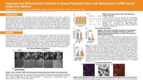 科学海报Expansion and Differentiation Potential of Human Pluripotent Stem Cells Maintained in cGMP Animal Origin-Free Medium

 沪公网安备31010102008431号