A. Plengpanich et al. (Mar 2026)
Mycopathologia 191 2
Development of a Vertical Flow Dot-Immunobinding Assay (Dot-Iba) for Rapid Detection of Neoscytalidium dimidiatum
Neoscytalidium dimidiatum is a non-dermatophyte mold that commonly causes skin and nail infections in tropical regions and often resists conventional antifungal therapies. Because its clinical and laboratory features often resemble dermatophyte infections,diagnosis is frequently delayed and treatment is sometimes inappropriate. We therefore developed a dot-immunobinding assay (Dot-Iba) to detect N. dimidiatum antigens. We generated a highly specific monoclonal antibody,3E6F7 (MAb 3E6F7),for antigen capture,and used goat anti-mouse Ig conjugated with alkaline phosphatase (AP) as the signal generator. The test pad comprised a test hole,a nitrocellulose membrane (NC),and water-absorbent pads in a vertical flow-through format to allow a rapid antigen–antibody reaction. The assembled system detected N. dimidiatum antigens in vitro with high specificity and yielded visible results within 2 h; its detection limit was 0.9 µg without cross-reactivity to dermatophyte or non-dermatophyte fungi. This rapid,specific,and easy-to-use assay shows strong potential as a diagnostic tool,particularly in settings with limited access to fungal culture or advanced molecular diagnostics,where early,accurate identification is crucial.
View Publication
Sun J et al. (APR 2009)
The Journal of biological chemistry 284 17 11039--47
The D816V mutation of c-Kit circumvents a requirement for Src family kinases in c-Kit signal transduction.
The receptor tyrosine kinase c-Kit plays a critical role in hematopoiesis,and gain-of-function mutations of the receptor are frequently seen in several malignancies,including acute myeloid leukemia,gastrointestinal stromal tumors,and testicular carcinoma. The most common mutation of c-Kit in these disorders is a substitution of the aspartic acid residue in position 816 to a valine (D816V),leading to constitutive activation of the receptor. In this study,we aimed to investigate the role of Src family kinases in c-Kit/D816V signaling. Src family kinases are necessary for the phosphorylation of wild-type c-Kit as well as of activation of downstream signaling pathways including receptor ubiquitination and the Ras/Mek/Erk pathway. Our data demonstrate that,unlike wild-type c-Kit,the phosphorylation of c-Kit/D816V is not dependent on Src family kinases. In addition,we found that neither receptor ubiquitination nor Erk activation by c-Kit/D816V required activation of Src family kinases. In vitro kinase assay using synthetic peptides revealed that c-Kit/D816V had an altered substrate specificity resembling Src and Abl tyrosine kinases. We further present evidence that,in contrast to wild-type c-Kit,Src family kinases are dispensable for c-Kit/D816V cell survival,proliferation,and colony formation. Taken together,we demonstrate that the signal transduction pathways mediated by c-Kit/D816V are markedly different from those activated by wild-type c-Kit and that altered substrate specificity of c-Kit circumvents a need for Src family kinases in signaling of growth and survival,thereby contributing to the transforming potential of c-Kit/D816V.
View Publication
Yanai A et al. ( 2016)
Methods in molecular biology (Clifton,N.J.) 1307 357--369
Efficient Production of Photoreceptor Precursor Cells from Human Embryonic Stem Cells.
Transplantation of photoreceptor precursor cells (PPCs) differentiated from human embryonic stem cells (hESCs) is a promising approach to treat common blinding diseases such as age-related macular degeneration and retinitis pigmentosa. However,existing PPC generation methods are inefficient. To enhance differentiation protocols for rapid and high-yield production of PPCs,we focused on optimizing the handling of the cells by including feeder-independent growth of hESCs,using size-controlled embryoid bodies (EBs),and addition of triiodothyronine (T3) and taurine to the differentiation medium,with subsequent removal of undifferentiated cells via negative cell-selection. Our novel protocol produces higher yields of PPCs than previously reported while reducing the time required for differentiation,which will help understand retinal diseases and facilitate large-scale preclinical trials.
View Publication
Lgr5-positive supporting cells generate new hair cells in the postnatal cochlea.
The prevalence of hearing loss after damage to the mammalian cochlea has been thought to be due to a lack of spontaneous regeneration of hair cells,the primary receptor cells for sound. Here,we show that supporting cells,which surround hair cells in the normal cochlear epithelium,differentiate into new hair cells in the neonatal mouse following ototoxic damage. Using lineage tracing,we show that new hair cells,predominantly outer hair cells,arise from Lgr5-expressing inner pillar and third Deiters cells and that new hair cell generation is increased by pharmacological inhibition of Notch. These data suggest that the neonatal mammalian cochlea has some capacity for hair cell regeneration following damage alone and that Lgr5-positive cells act as hair cell progenitors in the cochlea.
View Publication
产品类型:
产品号#:
72792
72794
产品名:
LY411575
LY411575
Li Y et al. (FEB 2016)
Journal of Immunology 196 4 1617--25
Hepatic Stellate Cells Directly Inhibit B Cells via Programmed Death-Ligand 1.
We demonstrated previously that mouse hepatic stellate cells (HSCs) suppress T cells via programmed death-ligand 1 (PD-L1),but it remains unknown whether they exert any effects on B cells,the other component of the adaptive immune system. In this study,we found that mouse HSCs directly inhibited B cells and that PD-L1 was also integrally involved. We found that HSCs inhibited the upregulation of activation markers on activated B cells,as well as the proliferation of activated B cells and their cytokine/Ig production in vitro,and that pharmaceutically or genetically blocking the interaction of PD-L1 with programmed cell death protein 1 impaired the ability of HSCs to inhibit B cells. To test the newly discovered B cell-inhibitory activity of HSCs in vivo,we developed a protocol of intrasplenic artery injection to directly deliver HSCs into the spleen. We found that local delivery of wild-type HSCs into the spleens of mice that had been immunized with 4-hydroxy-3-nitrophenylacetyl-Ficoll,a T cell-independent Ag,significantly suppressed Ag-specific IgM and IgG production in vivo,whereas splenic artery delivery of PD-L1-deficient HSCs failed to do so. In conclusion,in addition to inhibiting T cells,mouse HSCs concurrently inhibit B cells via PD-L1. This direct B cell-inhibitory activity of HSCs should contribute to the mechanism by which HSCs maintain the liver's immune homeostasis.
View Publication
产品类型:
产品号#:
19854
19854RF
产品名:
EasySep™小鼠B细胞分选试剂盒
RoboSep™ 小鼠B细胞分选试剂盒
Shigeharu G. YABE et al. (MAR 2016)
Journal of Diabetes n/a--n/a
Efficient Generation of Functional Pancreatic $$ Cells from Human iPS Cells.
BACKGROUND Many groups have generated insulin-secreting cells from hESCs/iPSCs in multiple differentiation stages by mimicking the developmental processes. However,these cells do not always secrete glucose responsive insulin,one of the most important characteristics of pancreatic $$ cells. We focused on the importance of endodermal differentiation from human iPSCs in order to obtain functional pancreatic $$ cells. METHODS We established a 6-stage protocol for the differentiation process from hiPSCs to pancreatic $$ cells using defined culture media without feeders or serum. We examined the effect of CHIR99021,the selective inhibitor of GSK-3$$,in the presence of Activin,FGF2,and BMP4 during definitive endodermal induction by immunostaining for SOX17 and FOXA2. We also compared the insulin secretion at the last stage between monolayer culture and spheroid culture conditions. Cultured cells were transplanted under the kidney capsules of STZ-induced diabetic NOD-SCID mice,and blood glucose levels were measured. Immunohistochemical analysis was performed 4 weeks and 12 weeks after transplantation. RESULTS Addition of CHIR99021 in the presence of Activin,FGF2,and BMP4 for 2 days improved the viability of the endodermal cells,keeping the high positive rate of SOX17. Spheroid formation after the endocrine progenitor stage showed more efficient insulin secretion than monolayer culture did. After cell transplantation,diabetic mice showed lowered blood glucose levels,and we detected islet-like structures in vivo. CONCLUSION We generated functional pancreatic $$ cells from human iPS cells. Induction of definitive endoderm and spheroid formation might be key steps for producing them.
View Publication
Krenning G et al. (MAR 2007)
Biomaterials 28 8 1470--9
Efficient differentiation of CD14+ monocytic cells into endothelial cells on degradable biomaterials.
Vascular tissue engineering aims at creating self-renewing,anti-thrombogenic,vascular grafts,which can be based on endothelial progenitor cells (EPC). EPC harbor essential features such as plasticity and longevity. Unfortunately,the archetype CD34(+) EPC is rare in peripheral blood. Monocytes,i.e. CD14(+) cells also have the ability to differentiate into endothelial-like cells and are by far more abundant in peripheral blood than are CD34(+) EPC. Therefore,CD14(+) cells would seem appropriate candidates for tissue engineering of small-diameter blood vessels. In this study,we investigated the differentiation of CD14(+) cells on three biodegradable biomaterials under angiogenic conditions. Morphological analyses,gene transcript analyses,endothelial marker (i.e. VE-Cadherin and eNOS) and macrophage marker (i.e. CD68 and CD163) expression analyses,revealed that a small fraction (15-25%) of cultured CD14(+) cells differentiated into macrophages after 21 days of culture. The majority of CD14(+) cells (textgreater75%) differentiated into endothelial-like cells (ELC) on all biomaterials used. The expression of endothelial markers was similar to their expression on HUVEC. Since CD14(+) cells are present in high numbers in adult peripheral blood,easy to isolate and because they easily differentiate into ELC on biomaterials,we conclude that CD14(+) cells are a suitable cell source for progenitor-based vascular tissue engineering.
View Publication

 EasySep™小鼠TIL(CD45)正选试剂盒


 技术公告The CFU Assay in Preclinical Toxicity Testing: An In Vitro Tool for Predicting In Vivo Cytopenia
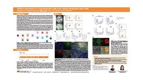 科学海报Efficient Generation of Lung Progenitor Cells From Human Pluripotent Stem Cells
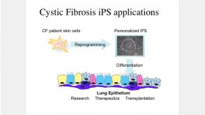 44:40
线上讲座Making Lung Cells from Pluripotent Stem Cells: Disease Modeling and Future Therapies发布日期: 08/12/2016
44:40
线上讲座Making Lung Cells from Pluripotent Stem Cells: Disease Modeling and Future Therapies发布日期: 08/12/2016


 沪公网安备31010102008431号