Yamazaki K et al. (DEC 2016)
Journal of Biomolecular Screening 21 10 1054--1064
Functional Comparison of Neuronal Cells Differentiated from Human Induced Pluripotent Stem CellDerived Neural Stem Cells under Different Oxygen and Medium Conditions
Because neurons are difficult to obtain from humans,generating functional neurons from human induced pluripotent stem cells (hiPSCs) is important for establishing physiological or disease-relevant screening systems for drug discovery. To examine the culture conditions leading to efficient differentiation of functional neural cells,we investigated the effects of oxygen stress (2% or 20% O2) and differentiation medium (DMEM/F12:Neurobasal-based [DN] or commercial [PhoenixSongs Biologicals; PS]) on the expression of genes related to neural differentiation,glutamate receptor function,and the formation of networks of neurons differentiated from hiPSCs (201B7) via long-term self-renewing neuroepithelial-like stem (lt-NES) cells. Expression of genes related to neural differentiation occurred more quickly in PS and/or 2% O2 than in DN and/or 20% O2,resulting in high responsiveness of neural cells to glutamate,N-methyl-d-aspartate (NMDA),α-amino-3-hydroxy-5-methyl-4-isoxazolepropionate (AMPA),and (S)-3,5-d...
View Publication
产品类型:
产品号#:
05832
产品名:
STEMdiff™ 神经花环选择试剂
Behar RZ et al. (NOV 2012)
Current protocols in stem cell biology 1 SUPPL.23 Unit 1C.13
Adaptation of stem cells to 96-well plate assays: use of human embryonic and mouse neural stem cells in the MTT assay.
Human embryonic stem cells (hESC) are difficult to adapt to 96-well plate assays,such as the MTT assay,because they survive best when plated as colonies,which are not easily counted and plated accurately. Two methods were developed to address this problem. In the first,ROCK inhibitor (ROCKi) was used,which allows accurate counting and plating of single hESC. In the second,small colonies were plated without ROCKi but with adaptations for accurate counting and plating. The MTT assay was also adapted for use with mouse neural stem cells. These methods allow the MTT assay to be conducted rapidly and accurately with high reproducibility between replicate experiments. When screening volatile chemicals in a 96-well plate,vapor effects may occur and dose ranges must be carefully defined. The methods were validated using the NIH assay guidance tool. These methodss could readily be translated to other 96-well plate assay.
View Publication
产品类型:
产品号#:
05850
05857
05870
05875
85850
85857
85870
85875
产品名:
mTeSR™1
mTeSR™1
Park S-W et al. (DEC 2010)
Blood 116 25 5762--72
Efficient differentiation of human pluripotent stem cells into functional CD34+ progenitor cells by combined modulation of the MEK/ERK and BMP4 signaling pathways.
Differentiation of human pluripotent stem cells (hPSCs) into functional cell types is a crucial step in cell therapy. In the present study,we demonstrate that functional CD34(+) progenitor cells can be efficiently produced from human embryonic stem cells (hESCs) and induced pluripotent stem cells (hiPSCs) by combined modulation of 2 signaling pathways. A higher proportion of CD34(+) cells (∼ 20%) could be derived from hPSCs by inhibition of mitogen-activated protein kinase (MAPK) extracellular signal-regulated protein kinase (MEK)/extracellular signal-regulated kinase (ERK) signaling and activation of bone morphogenic protein-4 (BMP4) signaling. hPSC-derived CD34(+) progenitor cells further developed to endothelial and smooth muscle cells with functionality. Moreover,they contributed directly to neovasculogenesis in ischemic mouse hind limbs,thereby resulting in improved blood perfusion and limb salvage. Our results suggest that combined modulation of signaling pathways may be an efficient means of differentiating hPSCs into functional CD34(+) progenitor cells.
View Publication
产品类型:
产品号#:
04434
04444
产品名:
MethoCult™ H4434 Classic
MethoCult™ H4434 Classic
Zeng J and Wang S (JAN 2014)
Stem cells translational medicine 3 1 69--80
Human dendritic cells derived from embryonic stem cells stably modified with CD1d efficiently stimulate antitumor invariant natural killer T cell response.
Invariant natural killer T (iNKT) cells are a unique lymphocyte subpopulation that mediates antitumor activities upon activation. A current strategy to harness iNKT cells for cancer treatment is endogenous iNKT cell activation using patient-derived dendritic cells (DCs). However,the limited number and functional defects of patient DCs are still the major challenges for this therapeutic approach. In this study,we investigated whether human embryonic stem cells (hESCs) with an ectopically expressed CD1d gene could be exploited to address this issue. Using a lentivector carrying an optimized expression cassette,we generated stably modified hESC lines that consistently overexpressed CD1d. These modified hESC lines were able to differentiate into DCs as efficiently as the parental line. Most importantly,more than 50% of such derived DCs were CD1d+. These CD1d-overexpressing DCs were more efficient in inducing iNKT cell response than those without modification,and their ability was comparable to that of DCs generated from monocytes of healthy donors. The iNKT cells expanded by the CD1d-overexpressing DCs were functional,as demonstrated by their ability to lyse iNKT cell-sensitive glioma cells. Therefore,hESCs stably modified with the CD1d gene may serve as a convenient,unlimited,and competent DC source for iNKT cell-based cancer immunotherapy.
View Publication
产品类型:
产品号#:
05850
05857
05870
05875
09600
09650
70024
70024.1
85850
85857
85870
85875
产品名:
StemSpan™ SFEM
StemSpan™ SFEM
冻存的人外周血Pan T细胞
冻存的人外周血Pan T细胞
mTeSR™1
mTeSR™1
R. Gélinas et al. (Apr 2024)
Frontiers in Genetics 15
Human induced pluripotent stem cells (hiPSCs) derived cells reflect tissue specificity found in patients with Leigh syndrome French Canadian variant (LSFC)
Leigh syndrome French Canadian type (LSFC) is a recessive neurodegenerative disease characterized by tissue-specific deficiency in cytochrome c oxidase (COX),the fourth complex in the oxidative phosphorylation system. LSFC is caused by mutations in the leucine rich pentatricopeptide repeat containing gene ( LRPPRC ). Most LSFC patients in Quebec are homozygous for an A354V substitution that causes a decrease in the expression of the LRPPRC protein. While LRPPRC is ubiquitously expressed and is involved in multiple cellular functions,tissue-specific expression of LRPPRC and COX activity is correlated with clinical features. In this proof-of-principle study,we developed human induced pluripotent stem cell (hiPSC)-based models from fibroblasts taken from a patient with LSFC,homozygous for the LRPPRC *354V allele,and from a control,homozygous for the LRPPRC *A354 allele. Specifically,for both of these fibroblast lines we generated hiPSC,hiPSC-derived cardiomyocytes (hiPSC-CMs) and hepatocyte-like cell (hiPSC-HLCs) lines,as well as the three germ layers. We observed that LRPPRC protein expression is reduced in all cell lines/layers derived from LSFC patient compared to control cells,with a reduction ranging from ∼70% in hiPSC-CMs to undetectable levels in hiPSC-HLC,reflecting tissue heterogeneity observed in patient tissues. We next performed exploratory analyses of these cell lines and observed that COX protein expression was reduced in all cell lines derived from LSFC patient compared to control cells. We also observed that mutant LRPPRC was associated with altered expression of key markers of endoplasmic reticulum stress response in hiPSC-HLCs but not in other cell types that were tested. While this demonstrates feasibility of the approach to experimentally study genotype-based differences that have tissue-specific impacts,this study will need to be extended to a larger number of patients and controls to not only validate the current observations but also to delve more deeply in the pathogenic mechanisms of LSFC.
View Publication
Chen AY et al. (DEC 2010)
Journal of virology 84 23 12385--96
Role of erythropoietin receptor signaling in parvovirus B19 replication in human erythroid progenitor cells.
Parvovirus B19 (B19V) infection is highly restricted to human erythroid progenitor cells. Although previous studies have led to the theory that the basis of this tropism is receptor expression,this has been questioned by more recent observation. In the study reported here,we have investigated the basis of this tropism,and a potential role of erythropoietin (Epo) signaling,in erythroid progenitor cells (EPCs) expanded ex vivo from CD34(+) hematopoietic cells in the absence of Epo (CD36(+)/Epo(-) EPCs). We show,first,that CD36(+)/Epo(-) EPCs do not support B19V replication,in spite of B19V entry,but Epo exposure either prior to infection or after virus entry enabled active B19V replication. Second,when Janus kinase 2 (Jak2) phosphorylation was inhibited using the inhibitor AG490,phosphorylation of the Epo receptor (EpoR) was also inhibited,and B19V replication in ex vivo-expanded erythroid progenitor cells exposed to Epo (CD36(+)/Epo(+) EPCs) was abolished. Third,expression of constitutively active EpoR in CD36(+)/Epo(-) EPCs led to efficient B19V replication. Finally,B19V replication in CD36(+)/Epo(+) EPCs required Epo,and the replication response was dose dependent. Our findings demonstrate that EpoR signaling is absolutely required for B19V replication in ex vivo-expanded erythroid progenitor cells after initial virus entry and at least partly accounts for the remarkable tropism of B19V infection for human erythroid progenitors.
View Publication
产品类型:
产品号#:
09600
09650
产品名:
StemSpan™ SFEM
StemSpan™ SFEM
Courtot A-M et al. (OCT 2014)
BioResearch open access 3 5 206--216
Morphological analysis of human induced pluripotent stem cells during induced differentiation and reverse programming.
The fine analysis of cell components during the generation of pluripotent cells and their comparison to bone fide human embryonic stem cells (hESCs) are valuable tools to understand their biological behavior. In this report,human mesenchymal cells (hMSCs) generated from the human ES cell line H9,were reprogrammed back to induced pluripotent state using Oct-4,Sox2,Nanog,and Lin28 transgenes. Human induced pluripotent stem cells (hIPSCs) were analyzed using electron microscopy and compared with regard to the original hESCs and the hMSCs from which they were derived. This analysis shows that hIPSCs and the original hESCs are morphologically undistinguishable but differ from the hMSCs with respect to the presence of several morphological features of undifferentiated cells at both the cytoplasmic (ribosomes,lipid droplets,glycogen,scarce reticulum) and nuclear levels (features of nuclear plasticity,presence of euchromatin,reticulated nucleoli). We show that hIPSC colonies generated this way presented epithelial aspects with specialized junctions highlighting morphological criteria of the mesenchymal-epithelial transition in cells engaged in a successful reprogramming process. Electron microscopic analysis revealed also specific morphological aspects of partially reprogrammed cells. These results highlight the valuable use of electron microscopy for a better knowledge of the morphological aspects of IPSC and cellular reprogramming.
View Publication
产品类型:
产品号#:
05850
05857
05870
05875
85850
85857
85870
85875
产品名:
mTeSR™1
mTeSR™1
Son M-Y et al. (JUL 2015)
Proteomics 15 13 2220--2229
Proteomic and network analysis of proteins regulated by REX1 in human embryonic stem cells.
Recent studies have suggested that REX1 (reduced expression 1) plays an important role in pluripotency,proliferation,and differentiation. However,the molecular mechanisms involved in REX1-dependent regulation of diverse cellular processes remain unclear. To elucidate the regulatory functions of REX1 in human embryonic stem cells (hESCs),comparative proteomic analysis was performed on REX1 RNAi specifically silenced hESCs. Analysis of the proteome via nano-LC-MS/MS identified 140 differentially expressed proteins (DEPs) displaying a textgreater2-fold difference in expression level between control and REX1 knockdown (KD) hESCs,which were then compared with transcriptome data and validated by quantitative real-time RT-PCR and Western blotting. These DEPs were analyzed by GO,pathway,and functional clustering analyses to determine the molecular functions of the proteins and pathways regulated by REX1. The REX1 KD-mediated DEPs mapped to major biological processes involved in the regulation of ribosome-mediated translation and mitochondrial function. Functional network analysis revealed a highly interconnected network among these DEPs and indicated that these interconnected proteins are predominantly involved in translation and the regulation of mitochondrial organization. These findings regarding REX1-mediated regulatory network have revealed the contributions of REX1 to maintaining the status of hESCs and have improved our understanding of the molecular events that underlie the fundamental properties of hESCs.
View Publication
产品类型:
产品号#:
05850
05857
05870
05875
85850
85857
85870
85875
产品名:
mTeSR™1
mTeSR™1
Radan L et al. ( 2016)
1341 133--142
Delivering antisense morpholino oligonucleotides to target telomerase splice variants in human embryonic stem cells
Morpholino oligonucleotides (MO) are an innovative tool that provides a means for examining and modifying gene expression outcomes by antisense interaction with targeted RNA transcripts. The site-specific nature of their binding facilitates focused modulation to alter splice variant expression patterns. Here we describe the steric-blocking of human telomerase reverse transcriptase (hTERT) $$$$ and $$$$ splice variants using MO to examine cellular outcomes related to pluripotency and differentiation in human embryonic stem cells.
View Publication
产品类型:
产品号#:
05850
05857
05870
05875
85850
85857
85870
85875
产品名:
mTeSR™1
mTeSR™1
Bogomazova AN et al. (JUN 2011)
Aging 3 6 584--596
Error-prone nonhomologous end joining repair operates in human pluripotent stem cells during late G2.
Genome stability of human embryonic stem cells (hESC) is an important issue because even minor genetic alterations can negatively impact cell functionality and safety. The incorrect repair of DNA double-stranded breaks (DSBs) is the ultimate cause of the formation of chromosomal aberrations. Using G2 radiosensitivity assay,we analyzed chromosomal aberrations in pluripotent stem cells and somatic cells. The chromatid exchange aberration rates in hESCs increased manifold 2 hours after irradiation as compared with their differentiated derivatives,but the frequency of radiation-induced chromatid breaks was similar. The rate of radiation-induced chromatid exchanges in hESCs and differentiated cells exhibited a quadratic dose response,revealing two-hit mechanism of exchange formation suggesting that a non-homologous end joining (NHEJ) repair may contribute to their formation. Inhibition of DNA-PK,a key NHEJ component,by NU7026 resulted in a significant decrease in radiation-induced chromatid exchanges in hESCs but not in somatic cells. In contrast,NU7026 treatment increased the frequency of radiation-induced breaks to a similar extent in pluripotent and somatic cells. Thus,DNA-PK dependent NHEJ efficiently participates in the elimination of radiation-induced chromatid breaks during the late G2 in both cell types and DNA-PK activity leads to a high level of misrejoining specifically in pluripotent cells.
View Publication
产品类型:
产品号#:
05850
05857
05870
05875
85850
85857
85870
85875
产品名:
mTeSR™1
mTeSR™1
Azarin SM et al. (MAR 2012)
Biomaterials 33 7 2041--2049
Modulation of Wnt/β-catenin signaling in human embryonic stem cells using a 3-D microwell array.
Intercellular interactions in the cell microenvironment play a critical role in determining cell fate,but the effects of these interactions on pathways governing human embryonic stem cell (hESC) behavior have not been fully elucidated. We and others have previously reported that 3-D culture of hESCs affects cell fates,including self-renewal and differentiation to a variety of lineages. Here we have used a microwell culture system that produces 3-D colonies of uniform size and shape to provide insight into the effect of modulating cell-cell contact on canonical Wnt/??-catenin signaling in hESCs. Canonical Wnt signaling has been implicated in both self-renewal and differentiation of hESCs,and competition for ??-catenin between the Wnt pathway and cadherin-mediated cell-cell interactions impacts various developmental processes,including the epithelial-mesenchymal transition. Our results showed that hESCs cultured in 3-D microwells exhibited higher E-cadherin expression than cells on 2-D substrates. The increase in E-cadherin expression in microwells was accompanied by a downregulation of Wnt signaling,as evidenced by the lack of nuclear ??-catenin and downregulation of Wnt target genes. Despite this reduction in Wnt signaling in microwell cultures,embryoid bodies (EBs) formed from hESCs cultured in microwells exhibited higher levels of Wnt signaling than EBs from hESCs cultured on 2-D substrates. Furthermore,the Wnt-positive cells within EBs showed upregulation of genes associated with cardiogenesis. These results demonstrate that modulation of intercellular interactions impacts Wnt/??-catenin signaling in hESCs. ?? 2011 Elsevier Ltd.
View Publication

 EasySep™小鼠TIL(CD45)正选试剂盒


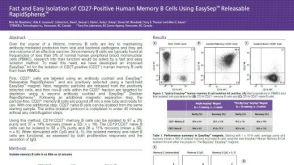 科学海报Fast and Easy Isolation of CD27-Positive Human Memory B Cells Using EasySep™ Releasable RapidSpheres™
 沪公网安备31010102008431号