P. N. Amartey et al. (Feb 2026)
Methods and Protocols 9 1
An Open-Source Automated Pipeline for Quantitative Morphological Analysis of 3D-Bioprinted Cancer Cell Spheroids
Three-dimensional (3D) culture systems that recapitulate the tumor microenvironment are essential for studying cancer cell behavior,drug response,and cell–matrix interactions. Here,we present a detailed protocol for generating 3D spheroid cultures from murine breast cancer cells using methacrylated gelatin (GelMA)-based bioink and a CELLINK BioX bioprinter. This method enables precise deposition of spheroid-laden GelMA droplets into low-attachment plates,facilitating high-throughput and reproducible 3D culture formation. The protocol includes steps for spheroid formation,GelMA preparation,bioprinting,and post-printing analysis using a customized CellProfiler pipeline. The analysis pipeline takes advantage of the functionality of CellProfiler and ImageJ software (version 2.14.0) packages to create a versatile and accessible analysis tool. This approach provides a robust and adaptable platform for in vitro cancer research,including studies of metastasis,drug resistance,cancer cell lipid metabolism,and TME-associated hypoxia.
View Publication
Physiological regulation of neuronal Wnt activity is essential for TDP-43 localization and function
Nuclear exclusion of the RNA- and DNA-binding protein TDP-43 can induce neurodegeneration in different diseases. Diverse processes have been implicated to influence TDP-43 mislocalization,including disrupted nucleocytoplasmic transport (NCT); however,the physiological pathways that normally ensure TDP-43 nuclear localization are unclear. The six-transmembrane enzyme glycerophosphodiester phosphodiesterase 2 (GDE2 or GDPD5) cleaves the glycosylphosphatidylinositol (GPI) anchor that tethers some proteins to the membrane. Here we show that GDE2 maintains TDP-43 nuclear localization by regulating the dynamics of canonical Wnt signaling. Ablation of GDE2 causes aberrantly sustained Wnt activation in adult neurons,which is sufficient to cause NCT deficits,nuclear pore abnormalities,and TDP-43 nuclear exclusion. Disruption of GDE2 coincides with TDP-43 abnormalities in postmortem tissue from patients with amyotrophic lateral sclerosis (ALS). Further,GDE2 deficits are evident in human neural cell models of ALS,which display erroneous Wnt activation that,when inhibited,increases mRNA levels of genes regulated by TDP-43. Our study identifies GDE2 as a critical physiological regulator of Wnt signaling in adult neurons and highlights Wnt pathway activation as an unappreciated mechanism contributing to nucleocytoplasmic transport and TDP-43 abnormalities in disease. Synopsis Nuclear exclusion of TDP-43 is observed in various pathologies,but the physiological mechanisms that ensure its nuclear localization are not well-known. This work shows that inhibition of persistent Wnt activation in neurons by GDE2 prevents TDP-43 nuclear exclusion. GDE2 inhibits canonical Wnt signaling in adult postmitotic neurons.Sustained activation of canonical Wnt signaling in neurons disrupts the nuclear pore complex,impairs nucleocytoplasmic transport,and results in TDP-43 nuclear exclusion.iPS neurons from patients with C9orf72 ALS show decreased GDE2 expression and increased activation of canonical Wnt signaling.Inhibition of Wnt activation mitigates TDP-43 dysfunction in C9orf72 iPS neurons. GDE2 maintains TDP-43 nuclear localization by inhibiting Wnt activation in neurons.
View Publication
产品类型:
产品号#:
100-0276
100-1130
产品名:
mTeSR™ Plus
mTeSR™ Plus
(Mar 2025)
Life Science Alliance 8 6
A novel human organoid model system reveals requirement of TCF4 for oligodendroglial differentiation
In this study,we developed a cell system to study TCF4 in human oligodendrocyte differentiation,showed that TCF4 regulates human oligodendroglial differentiation in a dose-dependent manner,and established a system to dissect TCF4 function in a human tissue–like context. Heterozygous mutations of TCF4 in humans cause Pitt–Hopkins syndrome,a neurodevelopmental disease associated with intellectual disability and brain malformations. Although most studies focus on the role of TCF4 in neural stem cells and neurons,we here set out to assess the implication of TCF4 for oligodendroglial differentiation. We discovered that both monoallelic and biallelic mutations in TCF4 result in a diminished capacity to differentiate human neural progenitor cells toward myelinating oligodendrocytes through the forced expression of the transcription factors SOX10,OLIG2,and NKX6.2. Using this experimental strategy,we established a novel organoid model,which generates oligodendroglial cells within a human neurogenic tissue–like context. Also,here we found a reduced ability of TCF4 heterozygous cells to differentiate toward oligodendroglial cells. In sum,we establish a role of human TCF4 in oligodendrocyte differentiation and provide a model system,which allows to dissect the disease etiology in a human tissue–like context.
View Publication
M. Mata Forsberg et al. (mar 2022)
Journal of leukocyte biology 111 3 597--609
Activation of human ?? T cells and NK cells by Staphylococcal enterotoxins requires both monocytes and conventional T cells.
Staphylococcal enterotoxins (SE) pose a great threat to human health due to their ability to bypass antigen presentation and activate large amounts of conventional T cells resulting in a cytokine storm potentially leading to toxic shock syndrome. Unconventional T- and NK cells are also activated by SE but the mechanisms remain poorly understood. In this study,the authors aimed to explore the underlying mechanism behind SE-mediated activation of MAIT-,?? T-,and NK cells in vitro. CBMC or PBMC were stimulated with the toxins SEA,SEH,and TSST-1,and cytokine and cytotoxic responses were analyzed with ELISA and flow cytometry. All toxins induced a broad range of cytokines,perforin and granzyme B,although SEH was not as potent as SEA and TSST-1. SE-induced IFN-$\gamma$ expression in MAIT-,?? T-,and NK cells was clearly reduced by neutralization of IL-12,while cytotoxic compounds were not affected at all. Kinetic assays showed that unconventional T cell and NK cell-responses are secondary to the response in conventional T cells. Furthermore,co-cultures of isolated cell populations revealed that the ability of SEA to activate ?? T- and NK cells was fully dependent on the presence of both monocytes and $\alpha$$\beta$ T cells. Lastly,it was found that SE provoked a reduced and delayed cytokine response in infants,particularly within the unconventional T and NK cell populations. This study provides novel insights regarding the activation of unconventional T- and NK cells by SE,which contribute to understanding the vulnerability of young children towards Staphylococcus aureus infections.
View Publication
产品类型:
产品号#:
17955
18000
19059
19059RF
17955RF
100-0960
产品名:
EasySep™人NK细胞分选试剂盒
EasySep™磁极
EasySep™人单核细胞富集试剂盒
RoboSep™ 人单核细胞富集试剂盒含滤芯吸头
RoboSep™ 人NK细胞分选试剂盒
EasySep™人NK细胞分离试剂盒
McPherson CA et al. (JUL 2011)
Brain,behavior,and immunity 25 5 850--62
Interleukin (IL)-1 and IL-6 regulation of neural progenitor cell proliferation with hippocampal injury: differential regulatory pathways in the subgranular zone (SGZ) of the adolescent and mature mouse brain.
Current data suggests an association between elevations in interleukin 1 (IL-1)α,IL-1β,and IL-6 and the proliferation of neural progenitor cells (NPCs) following brain injury. A limited amount of work implicates changes in these pro-inflammatory responses with diminished NPC proliferation observed as a function of aging. In the current study,adolescent (21day-old) and 1year-old CD-1 male mice were injected with trimethyltin (TMT,2.3mg/kg,i.p.) to produce acute apoptosis of hippocampal dentate granule cells. In this model,fewer 5-bromo-2'-deoxyuridine (BrdU)+ NPC were observed in both naive and injured adult hippocampus as compared to the corresponding number seen in adolescent mice. At 48h post-TMT,a similar level of neuronal death was observed across ages,yet activated ameboid microglia were observed in the adolescent and hypertrophic process-bearing microglia in the adult. IL-1α mRNA levels were elevated in the adolescent hippocampus; IL-6 mRNA levels were elevated in the adult. In subgranular zone (SGZ) isolated by laser-capture microdissection,IL-1β was detected but not elevated by TMT,IL-1a was elevated at both ages,while IL-6 was elevated only in the adult. Naïve NPCs isolated from the hippocampus expressed transcripts for IL-1R1,IL-6Rα,and gp130 with significantly higher levels of IL-6Rα mRNA in the adult. In vitro,IL-1α (150pg/ml) stimulated proliferation of adolescent NPCs; IL-6 (10ng/ml) inhibited proliferation of adolescent and adult NPCs. Microarray analysis of SGZ post-TMT indicated a prominence of IL-1a/IL-1R1 signaling in the adolescent and IL-6/gp130 signaling in the adult.
View Publication
产品类型:
产品号#:
05700
05701
05702
05707
05715
05740
产品名:
NeuroCult™ 基础培养基(小鼠和大鼠)
NeuroCult™ 扩增添加物(小鼠和大鼠)
NeuroCult™扩增试剂盒(小鼠和大鼠)
NeuroCult™化学解离试剂盒(小鼠)
NeuroCult™成年中枢神经系统(CNS)组织酶解试剂盒(小鼠和大鼠)
Naylor RW et al. ( 2016)
PloS one 11 10 e0165464
Derivation of Corneal Keratocyte-Like Cells from Human Induced Pluripotent Stem Cells.
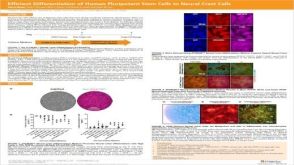
Corneal diseases such as keratoconus represent a relatively common disorder in the human population. However,treatment is restricted to corneal transplantation,which only occurs in the most advanced cases. Cell based therapies may offer an alternative approach given that the eye is amenable to such treatments and corneal diseases like keratoconus have been associated specifically with the death of corneal keratocytes. The ability to generate corneal keratocytes in vitro may enable a cell-based therapy to treat patients with keratoconus. Human induced pluripotent stem cells (hiPSCs) offer an abundant supply of cells from which any cell in the body can be derived. In the present study,hiPSCs were successfully differentiated into neural crest cells (NCCs),the embryonic precursor to keratocytes,and then cultured on cadaveric corneal tissue to promote keratocyte differentiation. The hiPSC-derived NCCs were found to migrate into the corneal stroma where they acquired a keratocyte-like morphology and an expression profile similar to corneal keratocytes in vivo. These results indicate that hiPSCs can be used to generate corneal keratocytes in vitro and lay the foundation for using these cells in cornea cell-based therapies.
View Publication
Hervé et al. (JUL 2007)
The Journal of experimental medicine 204 7 1583--93
CD40 ligand and MHC class II expression are essential for human peripheral B cell tolerance.
Hyper-IgM (HIGM) syndromes are primary immunodeficiencies characterized by defects of class switch recombination and somatic hypermutation. HIGM patients who carry mutations in the CD40-ligand (CD40L) gene expressed by CD4(+) T cells suffer from recurrent infections and often develop autoimmune disorders. To investigate the impact of CD40L-CD40 interactions on human B cell tolerance,we tested by ELISA the reactivity of recombinant antibodies isolated from single B cells from three CD40L-deficient patients. Antibody characteristics and reactivity from CD40L-deficient new emigrant B cells were similar to those from healthy donors,suggesting that CD40L-CD40 interactions do not regulate central B cell tolerance. In contrast,mature naive B cells from CD40L-deficient patients expressed a high proportion of autoreactive antibodies,including antinuclear antibodies. Thus,CD40L-CD40 interactions are essential for peripheral B cell tolerance. In addition,a patient with the bare lymphocyte syndrome who could not express MHC class II molecules failed to counterselect autoreactive mature naive B cells,suggesting that peripheral B cell tolerance also depends on major histocompatibility complex (MHC) class II-T cell receptor (TCR) interactions. The decreased frequency of MHC class II-restricted CD4(+) regulatory T cells in CD40L-deficient patients suggests that these T cells may mediate peripheral B cell tolerance through CD40L-CD40 and MHC class II-TCR interactions.
View Publication
产品类型:
产品号#:
15024
15064
产品名:
RosetteSep™人B细胞富集抗体混合物
RosetteSep™人B细胞富集抗体混合物
Glatigny S et al. (MAY 2016)
Journal of Immunology 196 9 3542--6
Cutting Edge: Integrin α4 Is Required for Regulatory B Cell Control of Experimental Autoimmune Encephalomyelitis.
The neutralization of integrin α4 (Itga4) is currently used as treatment in multiple sclerosis. Although most studies have focused on its function on lymphocyte migration to the CNS,we have uncovered the importance of Itga4 for the generation of regulatory B cells in peripheral immune organs and their control of pathogenic T cell response and CNS pathology. Our study underscores the importance of looking at the dual role of B cells in CNS autoimmunity and provides important perspectives regarding the efficacy and side effects associated with Itga4 neutralization and other B cell-targeting therapies.
View Publication


 EasySep™小鼠TIL(CD45)正选试剂盒





 科学海报Efficient Generation of Human Pluripotent Stem Cells to Neural Crest Cells


 沪公网安备31010102008431号