Higuchi A et al. (DEC 2015)
Scientific Reports 5 18136
Long-term xeno-free culture of human pluripotent stem cells on hydrogels with optimal elasticity
The tentative clinical application of human pluripotent stem cells (hPSCs),such as human embryonic stem cells and human induced pluripotent stem cells,is restricted by the possibility of xenogenic contamination resulting from the use of mouse embryonic fibroblasts (MEFs) as a feeder layer. Therefore,we investigated hPSC cultures on biomaterials with different elasticities that were grafted with different nanosegments. We prepared dishes coated with polyvinylalcohol-co-itaconic acid hydrogels grafted with an oligopeptide derived from vitronectin (KGGPQVTRGDVFTMP) with elasticities ranging from 10.3 to 30.4 kPa storage moduli by controlling the crosslinking time. The hPSCs cultured on the stiffest substrates (30.4 kPa) tended to differentiate after five days of culture,whereas the hPSCs cultured on the optimal elastic substrates (25 kPa) maintained their pluripotency for over 20 passages under xeno-free conditions. These results indicate that cell culture matrices with optimal elasticity can maintain the pluripotency of hPSCs in culture.
View Publication
产品类型:
产品号#:
05850
05857
05870
05875
85850
85857
85870
85875
产品名:
mTeSR™1
mTeSR™1
McCabe KL et al. (DEC 2015)
PloS one 10 12 e0145266
Efficient Generation of Human Embryonic Stem Cell-Derived Corneal Endothelial Cells by Directed Differentiation.
AIM To generate human embryonic stem cell derived corneal endothelial cells (hESC-CECs) for transplantation in patients with corneal endothelial dystrophies. MATERIALS AND METHODS Feeder-free hESC-CECs were generated by a directed differentiation protocol. hESC-CECs were characterized by morphology,expression of corneal endothelial markers,and microarray analysis of gene expression. RESULTS hESC-CECs were nearly identical morphologically to primary human corneal endothelial cells,expressed Zona Occludens 1 (ZO-1) and Na+/K+ATPase$\$1 (ATPA1) on the apical surface in monolayer culture,and produced the key proteins of Descemet's membrane,Collagen VIII$\$1 and VIII$\$2 (COL8A1 and 8A2). Quantitative PCR analysis revealed expression of all corneal endothelial pump transcripts. hESC-CECs were 96% similar to primary human adult CECs by microarray analysis. CONCLUSION hESC-CECs are morphologically similar,express corneal endothelial cell markers and express a nearly identical complement of genes compared to human adult corneal endothelial cells. hESC-CECs may be a suitable alternative to donor-derived corneal endothelium.
View Publication
产品类型:
产品号#:
05850
05857
05870
05875
85850
85857
85870
85875
产品名:
mTeSR™1
mTeSR™1
Freude KK et al. (JUL 2011)
Journal of Biological Chemistry 286 27 24264--24274
Soluble amyloid precursor protein induces rapid neural differentiation of human embryonic stem cells.
Human embryonic stem cells (hESCs) offer tremendous potential for not only treating neurological disorders but also for their ability to serve as vital reagents to model and investigate human disease. To further our understanding of a key protein involved in Alzheimer disease pathogenesis,we stably overexpressed amyloid precursor protein (APP) in hESCs. Remarkably,we found that APP overexpression in hESCs caused a rapid and robust differentiation of pluripotent stem cells toward a neural fate. Despite maintenance in standard hESC media,up to 80% of cells expressed the neural stem cell marker nestin,and 65% exhibited the more mature neural marker β-3 tubulin within just 5 days of passaging. To elucidate the mechanism underlying the effects of APP on neural differentiation,we examined the proteolysis of APP and performed both gain of function and loss of function experiments. Taken together,our results demonstrate that the N-terminal secreted soluble forms of APP (in particular sAPPβ) robustly drive neural differentiation of hESCs. Our findings not only reveal a novel and intriguing role for APP in neural lineage commitment but also identify a straightforward and rapid approach to generate large numbers of neurons from human embryonic stem cells. These novel APP-hESC lines represent a valuable tool to investigate the potential role of APP in development and neurodegeneration and allow for insights into physiological functions of this protein.
View Publication
产品类型:
产品号#:
05850
05857
05870
05875
85850
85857
85870
85875
产品名:
mTeSR™1
mTeSR™1
Lagadinou ED et al. (MAR 2013)
Cell stem cell 12 3 329--41
BCL-2 inhibition targets oxidative phosphorylation and selectively eradicates quiescent human leukemia stem cells.
Most forms of chemotherapy employ mechanisms involving induction of oxidative stress,a strategy that can be effective due to the elevated oxidative state commonly observed in cancer cells. However,recent studies have shown that relative redox levels in primary tumors can be heterogeneous,suggesting that regimens dependent on differential oxidative state may not be uniformly effective. To investigate this issue in hematological malignancies,we evaluated mechanisms controlling oxidative state in primary specimens derived from acute myelogenous leukemia (AML) patients. Our studies demonstrate three striking findings. First,the majority of functionally defined leukemia stem cells (LSCs) are characterized by relatively low levels of reactive oxygen species (termed ROS-low"). Second�
View Publication
产品类型:
产品号#:
07930
07931
07940
07955
07956
07959
07954
100-1061
07952
产品名:
CryoStor® CS10
CryoStor® CS10
CryoStor® CS10
CryoStor® CS10
CryoStor® CS10
CryoStor® CS10
CryoStor® CS10
R. Schmidt et al. (feb 2022)
Science (New York,N.Y.) 375 6580 eabj4008
CRISPR activation and interference screens decode stimulation responses in primary human T cells.
Regulation of cytokine production in stimulated T cells can be disrupted in autoimmunity,immunodeficiencies,and cancer. Systematic discovery of stimulation-dependent cytokine regulators requires both loss-of-function and gain-of-function studies,which have been challenging in primary human cells. We now report genome-wide CRISPR activation (CRISPRa) and interference (CRISPRi) screens in primary human T cells to identify gene networks controlling interleukin-2 (IL-2) and interferon-$\gamma$ (IFN-$\gamma$) production. Arrayed CRISPRa confirmed key hits and enabled multiplexed secretome characterization,revealing reshaped cytokine responses. Coupling CRISPRa screening with single-cell RNA sequencing enabled deep molecular characterization of screen hits,revealing how perturbations tuned T cell activation and promoted cell states characterized by distinct cytokine expression profiles. These screens reveal genes that reprogram critical immune cell functions,which could inform the design of immunotherapies.
View Publication
产品类型:
产品号#:
20144
产品名:
EasySep™缓冲液
(Feb 2024)
Genome Biology 25 12
HiHo-AID2: boosting homozygous knock-in efficiency enables robust generation of human auxin-inducible degron cells
Recent developments in auxin-inducible degron (AID) technology have increased its popularity for chemogenetic control of proteolysis. However,generation of human AID cell lines is challenging,especially in human embryonic stem cells (hESCs). Here,we develop HiHo-AID2,a streamlined procedure for rapid,one-step generation of human cancer and hESC lines with high homozygous degron-tagging efficiency based on an optimized AID2 system and homology-directed repair enhancers. We demonstrate its application for rapid and inducible functional inactivation of twelve endogenous target proteins in five cell lines,including targets with diverse expression levels and functions in hESCs and cells differentiated from hESCs.Supplementary InformationThe online version contains supplementary material available at 10.1186/s13059-024-03187-w.
View Publication
产品类型:
产品号#:
100-0276
100-1130
产品名:
mTeSR™ Plus
mTeSR™ Plus
M. Dastpak et al. (Dec 2025)
PLOS One 20 12
SF3B1K700E mutation in human embryonic stem cells causes aberrant expression of immune-related genes
SF3B1,a component of the U2 snRNP pre-mRNA splicing factor,plays a critical role in splicing and is frequently mutated in cancer,particularly hematologic malignancies. We investigated the effects of the most common SF3B1 mutation,heterozygous substitution of Lysine 700 to Glutamate (K700E),in human embryonic stem cells (hESC),using CRISPR-Cas9 to generate heterozygous SF3B1K700E clones. Interestingly,we observed the upregulation of several key transcription regulators associated with hematopoiesis and a broad range of immune genes in SF3B1K700E hESCs. Despite differences in the transcriptional and splicing profiles between hESC and myelodysplastic syndrome (MDS) cells harboring the SF3B1K700E mutation,several common immune gene programs were identified in both cell types. To elucidate the molecular mechanisms underlying dysregulated gene expression in SF3B1K700E hESCs,we mapped actively engaged RNA polymerase II (RNA Pol II) using Precision Run-On sequencing (PRO-seq). These analyses revealed that the SF3B1K700E mutation alters RNA Pol II elongation properties. Specifically,we observed a general increase in pause release in SF3B1K700E hESCs,consistent with recent work in leukemia cells suggesting that the SF3B1K700E mutation affects early transcription elongation. Taken together,our study identifies several candidate genes that could contribute to the SF3B1 mutated phenotype and clarifies the role for the U2 snRNP and pre-spliceosome assembly on transcription by RNA Pol II. Further,our data suggest that mutations of SF3B1 impact immune gene expression independent of cell type,providing new insights into the role of SF3B1K700E in hematologic malignancies.
View Publication
Non-integrating episomal plasmid-based reprogramming of human amniotic fluid stem cells into induced pluripotent stem cells in chemically defined conditions.
Amniotic fluid stem cells (AFSC) represent an attractive potential cell source for fetal and pediatric cell-based therapies. However,upgrading them to pluripotency confers refractoriness toward senescence,higher proliferation rate and unlimited differentiation potential. AFSC were observed to rapidly and efficiently reacquire pluripotency which together with their easy recovery makes them an attractive cell source for reprogramming. The reprogramming process as well as the resulting iPSC epigenome could potentially benefit from the unspecialized nature of AFSC. iPSC derived from AFSC also have potential in disease modeling,such as Down syndrome or $\$-thalassemia. Previous experiments involving AFSC reprogramming have largely relied on integrative vector transgene delivery and undefined serum-containing,feeder-dependent culture. Here,we describe non-integrative oriP/EBNA-1 episomal plasmid-based reprogramming of AFSC into iPSC and culture in fully chemically defined xeno-free conditions represented by vitronectin coating and E8 medium,a system that we found uniquely suited for this purpose. The derived AF-iPSC lines uniformly expressed a set of pluripotency markers Oct3/4,Nanog,Sox2,SSEA-1,SSEA-4,TRA-1-60,TRA-1-81 in a pattern typical for human primed PSC. Additionally,the cells formed teratomas,and were deemed pluripotent by PluriTest,a global expression microarray-based in-silico pluripotency assay. However,we found that the PluriTest scores were borderline,indicating a unique pluripotent signature in the defined condition. In the light of potential future clinical translation of iPSC technology,non-integrating reprogramming and chemically defined culture are more acceptable.
View Publication
产品类型:
产品号#:
05850
05857
05870
05875
05940
07180
07183
07190
27147
07191
07930
07931
07940
07955
07956
07959
07954
85850
85857
85870
85875
100-1061
07952
100-0763
产品名:
Vitronectin XF™
CellAdhere™ 稀释缓冲液
CryoStor® CS10
CryoStor® CS10
CryoStor® CS10
CryoStor® CS10
CryoStor® CS10
mTeSR™1
mTeSR™1
CryoStor® CS10
CryoStor® CS10
Vitronectin XF™
Zielske SP et al. (NOV 2003)
The Journal of clinical investigation 112 10 1561--70
In vivo selection of MGMT(P140K) lentivirus-transduced human NOD/SCID repopulating cells without pretransplant irradiation conditioning.
Infusion of transduced hematopoietic stem cells into nonmyeloablated hosts results in ineffective in vivo levels of transduced cells. To increase the proportion of transduced cells in vivo,selection based on P140K O6-methylguanine-DNA-methyltransferase (MGMT[P140K]) gene transduction and O6-benzylguanine/1,3-bis(2-chloroethyl)-1-nitrosourea (BG/BCNU) treatment has been devised. In this study,we transduced human NOD/SCID repopulating cells (SRCs) with MGMT(P140K) using a lentiviral vector and infused them into BG/BCNU-conditioned NOD/SCID mice before rounds of BG/BCNU treatment as a model for in vivo selection. Engraftment was not observed until the second round of BG/BCNU treatment,at which time human cells emerged to compose up to 20% of the bone marrow. Furthermore,99% of human CFCs derived from NOD/SCID mice were positive for provirus as measured by PCR,compared with 35% before transplant and 11% in untreated irradiation-preconditioned mice,demonstrating selection. Bone marrow showed BG-resistant O6-alkylguanine-DNA-alkyltransferase (AGT) activity,and CFUs were stained intensely for AGT protein,indicating high transgene expression. Real-time PCR estimates of the number of proviral insertions in individual CFUs ranged from 3 to 22. Selection resulted in expansion of one or more SRC clones containing similar numbers of proviral copies per mouse. To our knowledge,these results provide the first evidence of potent in vivo selection of MGMT(P140K) lentivirus-transduced human SRCs following BG/BCNU treatment.
View Publication
产品类型:
产品号#:
04434
04444
产品名:
MethoCult™ H4434 Classic
MethoCult™ H4434 Classic
Gupta R et al. (MAY 2012)
Molecular endocrinology (Baltimore,Md.) 26 5 859--72
Squelching of ETS2 transactivation by POU5F1 silences the human chorionic gonadotropin CGA subunit gene in human choriocarcinoma and embryonic stem cells.
The subunit genes encoding human chorionic gonadotropin,CGA,and CGB,are up-regulated in human trophoblast. However,they are effectively silenced in choriocarcinoma cells by ectopically expressed POU domain class 5 transcription factor 1 (POU5F1). Here we show that POU5F1 represses activity of the CGA promoter through its interactions with ETS2,a transcription factor required for both placental development and human chorionic gonadotropin subunit gene expression,by forming a complex that precludes ETS2 from interacting with the CGA promoter. Mutation of a POU5F1 binding site proximal to the ETS2 binding site does not alter the ability of POU5F1 to act as a repressor but causes a drop in basal promoter activity due to overlap with the binding site for DLX3. DLX3 has only a modest ability to raise basal CGA promoter activity,but its coexpression with ETS2 can up-regulate it 100-fold or more. The two factors form a complex,and both must bind to the promoter for the combination to be transcriptionally effective,a synergy compromised by POU5F1. Similarly,in human embryonic stem cells,which express ETS2 but not CGA,ETS2 does not occupy its binding site on the CGA promoter but is found instead as a soluble complex with POU5F1. When human embryonic stem cells differentiate in response to bone morphogenetic protein-4 and concentrations of POU5F1 fall and hCG and DLX3 rise,ETS2 then occupies its binding site on the CGA promoter. Hence,a squelching mechanism underpins the transcriptional silencing of CGA by POU5F1 and could have general relevance to how pluripotency is maintained and how the trophoblast lineage emerges from pluripotent precursor cells.
View Publication
产品类型:
产品号#:
05850
05857
05870
05875
85850
85857
85870
85875
产品名:
mTeSR™1
mTeSR™1
E. Yamashita et al. (Sep 2025)
The FASEB Journal 39 17
Red Blood Cell‐Mediated Enhancement of Hematopoietic Stem Cell Functions via a Hes1‐Dependent Pathway
In bone marrow,cell numbers are balanced between production and loss. After chemotherapy,blood cell counts decrease initially but later recover as hematopoietic progenitor cells expand,although the mechanisms underlying this recovery are still unclear. We investigated the influence of red blood cells (RBCs) on hematopoietic stem cell (HSC) function during bone marrow recovery. Following chemotherapy,RBC concentrations in bone marrow peaked on day 5 posttreatment,coinciding with the recovery of hematopoiesis. Coculture of HSCs with RBCs resulted in a significant increase in hematopoiesis. Direct contact between RBCs and HSCs was essential for enhancement of hematopoiesis,and HSCs precultured with RBCs resulted in greater numbers of donor‐derived mature hematopoietic cells after transplantation. RNA‐sequencing analysis showed that Hes1 was the most significantly upregulated transcription factor in RBC coculture,and the response to RBC‐induced hematopoiesis of Hes1‐deficient HSCs was reduced. These findings imply a role of RBCs and Hes1 in the enhancement of hematopoietic recovery following bone marrow stress.
View Publication

 EasySep™小鼠TIL(CD45)正选试剂盒


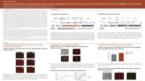 科学海报Generation, Maintenance and Cryopreservation of Neural Progenitor Cells Derived from Human Pluripotent Stem Cells Using the STEMdiff™ Neural System
 沪公网安备31010102008431号