Dendritic Cells Regulate Extrafollicular Autoreactive B Cells via T Cells Expressing Fas and Fas Ligand.
The extrafollicular (EF) plasmablast response to self-antigens that contain Toll-like receptor (TLR) ligands is prominent in murine lupus models and some bacterial infections,but the inhibitors and activators involved have not been fully delineated. Here,we used two conventional dendritic cell (cDC) depletion systems to investigate the role of cDCs on a classical TLR-dependent autoreactive EF response elicited in rheumatoid-factor B cells by DNA-containing immune complexes. Contrary to our hypothesis,cDC depletion amplified rather than dampened the EF response in Fas-intact but not Fas-deficient mice. Further,we demonstrated that cDC-dependent regulation requires Fas and Fas ligand (FasL) expression by T cells,but not Fas expression by B cells. Thus,cDCs activate FasL-expressing T cells that regulate Fas-expressing extrafollicular helper T (Tefh) cells. These studies reveal a regulatory role for cDCs in B cell plasmablast responses and provide a mechanistic explanation for the excess autoantibody production observed in Fas deficiency.
View Publication
产品类型:
产品号#:
19754
19754RF
产品名:
B. A. Jonas et al. ( 2016)
PloS one 11 7 e0159189
Alkylator-Induced and Patient-Derived Xenograft Mouse Models of Therapy-Related Myeloid Neoplasms Model Clinical Disease and Suggest the Presence of Multiple Cell Subpopulations with Leukemia Stem Cell Activity.
Acute myeloid leukemia (AML) is a heterogeneous group of aggressive bone marrow cancers arising from transformed hematopoietic stem and progenitor cells (HSPC). Therapy-related AML and MDS (t-AML/MDS) comprise a subset of AML cases occurring after exposure to alkylating chemotherapy and/or radiation and are associated with a very poor prognosis. Less is known about the pathogenesis and disease-initiating/leukemia stem cell (LSC) subpopulations of t-AML/MDS compared to their de novo counterparts. Here,we report the development of mouse models of t-AML/MDS. First,we modeled alkylator-induced t-AML/MDS by exposing wild type adult mice to N-ethyl-N-nitrosurea (ENU),resulting in several models of AML and MDS that have clinical and pathologic characteristics consistent with human t-AML/MDS including cytopenia,myelodysplasia,and shortened overall survival. These models were limited by their inability to transplant clinically aggressive disease. Second,we established three patient-derived xenograft models of human t-AML. These models led to rapidly fatal disease in recipient immunodeficient xenografted mice. LSC activity was identified in multiple HSPC subpopulations suggesting there is no canonical LSC immunophenotype in human t-AML. Overall,we report several new t-AML/MDS mouse models that could potentially be used to further define disease pathogenesis and test novel therapeutics.
View Publication
Cell-culture assays reveal the importance of retroviral vector design for insertional genotoxicity.
Retroviral vectors with long terminal repeats (LTRs),which contain strong enhancer/promoter sequences at both ends of their genome,are widely used for stable gene transfer into hematopoietic cells. However,recent clinical data and mouse models point to insertional activation of cellular proto-oncogenes as a dose-limiting side effect of retroviral gene delivery that potentially induces leukemia. Self-inactivating (SIN) retroviral vectors do not contain the terminal repetition of the enhancer/promoter,theoretically attenuating the interaction with neighboring cellular genes. With a new assay based on in vitro expansion of primary murine hematopoietic cells and selection in limiting dilution,we showed that SIN vectors using a strong internal retroviral enhancer/promoter may also transform cells by insertional mutagenesis. Most transformed clones,including those obtained after dose escalation of SIN vectors,showed insertions upstream of the third exon of Evi1 and in reverse orientation to its transcriptional orientation. Normalizing for the vector copy number,we found the transforming capacity of SIN vectors to be significantly reduced when compared with corresponding LTR vectors. Additional modifications of SIN vectors may further increase safety. Improved cell-culture assays will likely play an important role in the evaluation of insertional mutagenesis.
View Publication
产品类型:
产品号#:
09850
28600
产品名:
L-Calc™有限稀释软件
Jia Y-Y et al. (SEP 2016)
Cytometry. Part A : the journal of the International Society for Analytical Cytology 89 9 844--851
Sorting of chromosomes on FACSAria(TM) SORP for the preparation of painting probes.
High purity chromosome sorting can be performed on instruments such as MoFlo MLS and BD influx,which are stream-in-air sorters equipped with water-cooled high power lasers. The FACSAria is a true fixed alignment,low laser powered instrument with a quartz flow cell gel-coupled to the collection optics. However,whether high purity mouse and human chromosomes can be obtained by sorting on the BD FACSAria(TM) Special Order Research Product (FACSAria SORP) remains to be determined. Here,we report that the high resolution flow karyotype of mouse lymphocytes and normal male human peripheral blood mononuclear cells (hPBMCs) can be obtained on the FACSAria SORP using laser power settings of 50 mW for 355 nm and 20 mW for 444 nm excitation. Furthermore,the use of Fluorescence in situ hybridization (FISH) confirmed that chromosome paints prepared from the sorted chromosomes demonstrated high purity and signal specificity. Notably,human chromosome 12 was separated from the chromosome 9-12 cluster in the flow karyotype,and its identity was confirmed using FISH in trisomy 12 human ES cell lines B2-C7 and B2-B8. In addition,multicolor FISH (mFISH) with human chromosome painting probes to 13,18,21,and sex chromosomes X and Y showed high signal specificity in hPBMCs. Taken together,our findings demonstrated that high resolution flow karyotype can be obtained using FACSAria SORP. Moreover,a FISH analysis confirmed high purity of the sorted chromosomes. Additionally,in contrast to centromeric satellite probes,chromosome painting probes with high specificity are more suitable for detection of chromosome aberrations,such as deletions and translocations,in prenatal diagnosis. textcopyright 2016 International Society for Advancement of Cytometry.
View Publication
产品类型:
产品号#:
05850
05857
05870
05875
85850
85857
85870
85875
产品名:
mTeSR™1
mTeSR™1
Gray NS et al. (JUL 1998)
Science (New York,N.Y.) 281 5376 533--8
Exploiting chemical libraries, structure, and genomics in the search for kinase inhibitors.
Selective protein kinase inhibitors were developed on the basis of the unexpected binding mode of 2,6,9-trisubstituted purines to the adenosine triphosphate-binding site of the human cyclin-dependent kinase 2 (CDK2). By iterating chemical library synthesis and biological screening,potent inhibitors of the human CDK2-cyclin A kinase complex and of Saccharomyces cerevisiae Cdc28p were identified. The structural basis for the binding affinity and selectivity was determined by analysis of a three-dimensional crystal structure of a CDK2-inhibitor complex. The cellular effects of these compounds were characterized in mammalian cells and yeast. In the latter case the effects were characterized on a genome-wide scale by monitoring changes in messenger RNA levels in treated cells with high-density oligonucleotide probe arrays. Purine libraries could provide useful tools for analyzing a variety of signaling and regulatory pathways and may lead to the development of new therapeutics.
View Publication
产品类型:
产品号#:
73774
产品名:
Guan X et al. (MAR 2014)
Stem Cell Research 12 2 467--480
Dystrophin-deficient cardiomyocytes derived from human urine: New biologic reagents for drug discovery
The ability to extract somatic cells from a patient and reprogram them to pluripotency opens up new possibilities for personalized medicine. Induced pluripotent stem cells (iPSCs) have been employed to generate beating cardiomyocytes from a patient's skin or blood cells. Here,iPSC methods were used to generate cardiomyocytes starting from the urine of a patient with Duchenne muscular dystrophy (DMD). Urine was chosen as a starting material because it contains adult stem cells called urine-derived stem cells (USCs). USCs express the canonical reprogramming factors c-myc and klf4,and possess high telomerase activity. Pluripotency of urine-derived iPSC clones was confirmed by immunocytochemistry,RT-PCR and teratoma formation. Urine-derived iPSC clones generated from healthy volunteers and a DMD patient were differentiated into beating cardiomyocytes using a series of small molecules in monolayer culture. Results indicate that cardiomyocytes retain the DMD patient's dystrophin mutation. Physiological assays suggest that dystrophin-deficient cardiomyocytes possess phenotypic differences from normal cardiomyocytes. These results demonstrate the feasibility of generating cardiomyocytes from a urine sample and that urine-derived cardiomyocytes retain characteristic features that might be further exploited for mechanistic studies and drug discovery. ?? 2013.
View Publication
产品类型:
产品号#:
05850
05857
05870
05875
85850
85857
85870
85875
产品名:
mTeSR™1
mTeSR™1
Aflaki E et al. (JUN 2014)
Science translational medicine 6 240 240ra73
Macrophage models of Gaucher disease for evaluating disease pathogenesis and candidate drugs.
Gaucher disease is caused by an inherited deficiency of glucocerebrosidase that manifests with storage of glycolipids in lysosomes,particularly in macrophages. Available cell lines modeling Gaucher disease do not demonstrate lysosomal storage of glycolipids; therefore,we set out to develop two macrophage models of Gaucher disease that exhibit appropriate substrate accumulation. We used these cellular models both to investigate altered macrophage biology in Gaucher disease and to evaluate candidate drugs for its treatment. We generated and characterized monocyte-derived macrophages from 20 patients carrying different Gaucher disease mutations. In addition,we created induced pluripotent stem cell (iPSC)-derived macrophages from five fibroblast lines taken from patients with type 1 or type 2 Gaucher disease. Macrophages derived from patient monocytes or iPSCs showed reduced glucocerebrosidase activity and increased storage of glucocerebroside and glucosylsphingosine in lysosomes. These macrophages showed efficient phagocytosis of bacteria but reduced production of intracellular reactive oxygen species and impaired chemotaxis. The disease phenotype was reversed with a noninhibitory small-molecule chaperone drug that enhanced glucocerebrosidase activity in the macrophages,reduced glycolipid storage,and normalized chemotaxis and production of reactive oxygen species. Macrophages differentiated from patient monocytes or patient-derived iPSCs provide cellular models that can be used to investigate disease pathogenesis and facilitate drug development.
View Publication
产品类型:
产品号#:
05850
05857
05870
05875
19059
19059RF
85850
85857
85870
85875
27845
27945
27840
27865
27940
27965
产品名:
EasySep™人单核细胞富集试剂盒
RoboSep™ 人单核细胞富集试剂盒含滤芯吸头
mTeSR™1
mTeSR™1
Chen KG et al. (JUL 2014)
Journal of visualized experiments : JoVE 89 1--10
Alternative cultures for human pluripotent stem cell production, maintenance, and genetic analysis.
Human pluripotent stem cells (hPSCs) hold great promise for regenerative medicine and biopharmaceutical applications. Currently,optimal culture and efficient expansion of large amounts of clinical-grade hPSCs are critical issues in hPSC-based therapies. Conventionally,hPSCs are propagated as colonies on both feeder and feeder-free culture systems. However,these methods have several major limitations,including low cell yields and generation of heterogeneously differentiated cells. To improve current hPSC culture methods,we have recently developed a new method,which is based on non-colony type monolayer (NCM) culture of dissociated single cells. Here,we present detailed NCM protocols based on the Rho-associated kinase (ROCK) inhibitor Y-27632. We also provide new information regarding NCM culture with different small molecules such as Y-39983 (ROCK I inhibitor),phenylbenzodioxane (ROCK II inhibitor),and thiazovivin (a novel ROCK inhibitor). We further extend our basic protocol to cultivate hPSCs on defined extracellular proteins such as the laminin isoform 521 (LN-521) without the use of ROCK inhibitors. Moreover,based on NCM,we have demonstrated efficient transfection or transduction of plasmid DNAs,lentiviral particles,and oligonucleotide-based microRNAs into hPSCs in order to genetically modify these cells for molecular analyses and drug discovery. The NCM-based methods overcome the major shortcomings of colony-type culture,and thus may be suitable for producing large amounts of homogeneous hPSCs for future clinical therapies,stem cell research,and drug discovery.
View Publication
产品类型:
产品号#:
05850
05857
05870
05875
85850
85857
85870
85875
产品名:
mTeSR™1
mTeSR™1
Chen Y et al. (FEB 2011)
Biochemical and biophysical research communications 405 2 173--9
Aldehyde dehydrogenase 1B1 (ALDH1B1) is a potential biomarker for human colon cancer.
Aldehyde dehydrogenases (ALDHs) belong to a superfamily of NAD(P)+-dependent enzymes,which catalyze the oxidation of endogenous and exogenous aldehydes to their corresponding acids. Increased expression and/or activity of ALDHs,particularly ALDH1A1,have been reported to occur in human cancers. It is proposed that the metabolic function of ALDH1A1 confers the stemness" properties to normal and cancer stem cells. Nevertheless�
View Publication
产品类型:
产品号#:
01700
01705
01701
01702
产品名:
ALDEFLUOR™ 试剂盒
ALDEFLUOR™ DEAB试剂, 1.5 mM, 1 mL
ALDEFLUOR™检测缓冲液
Barbaric I et al. (JUL 2011)
Journal of biomolecular screening 16 6 603--17
High-content screening for chemical modulators of embryonal carcinoma cell differentiation and survival.
Disentangling the complex interactions that govern stem cell fate choices of self-renewal,differentiation,or death presents a formidable challenge. Image-based phenotype-driven screening meets this challenge by providing means for rapid testing of many small molecules simultaneously. Pluripotent embryonal carcinoma (EC) cells offer a convenient substitute for embryonic stem (ES) cells in such screens because they are simpler to maintain and control. The authors developed an image-based screening assay to identify compounds that affect survival or differentiation of the human EC stem cell line NTERA2 by measuring the effect on cell number and the proportion of cells expressing a pluripotency-associated marker SSEA3. A pilot screen of 80 kinase inhibitors identified several compounds that improved cell survival or induced differentiation. The survival compounds Y-27632,HA-1077,and H-8 all strongly inhibit the kinases ROCK and PRK2,highlighting the important role of these kinases in EC cell survival. Two molecules,GF109203x and rottlerin,induced EC differentiation. The effects of rottlerin were also investigated in human ES cells. Rottlerin inhibited the self-renewal ability of ES cells,caused the cell cycle arrest,and repressed the expression of pluripotency-associated genes.
View Publication
产品类型:
产品号#:
05850
05857
05870
05875
85850
85857
85870
85875
产品名:
mTeSR™1
mTeSR™1
Du A et al. (MAY 2012)
Developmental Biology 365 1 175--188
Arx is required for normal enteroendocrine cell development in mice and humans
Enteroendocrine cells of the gastrointestinal (GI) tract play a central role in metabolism,digestion,satiety and lipid absorption,yet their development remains poorly understood. Here we show that Arx,a homeodomain-containing transcription factor,is required for the normal development of mouse and human enteroendocrine cells. Arx expression is detected in a subset of Neurogenin3 (Ngn3)-positive endocrine progenitors and is also found in a subset of hormone-producing cells. In mice,removal of Arx from the developing endoderm results in a decrease of enteroendocrine cell types including gastrin-,glucagon/GLP-1-,CCK-,secretin-producing cell populations and an increase of somatostatin-expressing cells. This phenotype is also observed in mice with endocrine-progenitor-specific Arx ablation suggesting that Arx is required in the progenitor for enteroendocrine cell development. In addition,depletion of human ARX in developing human intestinal tissue results in a profound deficit in expression of the enteroendocrine cell markers CCK,secretin and glucagon while expression of a pan-intestinal epithelial marker,CDX2,and other non-endocrine markers remained unchanged. Taken together,our findings uncover a novel and conserved role of Arx in mammalian endocrine cell development and provide a potential cause for the chronic diarrhea seen in both humans and mice carrying Arx mutations.
View Publication

 EasySep™小鼠TIL(CD45)正选试剂盒


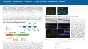 科学海报PneumaCult™: An Integrated Culture Medium System for in vitro Human Airway Modeling
 沪公网安备31010102008431号