Bilkovski R et al. (FEB 2010)
The Journal of biological chemistry 285 9 6170--8
Role of WNT-5a in the determination of human mesenchymal stem cells into preadipocytes.
Increasing adipocyte size as well as numbers is important in the development of obesity and type 2 diabetes,with adipocytes being generated from mesenchymal precursor cells. This process includes the determination of mesenchymal stem cells (MSC) into preadipocytes (PA) and the differentiation of PA into mature fat cells. Although the process of differentiation has been highly investigated,the determination in humans is poorly understood. In this study,we compared human MSC and human committed PA on a cellular and molecular level to gain further insights into the regulatory mechanisms in the determination process. Both cell types showed similar morphology and expression patterns of common mesenchymal and hematopoietic surface markers. However,although MSC were able to differentiate into adipocytes and osteocytes,PA were only able to undergo adipogenesis,indicating that PA lost their multipotency during determination. WNT-5a expression showed significantly higher levels in MSC compared with PA suggesting that WNT-5a down-regulation might be important in the determination process. Indeed,incubation of human MSC in medium containing neutralizing WNT-5a antibodies abolished their ability to undergo osteogenesis,although adipogenesis was still possible. An opposite effect was achieved using recombinant WNT-5a protein. On a molecular level,WNT-5a was found to promote c-Jun N-terminal kinase-dependent intracellular signaling in MSC. Activation of this noncanonical pathway resulted in the induction of osteopontin expression further indicating pro-osteogenic effects of WNT-5a. Our data suggest that WNT-5a is necessary to maintain osteogenic potential of MSC and that inhibition of WNT-5a signaling therefore plays a role in their determination into PA in humans.
View Publication
产品类型:
产品号#:
72642
产品名:
SP600125
Lagarkova MA et al. (MAR 2010)
Cell Cycle 9 5 937--46
Induction of pluripotency in human endothelial cells resets epigenetic profile on genome scale
Reprogramming of a limited number of human cell types has been achieved through ectopic expression of four transcription factors to yield induced pluripotent stem (iPS) cells that closely resemble human embryonic stem cells (ESCs). Here,we determined functional and epigenetic properties of iPS cells generated from human umbilical vein endothelial cells (HUVEC) by conventional method of direct reprogramming. Retroviral overexpression of four transcription factors resets HUVEC to the pluripotency. Human endothelial cell-derived iPS (endo-iPS) cells were similar to human ESCs in morphology,gene expression,in vitro and in vivo differentiation capacity. Endo-iPS cells were efficiently differentiated in vitro into endothelial cells. Using genome-wide methylation profiling we show that promoter elements of endothelial specific genes were methylated following reprogramming whereas pluripotency-related gene promoters were hypomethylated similar to levels observed in ESCs. Genome-wide methylation analysis of CpG sites located in the functional regions of over than 14,000 genes indicated that human endo-iPS cells were highly similar to human ES cells,although differences in methylation levels of 46 genes were found. Overall CpG methylation of promoter regions in the pluripotent cells was higher than in somatic. We also show that during reprogramming female human endo-iPS cells exhibited reactivation of the somatically silenced X chromosome. Our findings demonstrate that iPS cells can be generated from human endothelial cells and reprogramming resets epigenetic status of endothelial cells to pluripotency.
View Publication
产品类型:
产品号#:
05850
05857
05870
05875
85850
85857
85870
85875
产品名:
mTeSR™1
mTeSR™1
Tominaga S et al. (JAN 2005)
Biochemical and biophysical research communications 326 2 499--504
Negative regulation of adipogenesis from human mesenchymal stem cells by Jun N-terminal kinase.
Human mesenchymal stem cells (hMSCs) are capable of differentiating into several cell types including adipocytes,osteoblasts,and chondrocytes,under appropriate culture conditions. We found that SP600125,an inhibitor of Jun N-terminal kinase (JNK),promoted adipogenesis whereas it repressed osteogenesis from hMSCs. SP600125 increased the expression of adipogenic transcription factors,CCAAT/enhancer-binding proteins alpha and beta as well as peroxisome proliferator-activated receptor gamma2,which suggested that the chemical acted on the early steps of transcriptional regulatory cascade in adipogenesis. A gene reporter assay showed that SP600125 and a dominant negative JNK promoted a transcriptional activity dependent on the cAMP-response element (CRE). Thus,JNK represses adipogenesis from hMSCs probably by,at least in part,inhibiting the transactivating function of CRE-binding protein. Another action of JNK,phosphorylation at Ser(307) of insulin receptor substrate-1,was also predicted to contribute to the repression of adipogenesis.
View Publication
产品类型:
产品号#:
72642
产品名:
SP600125
Cho HH et al. (OCT 2005)
Journal of cellular biochemistry 96 3 533--42
Induction of osteogenic differentiation of human mesenchymal stem cells by histone deacetylase inhibitors.
Valproic acid (VPA) has been used as an anticonvulsant agent for the treatment of epilepsy,as well as a mood stabilizer for the treatment of bipolar disorder,for several decades. The mechanism of action for these effects remains to be elucidated and is most likely multifactorial. Recently,VPA has been reported to inhibit histone deacetylase (HDAC) and HDAC has been reported to play roles in differentiation of mammalian cells. In this study,the effects of HDAC inhibitors on differentiation and proliferation of human adipose tissue-derived stromal cells (hADSC) and bone marrow stromal cells (hBMSC) were determined. VPA increased osteogenic differentiation in a dose dependent manner. The pretreatment of VPA before induction of differentiation also showed stimulatory effects on osteogenic differentiation of hMSC. Trichostatin A (TSA),another HDAC inhibitor,also increased osteogenic differentiation,whereas valpromide (VPM),a structural analog of VPA which does not possess HDAC inhibitory effects,did not show any effect on osteogenic differentiation on hADSC. RT-PCR and Real-time PCR analysis revealed that VPA treatment increased osterix,osteopontin,BMP-2,and Runx2 expression. The addition of noggin inhibited VPA-induced potentiation of osteogenic differentiation. VPA inhibited proliferation of hADSC and hBMSC. Our results suggest that VPA enhance osteogenic differentiation,probably due to inhibition of HDAC,and could be useful for in vivo bone engineering using hMSC.
View Publication
产品类型:
产品号#:
72292
产品名:
丙戊酸(钠盐)
Vallier L et al. (OCT 2005)
Journal of cell science 118 Pt 19 4495--509
Activin/Nodal and FGF pathways cooperate to maintain pluripotency of human embryonic stem cells.
Maintenance of pluripotency is crucial to the mammalian embryo's ability to generate the extra-embryonic and embryonic tissues that are needed for intrauterine survival and foetal development. The recent establishment of embryonic stem cells from human blastocysts (hESCs) provides an opportunity to identify the factors supporting pluripotency at early stages of human development. Using this in vitro model,we have recently shown that Nodal can block neuronal differentiation,suggesting that TGFbeta family members are involved in cell fate decisions of hESCs,including preservation of their pluripotency. Here,we report that Activin/Nodal signalling through Smad2/3 activation is necessary to maintain the pluripotent status of hESCs. Inhibition of Activin/Nodal signalling by follistatin and by overexpression of Lefty or Cerberus-Short,or by the Activin receptor inhibitor SB431542,precipitates hESC differentiation. Nevertheless,neither Nodal nor Activin is sufficient to sustain long-term hESC growth in a chemically defined medium without serum. Recent studies have shown that FGF2 can also maintain long-term expression of pluripotency markers,and we find that inhibition of the FGF signalling pathway by the tyrosine kinase inhibitor SU5402 causes hESC differentiation. However,this effect of FGF on hESC pluripotency depends on Activin/Nodal signalling,because it is blocked by SB431542. Finally,long-term maintenance of in-vitro pluripotency can be achieved with a combination of Activin or Nodal plus FGF2 in the absence of feeder-cell layers,conditioned medium or Serum Replacer. These findings suggest that the Activin/Nodal pathway maintains pluripotency through mechanism(s) in which FGF acts as a competence factor and therefore provide further evidence of distinct mechanisms for preservation of pluripotency in mouse and human ESCs.
View Publication
产品类型:
产品号#:
72232
72234
100-1051
产品名:
SB431542(水合物)
SB431542(水合物)
SB431542(水合物)
Richards GR et al. ( 2006)
Journal of neurochemistry 97 1 201--210
The JAK3 inhibitor WHI-P154 prevents PDGF-evoked process outgrowth in human neural precursor cells.
The prospect of manipulating endogenous neural stem cells to replace damaged tissue and correct functional deficits offers a novel mechanism for treating a variety of CNS disorders. The aim of this study was to investigate pathways controlling neurite outgrowth in human neural precursor cells,in particular in response to platelet-derived growth factor (PDGF). PDGF-AA,-AB and -BB were found to initiate calcium signalling and produce robust increases in neurite outgrowth. PDGF-induced outgrowth of Tuj1-positive precursors was abolished by the addition of EGTA,suggesting that calcium entry is a critical part of the signalling pathway. Wortmannin and PD098059 failed to inhibit PDGF-induced outgrowth. Clostridium Toxin B increased the amount of PDGF-induced neurite branching but had no effect on basal levels. In contrast,WHI-P154,an inhibitor of Janus protein tyrosine kinase (JAK3),Hck and Syk,prevented PDGF-induced neurite outgrowth. PDGF activates multiple signalling pathways with considerable potential for cross-talk. This study has highlighted the complexity of the pathways leading to neurite outgrowth in human neural precursors,and provided initial evidence to suggest that calcium entry is critical in producing the morphological changes observed.
View Publication
Thayanithy V et al. (APR 2014)
Experimental Cell Research 323 1 178--188
Tumor exosomes induce tunneling nanotubes in lipid raft-enriched regions of human mesothelioma cells
Tunneling nanotubes (TnTs) are long,non-adherent,actin-based cellular extensions that act as conduits for transport of cellular cargo between connected cells. The mechanisms of nanotube formation and the effects of the tumor microenvironment and cellular signals on TnT formation are unknown. In the present study,we explored exosomes as potential mediators of TnT formation in mesothelioma and the potential relationship of lipid rafts to TnT formation. Mesothelioma cells co-cultured with exogenous mesothelioma-derived exosomes formed more TnTs than cells cultured without exosomes within 24-48. h; and this effect was most prominent in media conditions (low-serum,hyperglycemic medium) that support TnT formation (1.3-1.9-fold difference). Fluorescence and electron microscopy confirmed the purity of isolated exosomes and revealed that they localized predominantly at the base of and within TnTs,in addition to the extracellular environment. Time-lapse microscopic imaging demonstrated uptake of tumor exosomes by TnTs,which facilitated intercellular transfer of these exosomes between connected cells. Mesothelioma cells connected via TnTs were also significantly enriched for lipid rafts at nearly a 2-fold higher number compared with cells not connected by TnTs. Our findings provide supportive evidence of exosomes as potential chemotactic stimuli for TnT formation,and also lipid raft formation as a potential biomarker for TnT-forming cells. textcopyright 2014 Elsevier Inc.
View Publication
产品类型:
产品号#:
05850
05857
05870
05875
85850
85857
85870
85875
产品名:
mTeSR™1
mTeSR™1
Zhang K et al. (JAN 2014)
Protein and Cell 5 1 48--58
Direct conversion of human fibroblasts into retinal pigment epithelium-like cells by defined factors
The generation of functional retinal pigment epithelium (RPE) is of great therapeutic interest to the field of regenerative medicine and may provide possible cures for retinal degenerative diseases,including age-related macular degeneration (AMD). Although RPE cells can be produced from either embryonic stem cells or induced pluripotent stem cells,direct cell reprogramming driven by lineagedetermining transcription factors provides an immediate route to their generation. By monitoring a human RPE specific Best1::GFP reporter,we report the conversion of human fibroblasts into RPE lineage using defined sets of transcription factors. We found that Best1::GFP positive cells formed colonies and exhibited morphological and molecular features of early stage RPE cells. Moreover,they were able to obtain pigmentation upon activation of Retinoic acid (RA) and Sonic Hedgehog (SHH) signaling pathways. Our study not only established an ideal platform to investigate the transcriptional network regulating the RPE cell fate determination,but also provided an alternative strategy to generate functional RPE cells that complement the use of pluripotent stem cells for disease modeling,drug screening,and cell therapy of retinal degeneration.
View Publication
产品类型:
产品号#:
05850
05857
05870
05875
85850
85857
85870
85875
产品名:
mTeSR™1
mTeSR™1
Gordon DJ et al. (JUN 2015)
Oncogene 35 August 1--11
Modeling the initiation of Ewing sarcoma tumorigenesis in differentiating human embryonic stem cells.
Oncogenic transformation in Ewing sarcoma tumors is driven by the fusion oncogene EWS-FLI1. However,despite the well-established role of EWS-FLI1 in tumor initiation,the development of models of Ewing sarcoma in human cells with defined genetic elements has been challenging. Here,we report a novel approach to model the initiation of Ewing sarcoma tumorigenesis that exploits the developmental and pluripotent potential of human embryonic stem cells. The inducible expression of EWS-FLI1 in embryoid bodies,or collections of differentiating stem cells,generates cells with properties of Ewing sarcoma tumors,including characteristics of transformation. These cell lines exhibit anchorage-independent growth,a lack of contact inhibition and a strong Ewing sarcoma gene expression signature. Furthermore,these cells also demonstrate a requirement for the persistent expression of EWS-FLI1 for cell survival and growth,which is a hallmark of Ewing sarcoma tumors.Oncogene advance online publication,12 October 2015; doi:10.1038/onc.2015.368.
View Publication
产品类型:
产品号#:
05850
05857
05870
05875
07920
05893
85850
85857
85870
85875
07922
产品名:
ACCUTASE™
AggreWell™ EB形成培养基
mTeSR™1
mTeSR™1
ACCUTASE™
Janson C et al. (OCT 2015)
Cytogenetic and Genome Research 146 4 251--260
Replication Stress and Telomere Dysfunction Are Present in Cultured Human Embryonic Stem Cells
Replication stress causes DNA damage at fragile sites in the genome. DNA damage at telomeres can initiate breakage-fusion-bridge cycles and chromosome instability,which can result in replicative senescence or tumor formation. Little is known about the extent of replication stress or telomere dysfunction in human embryonic stem cells (hESCs). hESCs are grown in culture with the expectation of being used therapeutically in humans,making it important to minimize the levels of replication stress and telomere dysfunction. Here,the hESC line UCSF4 was cultured in a defined medium with growth factor Activin A,exogenous nucleosides,or DNA polymerase inhibitor aphidicolin. We used quantitative fluorescence in situ hybridization to analyze individual telomeres for dysfunction and observed that it can be increased by aphidicolin or Activin A. In contrast,adding exogenous nucleosides relieved dysfunction,suggesting that telomere dysfunction results from replication stress. Whether these findings can be applied to other hESC lines remains to be determined. However,because the loss of telomeres can lead to chromosome instability and cancer,we conclude that hESCs grown in culture for future therapeutic purposes should be routinely checked for replication stress and telomere dysfunction.
View Publication


 EasySep™小鼠TIL(CD45)正选试剂盒





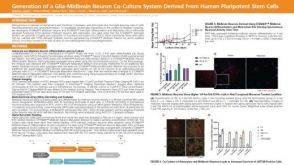 科学海报Generation of a Glia-Midbrain Neuron Co-Culture System Derived From Human Pluripotent Stem Cells
 新闻TeSR™-E8™ and STEMdiff™: New Products to Advance Research on Human Pluripotent Stem Cells
 沪公网安备31010102008431号