Leung HW et al. (FEB 2011)
Tissue engineering. Part C,Methods 17 2 165--72
Agitation can induce differentiation of human pluripotent stem cells in microcarrier cultures.
One of the factors that can impact human embryonic stem cell expansion in stirred microcarrier culture reactors is mechanical stress caused by agitation. Therefore,we have investigated the effects of agitation on human embryonic stem cell growth and expression of pluripotent markers. Agitation of HES-2 cell line in microcarrier cultures in stirred spinner and agitated six-well plates did not affect expression of pluripotent markers,cell viability,and cell doubling times even after seven passages. However,HES-3 cell line was found to be shear sensitive,showing downregulation of three pluripotent markers Oct-4,mAb 84,and Tra-1-60,and lower cell densities in agitated as compared with static cultures,even after one passage. Cell viability was unaffected. The HES-3-agitated cultures showed increased expression of genes and proteins of the three germ layers. We were unable to prevent loss of pluripotent markers or restore doubling times in agitated HES-3 microcarrier cultures by addition of five different known cell protective polymers. In addition,the human induced pluripotent cell line IMR90 was also shown to differentiate in agitated conditions. These results indicate that the effect of agitation on cell growth and differentiation is cell line specific. We assume that the changes in the growth and differentiation of the agitation-sensitive (HES-3) cell line do not result from the effect of shear stress directly on cell viability,but rather by signaling effects that influence the cells to differentiate resulting in slower growth.
View Publication
产品类型:
产品号#:
05850
05857
05870
05875
85850
85857
85870
85875
产品名:
mTeSR™1
mTeSR™1
Xu C et al. (JAN 2011)
Regenerative medicine 6 1 53--66
Efficient generation and cryopreservation of cardiomyocytes derived from human embryonic stem cells.
AIM Human embryonic stem cells (hESCs) represent a novel cell source to treat diseases such as heart failure and for use in drug screening. In this study,we aim to promote efficient generation of cardiomyocytes from hESCs by combining the current optimal techniques of controlled growth of undifferentiated cells and specific induction for cardiac differentiation. We also aim to examine whether these methods are scalable and whether the differentiated cells can be cryopreserved. METHODS & RESULTS hESCs were maintained without conditioned medium or feeders and were sequentially treated with activin A and bone morphogenetic protein-4 in a serum-free medium. This led to differentiation into cell populations containing high percentages of cardiomyocytes. The differentiated cells expressed appropriate cardiomyocyte markers and maintained contractility in culture,and the majority of the cells displayed working chamber (atrial and ventricular) type electrophysiological properties. In addition,the cell growth and differentiation process was adaptable to large culture formats. Moreover,the cardiomyocytes survived following cryopreservation,and viable cardiac grafts were detected after transplantation of cryopreserved cells into rat hearts following myocardial infarctions. CONCLUSION These results demonstrate that cardiomyocytes of high quality can be efficiently generated and cryopreserved using hESCs maintained in serum-free medium,a step forward towards the application of these cells to human clinical use or drug discovery.
View Publication
产品类型:
产品号#:
07930
07931
07940
07955
07956
07959
07954
100-1061
07952
产品名:
CryoStor® CS10
CryoStor® CS10
CryoStor® CS10
CryoStor® CS10
CryoStor® CS10
CryoStor® CS10
CryoStor® CS10
Mehta A et al. (SEP 2011)
Cardiovascular Research 91 4 577--86
Pharmacological response of human cardiomyocytes derived from virus-free induced pluripotent stem cells.
AIMS: Generation of human induced pluripotent stem cell (hiPSC) lines by reprogramming of fibroblast cells with virus-free methods offers unique opportunities for translational cardiovascular medicine. The aim of the study was to reprogramme fibroblast cells to hiPSCs and to study cardiomyogenic properties and ion channel characteristics of the virus-free hiPSC-derived cardiomyocytes. METHODS AND RESULTS: The hiPSCs generated by episomal vectors generated teratomas in severe combined immunodeficient mice,readily formed embryoid bodies,and differentiated into cardiomyocytes with comparable efficiency to human embryonic stem cells. Temporal gene expression of these hiPSCs indicated that differentiation of cardiomyocytes was initiated by increasing expression of cardio/mesodermal markers followed by cardiac-specific transcription factors,structural,and ion channel genes. Furthermore,the cardiomyocytes showed characteristic cross-striations of sarcomeric proteins and expressed calcium-handling and ion channel proteins,confirming their cardiac ontogeny. Microelectrode array recordings established the electrotonic development of a functional syncytium that responded predictably to pharmacologically active drugs. The cardiomyocytes showed a chronotropic dose-response (0.1-10 µM) to isoprenaline and Bay K 8644. Furthermore,carbamycholine (5 µM) suppressed the response to isoprenaline,while verapamil (2.5 µM) blocked Bay K 8644-induced inotropic activity. Moreover,verapamil (1 µM) reduced the corrected field potential duration by 45%,tetrodotoxin (10 µM) shortened the minimal field potential by 40%,and E-4031 (50 nM) prolonged field repolarization. CONCLUSION: Virus-free hiPSCs differentiate efficiently into cardiomyocytes with cardiac-specific molecular,structural,and functional properties that recapitulate the developmental ontogeny of cardiogenesis. These results,coupled with the potential to generate patient-specific hiPSC lines,hold great promise for the development of an in vitro platform for drug pharmacogenomics,disease modelling,and regenerative medicine.
View Publication
产品类型:
产品号#:
05850
05857
05870
05875
85850
85857
85870
85875
产品名:
mTeSR™1
mTeSR™1
Vilchez D et al. (SEP 2012)
Nature 489 7415 304--308
Increased proteasome activity in human embryonic stem cells is regulated by PSMD11
Embryonic stem cells can replicate continuously in the absence of senescence and,therefore,are immortal in culture. Although genome stability is essential for the survival of stem cells,proteome stability may have an equally important role in stem-cell identity and function. Furthermore,with the asymmetric divisions invoked by stem cells,the passage of damaged proteins to daughter cells could potentially destroy the resulting lineage of cells. Therefore,a firm understanding of how stem cells maintain their proteome is of central importance. Here we show that human embryonic stem cells (hESCs) exhibit high proteasome activity that is correlated with increased levels of the 19S proteasome subunit PSMD11 (known as RPN-6 in Caenorhabditis elegans) and a corresponding increased assembly of the 26S/30S proteasome. Ectopic expression of PSMD11 is sufficient to increase proteasome assembly and activity. FOXO4,an insulin/insulin-like growth factor-I (IGF-I) responsive transcription factor associated with long lifespan in invertebrates,regulates proteasome activity by modulating the expression of PSMD11 in hESCs. Proteasome inhibition in hESCs affects the expression of pluripotency markers and the levels of specific markers of the distinct germ layers. Our results suggest a new regulation of proteostasis in hESCs that links longevity and stress resistance in invertebrates to hESC function and identity.
View Publication
产品类型:
产品号#:
05850
05857
05870
05875
85850
85857
85870
85875
产品名:
mTeSR™1
mTeSR™1
T. Halegua et al. (Jan 2025)
Nature Communications 16
Delivery of Prime editing in human stem cells using pseudoviral NanoScribes particles
Prime Editing can rewrite genes in living cells by allowing point mutations,deletions,or insertion of small DNA sequences with high precision. However,its safe and efficient delivery into human stem cells remains a technical challenge. In this report,we engineer Nanoscribes,virus-like particles that encapsidate ribonucleoprotein complexes of the Prime Editing system and allow their delivery into recipient cells. We identify key features that unlock the potential of Nanoscribes,including the use of multiple fusogens,the improvement of pegRNAs structures,their encoding by a Pol II system and the optimization of Prime-Editors. Nanoscribes edit HEK293T with an efficiency of 68% at the HEK3 locus with increased fidelity over DNA-transfection and support pegRNA-multiplexing. Importantly,Nanoscribes permit editing of myoblasts,hiPSCs and hiPSCs-derived hematopoietic stem cells with an editing efficiency up to 25%. Nanoscribes is an asset for development of next generation genome editing approaches using VLPs. Subject terms: CRISPR-Cas9 genome editing,Genetic vectors,Nanoparticles
View Publication
Keung W et al. (SEP 2016)
Scientific reports 6 34154
Non-cell autonomous cues for enhanced functionality of human embryonic stem cell-derived cardiomyocytes via maturation of sarcolemmal and mitochondrial KATP channels.
Human embryonic stem cells (hESCs) is a potential unlimited ex vivo source of ventricular (V) cardiomyocytes (CMs),but hESC-VCMs and their engineered tissues display immature traits. In adult VCMs,sarcolemmal (sarc) and mitochondrial (mito) ATP-sensitive potassium (KATP) channels play crucial roles in excitability and cardioprotection. In this study,we aim to investigate the biological roles and use of sarcKATP and mitoKATP in hESC-VCM. We showed that SarcIK,ATP in single hESC-VCMs was dormant under baseline conditions,but became markedly activated by cyanide (CN) or the known opener P1075 with a current density that was ˜8-fold smaller than adult; These effects were reversible upon washout or the addition of GLI or HMR1098. Interestingly,sarcIK,ATP displayed a ˜3-fold increase after treatment with hypoxia (5% O2). MitoIK,ATP was absent in hESC-VCMs. However,the thyroid hormone T3 up-regulated mitoIK,ATP,conferring diazoxide protective effect on T3-treated hESC-VCMs. When assessed using a multi-cellular engineered 3D ventricular cardiac micro-tissue (hvCMT) system,T3 substantially enhanced the developed tension by 3-folds. Diazoxide also attenuated the decrease in contractility induced by simulated ischemia (1% O2). We conclude that hypoxia and T3 enhance the functionality of hESC-VCMs and their engineered tissues by selectively acting on sarc and mitoIK,ATP.
View Publication
Beloti M et al. (JUL 2005)
Cell biology international 29 7 537--41
Purmorphamine enhances osteogenic activity of human osteoblasts derived from bone marrow mesenchymal cells.
Purmorphamine is a novel small molecule with osteogenesis-inducing activity in multipotent mesenchymal progenitor cells,but there has been no evaluation of its effect on human cells to date. The aim of this study was to investigate the induction of osteogenic activity by purmorphamine in human osteoblasts differentiated from bone marrow mesenchymal cells. Cells were cultured in 24-well plates at a density of 2x10(4)/well in medium containing 1,2 or 3 microM purmorphamine,or vehicle. At 7,14 and 21 days,cell proliferation,viability,and alkaline phosphatase (ALP) activity were evaluated. Bone-like nodule formation was evaluated at 21 days. Purmorphamine did not affect cell proliferation or viability,but increased ALP activity and bone-like nodule formation. These results indicate that events related to osteoblast differentiation,including increased ALP activity and bone-like nodule formation,are enhanced by purmorphamine.
View Publication
产品类型:
产品号#:
72202
72204
100-1049
产品名:
Purmorphamine
Purmorphamine
Purmorphamine
Tsang JY-S et al. (JUL 2006)
Journal of leukocyte biology 80 1 145--51
Altered proximal T cell receptor (TCR) signaling in human CD4+CD25+ regulatory T cells.
CD4+CD25+ regulatory T cells play an important role in peripheral tolerance. Upon T cell receptor (TCR)-mediated activation,the cells fail to proliferate but are induced to have a suppressor function. The intracellular signaling events that lead to their responses have not been elucidated. In this study,we have examined the proximal TCR signaling events in freshly isolated human CD4+CD25+ regulatory T cells after TCR ligation. In contrast to CD4+CD25- T cells,TCR ligation of CD4+CD25+ regulatory T cells by anti-CD3 cross-linking resulted in a lower calcium influx and extracellular signal-regulated kinase 1/2 phosphorylation. Examination of the CD3zeta chain phosphorylation status indicated that CD4+CD25+ regulatory T cells have poor phosphorylation of the protein and consequently,reduced recruitment of zeta-associated protein-70 to the TCR immunoreceptor tyrosine motif. The adaptor protein,Src homology 2 domain-containing leukocyte phosphoprotein of 76 kDa,which relays signals to downstream signaling components,also showed reduced phosphorylation,which correlated with reduced VAV guanine nucleotide exchange factors association. Consistent with other findings,the defect is accompanied with impaired actin cap formation,implicating a failure of actin remodeling of the cells. Together,our results demonstrate that CD4+CD25+ regulatory T cells have altered TCR proximal signaling pathways,which could be critical for inducing the distinct behavior of these cells.
View Publication

 EasySep™小鼠TIL(CD45)正选试剂盒


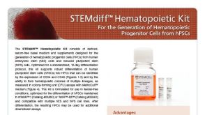 产品手册STEMdiff™ Hematopoietic Kit
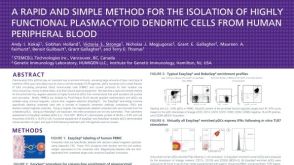 科学海报Rapid Cell Isolation of Highly Functional Plasmacytoid Dendritic Cells from Human Peripheral Blood

 沪公网安备31010102008431号