High capacity nanoporous silicon carrier for systemic delivery of gene silencing therapeutics.
Gene silencing agents such as small interfering RNA (siRNA) and microRNA offer the promise to modulate expression of almost every gene for the treatment of human diseases including cancer. However,lack of vehicles for effective systemic delivery to the disease organs has greatly limited their in vivo applications. In this study,we developed a high capacity polycation-functionalized nanoporous silicon (PCPS) platform comprised of nanoporous silicon microparticles functionalized with arginine-polyethyleneimine inside the nanopores for effective delivery of gene silencing agents. Incubation of MDA-MB-231 human breast cancer cells with PCPS loaded with STAT3 siRNA (PCPS/STAT3) or GRP78 siRNA (PCPS/GRP78) resulted in 91 and 83% reduction of STAT3 and GRP78 gene expression in vitro. Treatment of cells with a microRNA-18a mimic in PCPS (PCPS/miR-18) knocked down 90% expression of the microRNA-18a target gene ATM. Systemic delivery of PCPS/STAT3 siRNA in murine model of MDA-MB-231 breast cancer enriched particles in tumor tissues and reduced STAT3 expression in cancer cells,causing significant reduction of cancer stem cells in the residual tumor tissue. At the therapeutic dosage,PCPS/STAT3 siRNA did not trigger acute immune response in FVB mice,including changes in serum cytokines,chemokines,and colony-stimulating factors. In addition,weekly dosing of PCPS/STAT3 siRNA for four weeks did not cause signs of subacute toxicity based on changes in body weight,hematology,blood chemistry,and major organ histology. Collectively,the results suggest that we have developed a safe vehicle for effective delivery of gene silencing agents.
View Publication
产品类型:
产品号#:
05620
产品名:
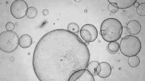
MammoCult™ 人源培养基套装
Gkountela S et al. (APR 2014)
Stem Cell Reviews and Reports 10 2 230--239
PRMT5 is required for human embryonic stem cell proliferation but not pluripotency.
Human pluripotent stem cells (PSCs) are critical in vitro tools forbackslashnunderstanding mechanisms that regulate lineage differentiation inbackslashnthe human embryo as well as a potentially unlimited supply of stembackslashncells for regenerative medicine. Pluripotent human and mouse embryonicbackslashnstem cells (ESCs) derived from the inner cell mass of blastocystsbackslashnshare a similar transcription factor network to maintain pluripotencybackslashnand self-renewal,yet there are considerable molecular differencesbackslashnreflecting the diverse environments in which mouse and human ESCsbackslashnare derived. In the current study we evaluated the role of Proteinbackslashnarginine methyltransferase 5 (PRMT5) in human ESC (hESC) self-renewalbackslashnand pluripotency given its critical role in safeguarding mouse ESCbackslashnpluripotency. Unlike the mouse,we discovered that PRMT5 has no rolebackslashnin hESC pluripotency. Using microarray analysis we discovered thatbackslashna significant depletion in PRMT5 RNA and protein from hESCs changedbackslashnthe expression of only 78 genes,with the majority being repressed.backslashnFunctionally,we discovered that depletion of PRMT5 had no effectbackslashnon expression of OCT4,NANOG or SOX2,and did not prevent teratomabackslashnformation. Instead,we show that PRMT5 functions in hESCs to regulatebackslashnproliferation in the self-renewing state by regulating the fractionbackslashnof cells in Gap 1 (G1) of the cell cycle and increasing expressionbackslashnof the G1 cell cycle inhibitor P57. Taken together our data unveilsbackslashna distinct role for PRMT5 in hESCs and identifies P57 as new target.
View Publication
产品类型:
产品号#:
05850
05857
05870
05875
85850
85857
85870
85875
产品名:
mTeSR™1
mTeSR™1
Date Y et al. ( 2014)
Analytical Chemistry 86 6 2989--96
Label-free impedimetric immunoassay for trace levels of polychlorinated biphenyls in insulating oil
A rapid,ultrasensitive,and practical label-free impedimetric immunoassay for measuring trace levels of total polychlorinated biphenyls (PCBs) in insulating oil was developed. First,we developed a novel monoclonal antibody (RU6F9) for PCBs by using a designed immunogen and characterized its binding affinity for a commercial mixtures of PCBs and its main congeners. A micro comblike gold electrode was fabricated,and the antibody was covalently immobilized on the electrode through a self-assembled monolayer formed by dithiobis-N-succinimidyl propionate. The antigen-binding event on the surface of the functionalized electrode was determined as the change in charge transfer resistance by using electrochemical impedance spectroscopy. The resulting impedimetric immunoassay in aqueous solution achieved a wide determination range (0.01-10 μg/L) and a low detection limit (LOD) of 0.001 μg/L,which was 100-fold more sensitive than a conventional flow-based immunoassay for PCBs. By combining the impedimetric immunoassay with a cleanup procedure for insulating oil utilizing a multilayer cleanup column followed by DMSO partitioning,an LOD of 0.052 mg/kg-oil was achieved,which satisfied the Japanese regulation criterion of 0.5 mg/kg-oil. Finally,the immunoassay was employed to determine total PCB levels in actual used insulating oils (n = 33) sampled from a used transformer containing trace levels of PCBs,and the results agreed well with the Japanese official method (HRGC/HRMS).
View Publication
产品类型:
产品号#:
03800
产品名:
ClonaCell™-HY杂交瘤试剂盒
Malara A et al. (FEB 2011)
Blood 117 8 2476--83
Megakaryocyte-matrix interaction within bone marrow: new roles for fibronectin and factor XIII-A.
The mechanisms by which megakaryocytes (MKs) differentiate and release platelets into the circulation are not well understood. However,growing evidence indicates that a complex regulatory mechanism involving MK-matrix interactions may contribute to the quiescent or permissive microenvironment related to platelet release within bone marrow. To address this hypothesis,in this study we demonstrate that human MKs express and synthesize cellular fibronectin (cFN) and transglutaminase factor XIII-A (FXIII-A). We proposed that these 2 molecules are involved in a new regulatory mechanism of MK-type I collagen interaction in the osteoblastic niche. In particular,we demonstrate that MK adhesion to type I collagen promotes MK spreading and inhibits pro-platelet formation through the release and relocation to the plasma membrane of cFN. This regulatory mechanism is dependent on the engagement of FN receptors at the MK plasma membrane and on transglutaminase FXIII-A activity. Consistently,the same mechanism regulated the assembly of plasma FN (pFN) by adherent MKs to type I collagen. In conclusion,our data extend the knowledge of the mechanisms that regulate MK-matrix interactions within the bone marrow environment and could serve as an important step for inquiring into the origins of diseases such as myelofibrosis and congenital thrombocytopenias that are still poorly understood.
View Publication
S. Chatterjee et al. (Apr 2024)
Cellular and Molecular Life Sciences: CMLS 81 1
Telomerase is essential for cardiac differentiation and sustained metabolism of human cardiomyocytes
Telomeres as the protective ends of linear chromosomes,are synthesized by the enzyme telomerase (TERT). Critically short telomeres essentially contribute to aging-related diseases and are associated with a broad spectrum of disorders known as telomeropathies. In cardiomyocytes,telomere length is strongly correlated with cardiomyopathies but it remains ambiguous whether short telomeres are the cause or the result of the disease. In this study,we employed an inducible CRISPRi human induced pluripotent stem cell (hiPSC) line to silence TERT expression enabling the generation of hiPSCs and hiPSC-derived cardiomyocytes with long and short telomeres. Reduced telomerase activity and shorter telomere lengths of hiPSCs induced global transcriptomic changes associated with cardiac developmental pathways. Consequently,the differentiation potential towards cardiomyocytes was strongly impaired and single cell RNA sequencing revealed a shift towards a more smooth muscle cell like identity in the cells with the shortest telomeres. Poor cardiomyocyte function and increased sensitivity to stress directly correlated with the extent of telomere shortening. Collectively our data demonstrates a TERT dependent cardiomyogenic differentiation defect,highlighting the CRISPRi TERT hiPSCs model as a powerful platform to study the mechanisms and consequences of short telomeres in the heart and also in the context of telomeropathies. The online version contains supplementary material available at 10.1007/s00018-024-05239-7.
View Publication
产品类型:
产品号#:
05230
产品名:
STEMdiff™ 三胚层分化试剂盒
(Apr 2025)
Communications Medicine 5
Drug and siRNA screens identify ROCK2 as a therapeutic target for ciliopathies
BackgroundPrimary cilia mediate vertebrate development and growth factor signalling. Defects in primary cilia cause inherited developmental conditions termed ciliopathies. Ciliopathies often present with cystic kidney disease,a major cause of early renal failure. Currently,only one drug,Tolvaptan,is licensed to slow the decline of renal function for the ciliopathy polycystic kidney disease. Novel therapeutic interventions are needed.MethodsWe screened clinical development compounds to identify those that reversed cilia loss due to siRNA knockdown. In parallel,we undertook a whole genome siRNA-based reverse genetics phenotypic screen to identify positive modulators of cilia formation.ResultsUsing a clinical development compound screen,we identify fasudil hydrochloride. Fasudil is a generic,off-patent drug that is a potent,broadly selective Rho-associated coiled-coil-containing protein kinase (ROCK) inhibitor. In parallel,the siRNA screen identifies ROCK2 and we demonstrate that ROCK2 is a key mediator of cilium formation and function through its possible effects on actin cytoskeleton remodelling.ConclusionsOur results indicate that specific ROCK2 inhibitors (e.g. belumosudil) could be repurposed for cystic kidney disease treatment. We propose that ROCK2 inhibition represents a novel,disease-modifying therapeutic approach for heterogeneous ciliopathies. Plain language summaryPrimary cilia are antennae-like structures on cells that are important for early development and healthy cell function. Defects in primary cilia can cause inherited diseases called ciliopathies. Ciliopathies often cause fluid-filled sacs,called cysts,that are a major cause of kidney disease and failure. There is currently one drug licensed to slow kidney disease progression,but it is poorly tolerated in patients. Therefore,new drugs are needed. In this study,we used screening assays to identify potential drugs and their targets that are effective in promoting the formation of primary cilia. Our results identified ROCK2 (Rho-associated coiled-coil-containing protein kinase 2),an inhibitor of protein signalling,as a key mediator of cilium function. These findings suggest that drugs that specifically target ROCK2 could be a potential treatment option for cystic kidney disease. Smith et al. use clinical development screen and whole genome siRNA-reverse genetics phenotypic screen to identify ROCK2,as a modulator of cilia formation and function via its effects on actin cytoskeleton remodelling. Repurposing ROCK2 is a viable treatment for ciliopathies,for which a limited therapeutic option is available.
View Publication
产品类型:
产品号#:
100-0276
100-1130
产品名:
mTeSR™ Plus
mTeSR™ Plus
(Jul 2024)
Molecular Therapy Oncology 32 3
T cell receptor-directed antibody-drug conjugates for the treatment of T cell-derived cancers
T cell-derived cancers are hallmarked by heterogeneity,aggressiveness,and poor clinical outcomes. Available targeted therapies are severely limited due to a lack of target antigens that allow discrimination of malignant from healthy T cells. Here,we report a novel approach for the treatment of T cell diseases based on targeting the clonally rearranged T cell receptor displayed by the cancerous T cell population. As a proof of concept,we identified an antibody with unique specificity toward a distinct T cell receptor (TCR) and developed antibody-drug conjugates,precisely recognizing and eliminating target T cells while preserving overall T cell repertoire integrity and cellular immunity. Our anti-TCR antibody-drug conjugates demonstrated effective receptor-mediated cell internalization,associated with induction of cancer cell death with strong signs of apoptosis. Furthermore,cell proliferation-inhibiting bystander effects observed on target-negative cells may contribute to the molecules’ anti-tumor properties precluding potential tumor escape mechanisms. To our knowledge,this represents the first anti-TCR antibody-drug conjugate designed as custom-tailored immunotherapy for T cell-driven pathologies. Graphical abstract Harald Kolmar and colleagues report a novel approach for the treatment of the difficult-to-treat T cell lymphoma/leukemia based on targeting the clonally rearranged T cell receptor expressed by the malignant T cell population. The developed antibody-drug conjugates precisely eliminate target T cells while preserving the integrity of the T cell repertoire and cellular immunity.
View Publication
产品类型:
产品号#:
17851
17851RF
100-0692
产品名:
EasySep™人CD3正选试剂盒II
RoboSep™ 人CD3正选试剂盒II
EasySep™人CD3正选试剂盒II
E. Park et al. (Jan 2026)
Light,Science & Applications 15
Label-free mid-infrared dichroism-sensitive photoacoustic microscopy for histostructural analysis of engineered heart tissues
Many biological tissues,such as cardiac muscle,tendons,and the cornea,exhibit highly organized microstructural alignment that is critical for mechanical and physiological functions. Disruptions in this structural organization are commonly associated with pathological conditions such as fibrosis,infarction,and cancer. However,conventional histological imaging techniques rely on immunofluorescence or histochemical staining,and they evaluate tissue alignment via non-physical 2D gradient-based calculation,which is labor-intensive,antibody-dependent,and prone to variability. Here,we demonstrate label-free mid-infrared dichroism-sensitive photoacoustic microscopy (MIR-DS-PAM),an analytical imaging system for cardiac tissue assessments. By combining molecular specificity with polarization sensitivity,this method selectively visualizes protein-rich engineered heart tissue (EHT) and quantifies the extracellular matrix (ECM) alignment without any labeling. The extracted dichroism-sensitive parameters,such as the degree of dichroism and the orientation angle,enable histostructural evaluation of tissue integrity and reveal diagnostic cues in fibrotic EHT. This technique offers a label-free analytical tool for fibrosis research and tissue engineering applications. Mid-infrared dichroism-sensitive photoacoustic microscopy enables label-free,quantitative histostructural analysis by combining spectral specificity and polarization sensitivity to visualize protein-rich components and evaluate anisotropic tissue alignment.
View Publication
E. Vokali et al. (jan 2020)
Nature communications 11 1 538
Lymphatic endothelial cells prime na\ive CD8+ T cells into memory cells under steady-state conditions."
Lymphatic endothelial cells (LECs) chemoattract na{\{i}}ve T cells and promote their survival in the lymph nodes and can cross-present antigens to na{\"{i}}ve CD8+ T cells to drive their proliferation despite lacking key costimulatory molecules. However the functional consequence of LEC priming of CD8+ T cells is unknown. Here we show that while many proliferating LEC-educated T cells enter early apoptosis the remainders comprise a long-lived memory subset with transcriptional metabolic and phenotypic features of central memory and stem cell-like memory T cells. In vivo these memory cells preferentially home to lymph nodes and display rapid proliferation and effector differentiation following memory recall and can protect mice against a subsequent bacterial infection. These findings introduce a new immunomodulatory role for LECs in directly generating a memory-like subset of quiescent yet antigen-experienced CD8+ T cells that are long-lived and can rapidly differentiate into effector cells upon inflammatory antigenic challenge."""
View Publication
产品类型:
产品号#:
19853
19853RF
产品名:
EasySep™小鼠CD8+ T细胞分选试剂盒
RoboSep™ 小鼠CD8+ T细胞分选试剂盒
Lei IL et al. (JAN 2015)
Journal of visualized experiments : JoVE January 52047. doi: 10.3791/52047.
Derivation of cardiac progenitor cells from embryonic stem cells.
Cardiac progenitor cells (CPCs) have the capacity to differentiate into cardiomyocytes,smooth muscle cells (SMC),and endothelial cells and hold great promise in cell therapy against heart disease. Among various methods to isolate CPCs,differentiation of embryonic stem cell (ESC) into CPCs attracts great attention in the field since ESCs can provide unlimited cell source. As a result,numerous strategies have been developed to derive CPCs from ESCs. In this protocol,differentiation and purification of embryonic CPCs from both mouse and human ESCs is described. Due to the difficulty of using cell surface markers to isolate embryonic CPCs,ESCs are engineered with fluorescent reporters activated by CPC-specific cre recombinase expression. Thus,CPCs can be enriched by fluorescence-activated cell sorting (FACS). This protocol illustrates procedures to form embryoid bodies (EBs) from ESCs for CPC specification and enrichment. The isolated CPCs can be subsequently cultured for cardiac lineage differentiation and other biological assays. This protocol is optimized for robust and efficient derivation of CPCs from both mouse and human ESCs.
View Publication

 EasySep™小鼠TIL(CD45)正选试剂盒


 实验方案How to Process Epithelial Organoids and Organoid-Derived Epithelial Monolayers for RNA Isolation
 28:34
线上讲座Key Regulatory Considerations for Moving Your Cell Therapy Research to the Clinic发布日期: 08/27/2024
28:34
线上讲座Key Regulatory Considerations for Moving Your Cell Therapy Research to the Clinic发布日期: 08/27/2024
 沪公网安备31010102008431号