Amelioration of murine beta-thalassemia through drug selection of hematopoietic stem cells transduced with a lentiviral vector encoding both gamma-globin and the MGMT drug-resistance gene.
Correction of murine models of beta-thalassemia has been achieved through high-level globin lentiviral vector gene transfer into mouse hematopoietic stem cells (HSCs). However,transduction of human HSCs is less robust and may be inadequate to achieve therapeutic levels of genetically modified erythroid cells. We therefore developed a double gene lentiviral vector encoding both human gamma-globin under the transcriptional control of erythroid regulatory elements and methylguanine methyltransferase (MGMT),driven by a constitutive cellular promoter. MGMT expression provides cellular resistance to alkylator drugs,which can be administered to kill residual untransduced,diseased HSCs,whereas transduced cells are protected. Mice transplanted with beta-thalassemic HSCs transduced with a gamma-globin/MGMT vector initially had subtherapeutic levels of red cells expressing gamma-globin. To enrich gamma-globin-expressing cells,transplanted mice were treated with the alkylator agent 1,3-bis-chloroethyl-1-nitrosourea. This resulted in significant increases in the number of gamma-globin-expressing red cells and the amount of fetal hemoglobin,leading to resolution of anemia. Selection of transduced HSCs was also obtained when cells were drug-treated before transplantation. Mice that received these cells demonstrated reconstitution with therapeutic levels of gamma-globin-expressing cells. These data suggest that MGMT-based drug selection holds promise as a modality to improve gene therapy for beta-thalassemia.
View Publication
产品类型:
产品号#:
09600
09650
产品名:
StemSpan™ SFEM
StemSpan™ SFEM
Bianco C et al. (JUN 2013)
Journal of cellular physiology 228 6 1174--1188
Regulation of human Cripto-1 expression by nuclear receptors and DNA promoter methylation in human embryonal and breast cancer cells.
Human Cripto-1 (CR-1) plays an important role in regulating embryonic development while also regulating various stages of tumor progression. However,mechanisms that regulate CR-1 expression during embryogenesis and tumorigenesis are still not well defined. In the present study,we investigated the effects of two nuclear receptors,liver receptor homolog (LRH)-1 and germ cell nuclear factor receptor (GCNF) and epigenetic modifications on CR-1 gene expression in NTERA-2 human embryonal carcinoma cells and in breast cancer cells. CR-1 expression in NTERA-2 cells was positively regulated by LRH-1 through direct binding to a DR0 element within the CR-1 promoter,while GCNF strongly suppressed CR-1 expression in these cells. In addition,the CR-1 promoter was unmethylated in NTERA-2 cells,while T47D,ZR75-1,and MCF7 breast cancer cells showed high levels of CR-1 promoter methylation and low CR-1 mRNA and protein expression. Treatment of breast cancer cells with a demethylating agent and histone deacetylase inhibitors reduced methylation of the CR-1 promoter and reactivated CR-1 mRNA and protein expression in these cells,promoting migration and invasion of breast cancer cells. Analysis of a breast cancer tissue array revealed that CR-1 was highly expressed in the majority of human breast tumors,suggesting that CR-1 expression in breast cancer cell lines might not be representative of in vivo expression. Collectively,these findings offer some insight into the transcriptional regulation of CR-1 gene expression and its critical role in the pathogenesis of human cancer.
View Publication
产品类型:
产品号#:
05620
产品名:
MammoCult™ 人源培养基套装
I. Elcheva et al. (jul 2014)
Nature communications 5 164 4372
Direct induction of haematoendothelial programs in human pluripotent stem cells by transcriptional regulators.
Advancing pluripotent stem cell technologies for modelling haematopoietic stem cell development and blood therapies requires identifying key regulators of haematopoietic commitment from human pluripotent stem cells (hPSCs). Here,by screening the effect of 27 candidate factors,we reveal two groups of transcriptional regulators capable of inducing distinct haematopoietic programs from hPSCs: pan-myeloid (ETV2 and GATA2) and erythro-megakaryocytic (GATA2 and TAL1). In both cases,these transcription factors directly convert hPSCs to endothelium,which subsequently transform into blood cells with pan-myeloid or erythro-megakaryocytic potential. These data demonstrate that two distinct genetic programs regulate the haematopoietic development from hPSCs and that both of these programs specify hPSCs directly to haemogenic endothelial cells. In addition,this study provides a novel method for the efficient induction of blood and endothelial cells from hPSCs via the overexpression of modified mRNA for the selected transcription factors.
View Publication
产品类型:
产品号#:
02625
05850
05857
05870
05875
78012
78012.1
78012.2
78012.3
78015
78015.1
78015.2
78015.3
78062
78062.1
78062.2
85850
85857
85870
85875
产品名:
重组人 G-CSF(E. coli表达)
重组人 G-CSF(E. coli表达)
重组人 G-CSF(E. coli表达)
Hu Recom G-CSF, 500 µg
重组人 GM-CSF(E. coli表达)
重组人 GM-CSF(E. coli表达)
重组人 GM-CSF(E. coli表达)
重组人 GM-CSF(E. coli表达)
重组人SCF(大肠杆菌表达)
重组人SCF(大肠杆菌表达)
重组人SCF(大肠杆菌表达)
mTeSR™1
mTeSR™1
Buono M et al. (AUG 2010)
The Journal of experimental medicine 207 8 1647--60
Self-renewal and differentiation of hematopoietic stem cells (HSCs) are balanced by the concerted activities of the fibroblast growth factor (FGF),Wnt,and Notch pathways,which are tuned by enzyme-mediated remodeling of heparan sulfate proteoglycans (HSPGs). Sulfatase modifying factor 1 (SUMF1) activates the Sulf1 and Sulf2 sulfatases that remodel the HSPGs,and is mutated in patients with multiple sulfatase deficiency. Here,we show that the FGF signaling pathway is constitutively activated in Sumf1(-/-) HSCs and hematopoietic stem progenitor cells (HSPCs). These cells show increased p-extracellular signal-regulated kinase levels,which in turn promote beta-catenin accumulation. Constitutive activation of FGF signaling results in a block in erythroid differentiation at the chromatophilic erythroblast stage,and of B lymphocyte differentiation at the pro-B cell stage. A reduction in mature myeloid cells and an aberrant development of T lymphocytes are also seen. These defects are rescued in vivo by blocking the FGF pathway in Sumf1(-/-) mice. Transplantation of Sumf1(-/-) HSPCs into wild-type mice reconstituted the phenotype of the donors,suggesting a cell autonomous defect. These data indicate that Sumf1 controls HSPC differentiation and hematopoietic lineage development through FGF and Wnt signaling.
View Publication
产品类型:
产品号#:
09600
09650
产品名:
StemSpan™ SFEM
StemSpan™ SFEM
Palmer DJ et al. ( 2016)
Molecular therapy. Nucleic acids 5 e372
Homology Requirements for Efficient, Footprintless Gene Editing at the CFTR Locus in Human iPSCs with Helper-dependent Adenoviral Vectors.
Helper-dependent adenoviral vectors mediate high efficiency gene editing in induced pluripotent stem cells without needing a designer nuclease thereby avoiding off-target cleavage. Because of their large cloning capacity of 37 kb,helper-dependent adenoviral vectors with long homology arms are used for gene editing. However,this makes vector construction and recombinant analysis difficult. Conversely,insufficient homology may compromise targeting efficiency. Thus,we investigated the effect of homology length on helper-dependent adenoviral vector targeting efficiency at the cystic fibrosis transmembrane conductance regulator locus in induced pluripotent stem cells and found a positive correlation. With 23.8 and 21.4 kb of homology,the frequencies of targeted recombinants were 50-64.6% after positive selection for vector integration,and 97.4-100% after negative selection against random integrations. With 14.8 kb,the frequencies were 26.9-57.1% after positive selection and 87.5-100% after negative selection. With 9.6 kb,the frequencies were 21.4 and 75% after positive and negative selection,respectively. With only 5.6 kb,the frequencies were 5.6-16.7% after positive selection and 50% after negative selection,but these were more than high enough for efficient identification and isolation of targeted clones. Furthermore,we demonstrate helper-dependent adenoviral vector-mediated footprintless correction of cystic fibrosis transmembrane conductance regulator mutations through piggyBac excision of the selectable marker. However,low frequencies (≤ 1 × 10(-3)) necessitated negative selection for piggyBac-excision product isolation.
View Publication
TLR7/8 signaling activation enhances the potency of human pluripotent stem cell-derived eosinophils in cancer immunotherapy for solid tumors
Efficient tumor T-cell infiltration is crucial for the effectiveness of T-cell-based therapies against solid tumors. Eosinophils play crucial roles in recruiting T cells in solid tumors. Our group has previously generated induced eosinophils (iEOs) from human pluripotent stem cells and exhibited synergistic efficacy with CAR-T cells in solid tumor inhibition. However,administrated eosinophils might influx into inflammatory lungs,posing a potential safety risk. Mitigating the safety concern and enhancing efficacy is a promising development direction for further application of eosinophils.MethodsWe developed a new approach to generate eosinophils with enhanced potency from human chemically reprogrammed induced pluripotent stem cells (hCiPSCs) with the Toll-like receptor (TLR) 7/8 signaling agonist R848.ResultsR848-activated iEOs (R-iEOs) showed significantly decreased influx to the inflamed lungs,indicating a lower risk of causing airway disorders. Furthermore,these R-iEOs had enhanced anti-tumor functions,preferably accumulated at tumor sites,and further increased T-cell infiltration. The combination of R-iEOs and CAR-T cells suppressed tumor growth in mice. Moreover,the chemo-trafficking signaling increased in R-iEOs,which may contribute to the decreased lung influx of R-iEOs and the increased tumor recruitment of T cells.ConclusionOur study provides a novel approach to alleviate the potential safety concerns associated with eosinophils while increasing T-cell infiltration in solid tumors. This finding offers a prospective strategy for incorporating eosinophils to improve CAR-T-cell immunotherapy for solid tumors in the future.
View Publication
产品类型:
产品号#:
100-0483
100-0484
100-0956
100-0276
100-1130
产品名:
Hausser Scientificᵀᴹ 明线血球计数板
ReLeSR™
ImmunoCult™ XF培养基
mTeSR™ Plus
mTeSR™ Plus
P. A. De Sousa et al. (APR 2017)
Stem cell research 20 105--114
Rapid establishment of the European Bank for induced Pluripotent Stem Cells (EBiSC) - the Hot Start experience.
A fast track Hot Start" process was implemented to launch the European Bank for Induced Pluripotent Stem Cells (EBiSC) to provide early release of a range of established control and disease linked human induced pluripotent stem cell (hiPSC) lines. Established practice amongst consortium members was surveyed to arrive at harmonised and publically accessible Standard Operations Procedures (SOPs) for tissue procurement
View Publication
产品类型:
产品号#:
05850
05857
05870
05875
05940
07930
07931
07940
07955
07959
07952
85850
85857
85870
85875
05270
05275
100-1061
产品名:
CryoStor® CS10
CryoStor® CS10
CryoStor® CS10
CryoStor® CS10
CryoStor® CS10
CryoStor® CS10
mTeSR™1
mTeSR™1
STEMdiff™ APEL™2 培养基
STEMdiff™ APEL™2 培养基
CryoStor® CS10
L. S. Cruz et al. (Oct 2024)
Cancer Research Communications 4 10
Chemotherapy Enriches for Proinflammatory Macrophage Phenotypes that Support Cancer Stem-Like Cells and Disease Progression in Ovarian Cancer
High-grade serous ovarian cancer remains a poorly understood disease with a high mortality rate. Although most patients respond to cytotoxic therapies,a majority will experience recurrence. This may be due to a minority of drug-resistant cancer stem-like cells (CSC) that survive chemotherapy and are capable of repopulating heterogeneous tumors. It remains unclear how CSCs are supported in the tumor microenvironment (TME) particularly during chemotherapy exposure. Tumor-associated macrophages (TAM) make up half of the immune population of the ovarian TME and are known to support CSCs and contribute to cancer progression. TAMs are plastic cells that alter their phenotype in response to environmental stimuli and thus may influence CSC maintenance during chemotherapy. Given the plasticity of TAMs,we studied the effects of carboplatin on macrophage phenotypes using both THP1- and peripheral blood mononuclear cell (PBMC)–derived macrophages and whether this supports CSCs and ovarian cancer progression following treatment. We found that carboplatin exposure induces an M1-like proinflammatory phenotype that promotes SOX2 expression,spheroid formation,and CD117 + ovarian CSCs,and that macrophage-secreted CCL2/MCP-1 is at least partially responsible for this effect. Depletion of TAMs during carboplatin exposure results in fewer CSCs and prolonged survival in a xenograft model of ovarian cancer. This study supports a role for platinum-based chemotherapies in promoting a transient proinflammatory M1-like TAM that enriches for CSCs during treatment. Improving our understanding of TME responses to cytotoxic drugs and identifying novel mechanisms of CSC maintenance will enable the development of better therapeutic strategies for high-grade serous ovarian cancer. Significance: We show that chemotherapy enhances proinflammatory macrophage phenotypes that correlate with ovarian cancer progression. Given that macrophages are the most prominent immune cell within these tumors,this work provides the foundation for future translational studies targeting specific macrophage populations during chemotherapy,a promising approach to prevent relapse in ovarian cancer.
View Publication
产品类型:
产品号#:
01700
产品名:
ALDEFLUOR™ 试剂盒
C. Zhang et al. (jan 2020)
Cell metabolism 31 1 148--161.e5
STAT3 Activation-Induced Fatty Acid Oxidation in CD8+ T Effector Cells Is Critical for Obesity-Promoted Breast Tumor Growth.
Although obesity is known to be critical for cancer development,how obesity negatively impacts antitumor immune responses remains largely unknown. Here,we show that increased fatty acid oxidation (FAO) driven by activated STAT3 in CD8+ T effector cells is critical for obesity-associated breast tumor progression. Ablating T cell Stat3 or treatment with an FAO inhibitor in obese mice spontaneously developing breast tumor reduces FAO,increases glycolysis and CD8+ T effector cell functions,leading to inhibition of breast tumor development. Moreover,PD-1 ligation in CD8+ T cells activates STAT3 to increase FAO,inhibiting CD8+ T effector cell glycolysis and functions. Finally,leptin enriched in mammary adipocytes and fat tissues downregulates CD8+ T cell effector functions through activating STAT3-FAO and inhibiting glycolysis. We identify a critical role of increased oxidation of fatty acids driven by leptin and PD-1 through STAT3 in inhibiting CD8+ T effector cell glycolysis and in promoting obesity-associated breast tumorigenesis.
View Publication
Meng G et al. (APR 2011)
Stem cells and development 20 4 583--91
Rapid isolation of undifferentiated human pluripotent stem cells from extremely differentiated colonies
Conventionally,researchers remove spontaneously differentiated areas in human pluripotent stem cell (hPSC) colonies by using a finely drawn glass pipette or a commercially available syringe needle. However,when extreme differentiation occurs,it is inefficient to purify the remaining undifferentiated cells,as these undifferentiated areas are too small to be isolated completely with the mechanical method. Antibodies can be utilized to purify the rare undifferentiated cells; however,this type of purification cannot be used in xeno-free culture systems. To avoid the loss of valuable hPSCs,we developed a novel method to isolate undifferentiated hPSCs from extremely differentiated colonies that could be easily adapted to xeno-free culture conditions. This protocol involves dissecting away differentiated areas,dissociating the remaining colony into clumps,seeding small clumps into new dishes,and picking undifferentiated colonies for expansion. Using this method,we routinely achieve completely undifferentiated colonies in one passage without the use of antibody-based purification.
View Publication
产品类型:
产品号#:
05850
05857
05870
05875
07923
85850
85857
85870
85875
产品名:
Dispase (1 U/mL)
mTeSR™1
mTeSR™1
Piccirillo SGM et al. (DEC 2006)
Nature 444 7120 761--5
Bone morphogenetic proteins inhibit the tumorigenic potential of human brain tumour-initiating cells.
Transformed,oncogenic precursors,possessing both defining neural-stem-cell properties and the ability to initiate intracerebral tumours,have been identified in human brain cancers. Here we report that bone morphogenetic proteins (BMPs),amongst which BMP4 elicits the strongest effect,trigger a significant reduction in the stem-like,tumour-initiating precursors of human glioblastomas (GBMs). Transient in vitro exposure to BMP4 abolishes the capacity of transplanted GBM cells to establish intracerebral GBMs. Most importantly,in vivo delivery of BMP4 effectively blocks the tumour growth and associated mortality that occur in 100% of mice after intracerebral grafting of human GBM cells. We demonstrate that BMPs activate their cognate receptors (BMPRs) and trigger the Smad signalling cascade in cells isolated from human glioblastomas (GBMs). This is followed by a reduction in proliferation,and increased expression of markers of neural differentiation,with no effect on cell viability. The concomitant reduction in clonogenic ability,in the size of the CD133+ population and in the growth kinetics of GBM cells indicates that BMP4 reduces the tumour-initiating cell pool of GBMs. These findings show that the BMP-BMPR signalling system--which controls the activity of normal brain stem cells--may also act as a key inhibitory regulator of tumour-initiating,stem-like cells from GBMs and the results also identify BMP4 as a novel,non-cytotoxic therapeutic effector,which may be used to prevent growth and recurrence of GBMs in humans.
View Publication

 EasySep™小鼠TIL(CD45)正选试剂盒


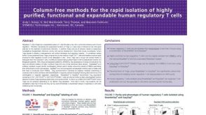 科学海报Rapid Isolation of Highly Purified, Functional and Expandable Human Regulatory T Cells
 沪公网安备31010102008431号