Lee WJ et al. (OCT 2005)
Molecular pharmacology 68 4 1018--30
Mechanisms for the inhibition of DNA methyltransferases by tea catechins and bioflavonoids.
In the present investigation,we studied the modulating effects of several tea catechins and bioflavonoids on DNA methylation catalyzed by prokaryotic SssI DNA methyltransferase (DNMT) and human DNMT1. We found that each of the tea polyphenols [catechin,epicatechin,and (-)-epigallocatechin-3-O-gallate (EGCG)] and bioflavonoids (quercetin,fisetin,and myricetin) inhibited SssI DNMT- and DNMT1-mediated DNA methylation in a concentration-dependent manner. The IC(50) values for catechin,epicatechin,and various flavonoids ranged from 1.0 to 8.4 microM,but EGCG was a more potent inhibitor,with IC(50) values ranging from 0.21 to 0.47 microM. When epicatechin was used as a model inhibitor,kinetic analyses showed that this catechol-containing dietary polyphenol inhibited enzymatic DNA methylation in vitro largely by increasing the formation of S-adenosyl-L-homocysteine (a potent noncompetitive inhibitor of DNMTs) during the catechol-O-methyltransferase-mediated O-methylation of this dietary catechol. In comparison,the strong inhibitory effect of EGCG on DNMT-mediated DNA methylation was independent of its own methylation and was largely due to its direct inhibition of the DNMTs. This inhibition is strongly enhanced by Mg(2+). Computational modeling studies showed that the gallic acid moiety of EGCG plays a crucial role in its high-affinity,direct inhibitory interaction with the catalytic site of the human DNMT1,and its binding with the enzyme is stabilized by Mg(2+). The modeling data on the precise molecular mode of EGCG's inhibitory interaction with human DNMT1 agrees perfectly with our experimental finding.
View Publication
产品类型:
产品号#:
73642
73644
产品名:
(-)-Epigallocatechin Gallate, 50 mg
(-)-Epigallocatechin Gallate
Kubicek S et al. (FEB 2007)
Molecular cell 25 3 473--81
Reversal of H3K9me2 by a small-molecule inhibitor for the G9a histone methyltransferase.
Histone lysine methylation has important roles in the organization of chromatin domains and the regulation of gene expression. To analyze its function and modulate its activity,we screened for specific inhibitors against histone lysine methyltransferases (HMTases) using recombinant G9a as the target enzyme. From a chemical library comprising 125,000 preselected compounds,seven hits were identified. Of those,one inhibitor,BIX-01294 (diazepin-quinazolin-amine derivative),does not compete with the cofactor S-adenosyl-methionine,and selectively impairs the G9a HMTase and the generation of H3K9me2 in vitro. In cellular assays,transient incubation of several cell lines with BIX-01294 lowers bulk H3K9me2 levels that are restored upon removal of the inhibitor. Importantly,chromatin immunoprecipitation at several G9a target genes demonstrates reversible reduction of promoter-proximal H3K9me2 in inhibitor-treated mouse ES cells and fibroblasts. Our data identify a biologically active HMTase inhibitor that allows for the transient modulation of H3K9me2 marks in mammalian chromatin.
View Publication
产品类型:
产品号#:
72042
72044
产品名:
BIX01294 (Trihydrochloride Hydrate)
BIX01294 (Trihydrochloride Hydrate)
An MC et al. ( 2014)
PLoS currents 6 1--19
Polyglutamine Disease Modeling: Epitope Based Screen for Homologous Recombination using CRISPR/Cas9 System.
We have previously reported the genetic correction of Huntington's disease (HD) patient-derived induced pluripotent stem cells using traditional homologous recombination (HR) approaches. To extend this work,we have adopted a CRISPR-based genome editing approach to improve the efficiency of recombination in order to generate allelic isogenic HD models in human cells. Incorporation of a rapid antibody-based screening approach to measure recombination provides a powerful method to determine relative efficiency of genome editing for modeling polyglutamine diseases or understanding factors that modulate CRISPR/Cas9 HR.
View Publication
产品类型:
产品号#:
05850
05857
05870
05875
85850
85857
85870
85875
产品名:
mTeSR™1
mTeSR™1
Mormone E et al. (NOV 2014)
Stem cells and development 23 21 2626--36
Footprint-free" human induced pluripotent stem cell-derived astrocytes for in vivo cell-based therapy."
The generation of human induced pluripotent stem cells (hiPSC) from somatic cells has enabled the possibility to provide patient-specific hiPSC for cell-based therapy,drug discovery,and other translational applications. Two major obstacles in using hiPSC for clinical application reside in the risk of genomic modification when they are derived with viral transgenes and risk of teratoma formation if undifferentiated cells are engrafted. In this study,we report the generation of footprint-free" hiPSC-derived astrocytes. These are efficiently generated�
View Publication
产品类型:
产品号#:
05850
05857
05870
05875
85850
85857
85870
85875
产品名:
mTeSR™1
mTeSR™1
Baatz JE et al. (JUL 2014)
In vivo (Athens,Greece) 28 4 411--423
Cryopreservation of viable human lung tissue for versatile post-thaw analyses and culture.
Clinical trials are currently used to test therapeutic efficacies for lung cancer,infections and diseases. Animal models are also used as surrogates for human disease. Both approaches are expensive and time-consuming. The utility of human biospecimens as models is limited by specialized tissue processing methods that preserve subclasses of analytes (e.g. RNA,protein,morphology) at the expense of others. We present a rapid and reproducible method for the cryopreservation of viable lung tissue from patients undergoing lobectomy or transplant. This method involves the pseudo-diaphragmatic expansion of pieces of fresh lung tissue with cryoprotectant formulation (pseudo-diaphragmatic expansion-cryoprotectant perfusion or PDX-CP) followed by controlled-rate freezing in cryovials. Expansion-perfusion rates,volumes and cryoprotectant formulation were optimized to maintain tissue architecture,decrease crystal formation and increase long-term cell viability. Rates of expansion of 4 cc/min or less and volumes ranging from 0.8-1.2 × tissue volume were well-tolerated by lung tissue obtained from patients with chronic obstructive pulmonary disease or idiopathic pulmonary fibrosis,showing minimal differences compared to standard histopathology. Morphology was greatly improved by the PDX-CP procedure compared to simple fixation. Fresh versus post-thawed lung tissue showed minimal differences in histology,RNA integrity numbers and post-translational modified protein integrity (2-dimensional differential gel electrophoresis). It was possible to derive numerous cell types,including alveolar epithelial cells,fibroblasts and stem cells,from the tissue for at least three months after cryopreservation. This new method should provide a uniform,cost-effective approach to the banking of biospecimens,with versatility to be amenable to any post-acquisition process applicable to fresh tissue samples.
View Publication
产品类型:
产品号#:
05850
05857
05870
05875
85850
85857
85870
85875
产品名:
mTeSR™1
mTeSR™1
Gorman BR et al. (DEC 2014)
PLoS ONE 9 12 e116037
Multi-scale imaging and informatics pipeline for in situ pluripotent stem cell analysis
Human pluripotent stem (hPS) cells are a potential source of cells for medical therapy and an ideal system to study fate decisions in early development. However,hPS cells cultured in vitro exhibit a high degree of heterogeneity,presenting an obstacle to clinical translation. hPS cells grow in spatially patterned colony structures,necessitating quantitative single-cell image analysis. We offer a tool for analyzing the spatial population context of hPS cells that integrates automated fluorescent microscopy with an analysis pipeline. It enables high-throughput detection of colonies at low resolution,with single-cellular and sub-cellular analysis at high resolutions,generating seamless in situ maps of single-cellular data organized by colony. We demonstrate the tool's utility by analyzing inter- and intra-colony heterogeneity of hPS cell cycle regulation and pluripotency marker expression. We measured the heterogeneity within individual colonies by analyzing cell cycle as a function of distance. Cells loosely associated with the outside of the colony are more likely to be in G1,reflecting a less pluripotent state,while cells within the first pluripotent layer are more likely to be in G2,possibly reflecting a G2/M block. Our multi-scale analysis tool groups colony regions into density classes,and cells belonging to those classes have distinct distributions of pluripotency markers and respond differently to DNA damage induction. Lastly,we demonstrate that our pipeline can robustly handle high-content,high-resolution single molecular mRNA FISH data by using novel image processing techniques. Overall,the imaging informatics pipeline presented offers a novel approach to the analysis of hPS cells that includes not only single cell features but also colony wide,and more generally,multi-scale spatial configuration.
View Publication
K. Tanaka et al. (Apr 2025)
Scientific Reports 15 23
Robust and reproducible human intestinal organoid-derived monolayer model for analyzing drug absorption
Predicting the absorption of orally administered drugs is crucial to drug development. Current in vitro models lack physiological relevance,robustness,and reproducibility,thus hindering reliable predictions. In this study,we developed a reproducible and robust culture method to generate a human intestinal organoid-derived monolayer model that can be applied to study drug absorption through a step-by-step approach. Our model showed similarity to primary enterocytes in terms of the drug absorption-related gene expression profile,tight barrier function,tolerability toward artificial bile juice,drug transporter and metabolizing enzyme function,and nuclear receptor activity. This method can be applied to organoids derived from multiple donors. The permeability of launched 19 drugs in our model demonstrated a correlation with human Fa values,with an R 2 value of 0.88. Additionally,by combining the modeling and simulation approaches,the estimated FaFg values for seven out of nine drugs,including CYP3A substrates,fell within 1.5 times the range of the human FaFg values. Applying this method to the drug discovery process might bridge the gap between preclinical and clinical research and increase the success rates of drug development.
View Publication

 EasySep™小鼠TIL(CD45)正选试剂盒


 1:07:14
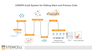
线上讲座Optimized Workflows for High-Efficiency Genome Editing in Stem and Primary Cell Types发布日期: 09/09/2019
1:07:14
线上讲座Optimized Workflows for High-Efficiency Genome Editing in Stem and Primary Cell Types发布日期: 09/09/2019 实验方案How to Coat Cultureware with Vitronectin XF™ for Pluripotent Stem Cell Culture


 沪公网安备31010102008431号