Differentiation of Human Pluripotent Stem Cells to Cardiomyocytes Under Defined Conditions.
Human embryonic stem cells (hESCs) and induced pluripotent stem cells (hiPSCs) can differentiate to cardiomyocytes in vitro,offering unique opportunities to investigate cardiac development and disease as well as providing a platform to perform drug and toxicity tests. Initial cardiac differentiation methods were based on either inductive co-culture or aggregation as embryoid bodies,often in the presence of fetal calf serum. More recently,monolayer differentiation protocols have evolved as feasible alternatives and are often performed in completely defined culture medium and substrates. Thus,our ability to efficiently and reproducibly generate cardiomyocytes from multiple different hESC and hiPSC lines has improved significantly.We have developed a directed differentiation monolayer protocol that can be used to generate cultures comprising ˜50% cardiomyocytes,in which both the culture of the undifferentiated human pluripotent stem cells (hPSCs) and the differentiation procedure itself are defined and serum-free. The differentiation method is also effective for hPSCs maintained in other culture systems. In this chapter,we outline the differentiation protocol and describe methods to assess cardiac differentiation efficiency as well as to identify and quantify the yield of cardiomyocytes.
View Publication
产品类型:
产品号#:
05850
05857
05870
05875
85850
85857
85870
85875
产品名:
mTeSR™1
mTeSR™1
Zhu S et al. (DEC 2010)
Cell stem cell 7 6 651--5
Reprogramming of human primary somatic cells by OCT4 and chemical compounds.
N. C. Leite et al. (jul 2020)
Cell reports 32 2 107894
Modeling Type 1 Diabetes In Vitro Using Human Pluripotent Stem Cells.
Understanding the root causes of autoimmune diseases is hampered by the inability to access relevant human tissues and identify the time of disease onset. To examine the interaction of immune cells and their cellular targets in type 1 diabetes,we differentiated human induced pluripotent stem cells into pancreatic endocrine cells,including $\beta$ cells. Here,we describe an in vitro platform that models features of human type 1 diabetes using stress-induced patient-derived endocrine cells and autologous immune cells. We demonstrate a cell-type-specific response by autologous immune cells against induced pluripotent stem cell-derived $\beta$ cells,along with a reduced effect on $\alpha$ cells. This approach represents a path to developing disease models that use patient-derived cells to predict the outcome of an autoimmune response.
View Publication
Ayasoufi K et al. (APR 2016)
Journal of Immunology 196 7 3180--90
CD4 T Cell Help via B Cells Is Required for Lymphopenia-Induced CD8 T Cell Proliferation.
Ab-mediated lymphoablation is commonly used in solid organ and hematopoietic cell transplantation. However,these strategies fail to control pathogenic memory T cells efficiently and to improve long-term transplant outcomes significantly. Understanding the mechanisms of T cell reconstitution is critical for enhancing the efficacy of Ab-mediated depletion in sensitized recipients. Using a murine analog of anti-thymocyte globulin (mATG) in a mouse model of cardiac transplantation,we previously showed that peritransplant lymphocyte depletion induces rapid memory T cell proliferation and only modestly prolongs allograft survival. We now report that T cell repertoire following depletion is dominated by memory CD4 T cells. Additional depletion of these residual CD4 T cells severely impairs the recovery of memory CD8 T cells after mATG treatment. The CD4 T cell help during CD8 T cell recovery depends on the presence of B cells expressing CD40 and intact CD40/CD154 interactions. The requirement for CD4 T cell help is not limited to the use of mATG in heart allograft recipients,and it is observed in nontransplanted mice and after CD8 T cell depletion with mAb instead of mATG. Most importantly,limiting helper signals increases the efficacy of mATG in controlling memory T cell expansion and significantly extends heart allograft survival in sensitized recipients. Our findings uncover the novel role for helper memory CD4 T cells during homeostatic CD8 T cell proliferation and open new avenues for optimizing lymphoablative therapies in allosensitized patients.
View Publication
产品类型:
产品号#:
19851
19851RF
产品名:
EasySep™小鼠T细胞分选试剂盒
RoboSep™ 小鼠T细胞分选试剂盒
He H et al. (OCT 2012)
Blood 120 15 3152--62
Endothelial cells provide an instructive niche for the differentiation and functional polarization of M2-like macrophages.
Endothelial cells and macrophages are known to engage in tight and specific interactions that contribute to the modulation of vascular function. Here we show that adult endothelial cells provide critical signals for the selective growth and differentiation of macrophages from several hematopoietic progenitors. The process features the formation of well-organized colonies that exhibit progressive differentiation from the center to the periphery and toward an M2-like phenotype,characterized by enhanced expression of Tie2 and CD206/Mrc1. These colonies are long-lived depending on the contact with the endothelium; removal of the endothelial monolayer results in rapid colony dissolution. We further found that Csf1 produced by the endothelium is critical for the expansion of the macrophage colonies and that blockade of Csf1 receptor impairs colony growth. Functional analyses indicate that these macrophages are capable of accelerating angiogenesis,promoting tumor growth,and effectively engaging in tight associations with endothelial cells in vivo. These findings uncover a critical role of endothelial cells in the induction of macrophage differentiation and their ability to promote further polarization toward a proangiogenic phenotype. This work also highlights some of the molecules underlying the M2-like differentiation,a process that is relevant to the progression of both developmental and pathologic angiogenesis.
View Publication
产品类型:
产品号#:
72472
72474
产品名:
GW2580
GW2580
Sequiera GL et al. (JAN 2013)
Life Sciences 92 1 63--71
Ontogenic development of cardiomyocytes derived from transgene-free human induced pluripotent stem cells and its homology with human heart
Aim: Reprogramming of somatic cells utilizing viral free methods provide a remarkable method to generate human induced pluripotent stem cells (hiPSCs) for regenerative medicine. In this study,we evaluate developmental ontogeny of cardiomyocytes following induced differentiation of hiPSCs. Main Methods: Fibroblasts were reprogrammed with episomal vectors to generate hiPSC and were subsequently differentiated to cardiomyocytes. Ontogenic development of cardiomyocytes was studied by real-time PCR. Key findings: Human iPSCs derived from episomal based vectors maintain classical pluripotency markers,generate teratomas and spontaneously differentiate into three germ layers in vitro. Cardiomyogenic induction of these hiPSCs efficiently generated cardiomyocytes. Ontogenic gene expression studies demonstrated that differentiation of cardiomyocytes was initiated by increased expression of mesodermal markers,followed by early cardiac committed markers,structural and ion channel genes. Furthermore,our correlation analysis of gene expression studies with human heart demonstrated that pivotal structural genes like cardiac troponin,actinin,myosin light chain maintained a high correlation with ion channel genes indicating coordinated activation of cardiac transcriptional machinery. Finally,microelectrode recordings show that these cardiomyocytes could respond aptly to pharmacologically active drugs. Cardiomyocytes showed a chronotropic response to isoproterenol,reduced Na+ influx with quinidine,prolongation of beating rate corrected field potential duration (cFPD) with E-4031 and reduced beating frequency and shortened cFPD with verapamil. Significance: Our study shows that viral free hiPSCs efficiently differentiate into cardiomyocytes with cardiac-specific molecular,structural,and functional properties that recapitulate developmental ontogeny of cardiogenesis. These results,coupled with the potential to generate patient-specific hiPSC lines hold great promise for the development of in vitro platform for drug pharmacogenomics; disease modeling and regenerative medicine. textcopyright 2012 Elsevier Inc. All rights reserved.
View Publication
产品类型:
产品号#:
05850
05857
05870
05875
85850
85857
85870
85875
产品名:
mTeSR™1
mTeSR™1
Garnache-Ottou F et al. (FEB 2005)
Blood 105 3 1256--64
Expression of the myeloid-associated marker CD33 is not an exclusive factor for leukemic plasmacytoid dendritic cells.
A new entity of acute leukemia coexpressing CD4(+)CD56(+) markers without any other lineage-specific markers has been identified recently as arising from lymphoid-related plasmacytoid dendritic cells (pDCs). In our laboratory,cells from a patient with such CD4(+)CD56(+) lineage-negative leukemia were unexpectedly found to also express the myeloid marker CD33. To confirm the diagnosis of pDC leukemia despite the CD33 expression,we demonstrated that the leukemic cells indeed exhibited pDC phenotypic and functional properties. In 7 of 8 other patients with CD4(+)CD56(+) pDC malignancies,we were able to confirm that the tumor cells expressed CD33 although with variable expression levels. CD33 expression was shown by flow cytometry,reverse transcriptase-polymerase chain reaction,and immunoblot analysis. Furthermore,CD33 monoclonal antibody stimulation of purified CD4(+)CD56(+) leukemic cells led to cytokine secretion,thus confirming the presence of a functional CD33 on these leukemic cells. Moreover,we found that circulating pDCs in healthy individuals also weakly express CD33. Overall,our results demonstrate that the expression of CD33 on CD4(+)CD56(+) lineage-negative cells should not exclude the diagnosis of pDC leukemia and underline that pDC-specific markers should be used at diagnosis for CD4(+)CD56(+) malignancies.
View Publication
产品类型:
产品号#:
15028
15068
产品名:
RosetteSep™人单核细胞富集抗体混合物
RosetteSep™人单核细胞富集抗体混合物
Ortega V et al. (MAR 2016)
Cancer genetics 209 3 82--6
Optimal strategy for obtaining routine chromosome analysis by using negative fractions of CD138 enriched plasma cells.
Fluorescence in situ hybridization (FISH) is superior to routine chromosome analysis (RCA) in detecting important prognostic genetic abnormalities in plasma cell dyscrasia (PCD); however,its sensitivity is hampered due to paucity of plasma cells (PC) in whole bone marrow (BM). Studies showed that the abnormality detection rate in enriched plasma cells (EPC) is greater than unselected plasma cells (UPC),but purification techniques are limiting to only FISH when sample volumes are inadequate. Not performing RCA may compromise patient care since RCA is equally important for detecting non-PC related abnormalities when the diagnosis is undefined. To resolve this critical issue,we designed a study where an immuno-magnetic CD138 enriched positive selection was used for FISH while the negative fraction (NF) was used to retrieve other myeloid elements for RCA. Parallel FISH studies were performed using UPC and CD138 EPC,while karyotyping was achieved using whole BM and discarded myeloid elements from the NF. Results showed that the abnormality rate of EPC was doubled compared to UPC for FISH,and CA displayed 100% success rate using the NF. PCD related chromosome abnormalities were confined to whole BM while non-PCD related abnormalities were found in both whole BM and NF. Our results demonstrate the feasibility of using the NF for RCA.
View Publication
产品类型:
产品号#:
21000
20119
20155
18387
18387RF
产品名:
RoboSep™- S
RoboSep™ 吸头组件抛光剂
RoboSep™分选管套装(9个塑料管)
Pollak J et al. (MAR 2017)
PLOS ONE 12 3 e0172884
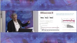
Ion channel expression patterns in glioblastoma stem cells with functional and therapeutic implications for malignancy
Ion channels and transporters have increasingly recognized roles in cancer progression through the regulation of cell proliferation,migration,and death. Glioblastoma stem-like cells (GSCs) are a source of tumor formation and recurrence in glioblastoma multiforme,a highly aggressive brain cancer,suggesting that ion channel expression may be perturbed in this population. However,little is known about the expression and functional relevance of ion channels that may contribute to GSC malignancy. Using RNA sequencing,we assessed the enrichment of ion channels in GSC isolates and non-tumor neural cell types. We identified a unique set of GSC-enriched ion channels using differential expression analysis that is also associated with distinct gene mutation signatures. In support of potential clinical relevance,expression of selected GSC-enriched ion channels evaluated in human glioblastoma databases of The Cancer Genome Atlas and Ivy Glioblastoma Atlas Project correlated with patient survival times. Finally,genetic knockdown as well as pharmacological inhibition of individual or classes of GSC-enriched ion channels constrained growth of GSCs compared to normal neural stem cells. This first-in-kind global examination characterizes ion channels enriched in GSCs and explores their potential clinical relevance to glioblastoma molecular subtypes,gene mutations,survival outcomes,regional tumor expression,and experimental responses to loss-of-function. Together,the data support the potential biological and therapeutic impact of ion channels on GSC malignancy and provide strong rationale for further examination of their mechanistic and therapeutic importance.
View Publication
产品类型:
产品号#:
05751
70913
产品名:
NeuroCult™ NS-A 扩增试剂盒(人)
Artyukhov AS et al. (MAY 2017)
Gene
New genes for accurate normalization of qRT-PCR results in study of iPS and iPS-derived cells.
iPSC-derived cells (from induced pluripotent stem cells) are a useful source that provide a powerful and widely accepted tool for the study of various types of human cells in vitro. Indeed,iPSC-derived cells from patients with hereditary diseases have been shown to reproduce the hallmarks of these diseases in vitro,phenotypes that can then also be manipulated in vitro. Quantitative reverse transcription PCR (qRT-PCR) is often used to characterize the progress of iPSC differentiation,validate mature cell types and to determine levels of pathological markers. Quantitative reverse transcription PCR (qRT-PCR) is used to quantify mRNA levels. This method requires some way of normalizing the data,typically by relating the obtained levels of gene expression to the levels of expression of a house keeping gene"�
View Publication
产品类型:
产品号#:
05850
05857
05870
05875
85850
85857
85870
85875
产品名:
mTeSR™1
mTeSR™1
Nie S et al. (FEB 2015)
Journal of proteome research 14 2 814--22
Tenascin-C: a novel candidate marker for cancer stem cells in glioblastoma identified by tissue microarrays.
Glioblastoma multiforme (GBM) is a highly aggressive brain tumor,with dismal survival outcomes. Recently,cancer stem cells (CSCs) have been demonstrated to play a role in therapeutic resistance and are considered to be the most likely cause of cancer relapse. The identification of CSCs is an important step toward finding new and effective ways to treat GBM. Tenascin-C (TNC) protein has been identified as a potential marker for CSCs in gliomas based on previous work. Here,we have investigated the expression of TNC in tissue microarrays including 17 GBMs,18 WHO grade III astrocytomas,15 WHO grade II astrocytomas,4 WHO grade I astrocytomas,and 7 normal brain tissue samples by immunohistochemical staining. TNC expression was found to be highly associated with the grade of astrocytoma. It has a high expression level in most of the grade III astrocytomas and GBMs analyzed and a very low expression in most grade II astrocytomas,whereas it is undetectable in grade I astrocytomas and normal brain tissues. Double-immunofluorescence staining for TNC and CD133 in GBM tissues revealed that there was a high overlap between theses two positive populations. The results were further confirmed by flow cytometry analysis of TNC and CD133 in GBM-derived stem-like neurospheres in vitro. A limiting dilution assay demonstrated that the sphere formation ability of CD133(+)/TNC(+) and CD133(-)/TNC(+) cell populations is much higher than that of the CD133(+)/TNC(-) and CD133(-)/TNC(-) populations. These results suggest that TNC is not only a potential prognostic marker for GBM but also a potential marker for glioma CSCs,where the TNC(+) population is identified as a CSC population overlapping with part of the CD133(-) cell population.
View Publication

 EasySep™小鼠TIL(CD45)正选试剂盒


 沪公网安备31010102008431号