
 EasySep™小鼠TIL(CD45)正选试剂盒


搜索结果: 'methocult media formulations for mouse hematopoietic cells serum containing'
-

产品类型:
产品号#:
18000
19853
19853RF
产品名:
EasySep™磁极
EasySep™小鼠CD8+ T细胞分选试剂盒
RoboSep™ 小鼠CD8+ T细胞分选试剂盒
-
产品类型:
产品号#:
19844
19844RF
19849
19851
19851RF
19762
19762RF
产品名:
EasySep™小鼠Pan-B细胞分选试剂盒
RoboSep™ 小鼠Pan-B细胞分选试剂盒
EasySep™小鼠/人嵌合体分选试剂盒
EasySep™小鼠T细胞分选试剂盒
RoboSep™ 小鼠T细胞分选试剂盒
EasySep™小鼠中性粒细胞富集试剂盒
RoboSep™ 小鼠中性粒细胞富集试剂盒含滤芯吸头
-
产品类型:
产品号#:
09600
09650
100-0785
10970
10990
产品名:
StemSpan™ SFEM
StemSpan™ SFEM
ImmunoCult™ 人CD3/CD28/CD2 T细胞激活剂
ImmunoCult™ 人CD3/CD28/CD2 T细胞激活剂
ImmunoCult™ 人CD3/CD28/CD2 T细胞激活剂
-
产品类型:
产品号#:
85850
85857
产品名:
mTeSR™1
mTeSR™1
-
产品类型:
产品号#:
27845
27945
27840
27865
27940
27965
产品名:
-
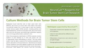 产品手册NeuroCult™: Reagents for Brain Tumor Stem Cell Research
产品类型:
品牌:
NeuroCult
产品号#:
01700
01705
01701
01702
05700
05702
05704
05707
05715
05740
05742
05751
05752
05761
05771
05772
07920
产品名:
ALDEFLUOR™ 试剂盒
ALDEFLUOR™ DEAB试剂, 1.5 mM, 1 mL
ALDEFLUOR™检测缓冲液
NeuroCult™ 基础培养基(小鼠和大鼠)
NeuroCult™扩增试剂盒(小鼠和大鼠)
NeuroCult™ 分化试剂盒(小鼠和大鼠)
NeuroCult™化学解离试剂盒(小鼠)
NeuroCult™成年中枢神经系统(CNS)组织酶解试剂盒(小鼠和大鼠)
NeuroCult™ NS-A 扩增试剂盒(人)
NeuroCult™ NS-A 分化试剂盒(人)
用于小鼠和大鼠神经干细胞和祖细胞分化培养的试剂盒
ACCUTASE™
-
产品类型:
产品号#:
05850
05857
05870
05875
85850
85857
85870
85875
产品名:
mTeSR™1
mTeSR™1
-
产品类型:
产品号#:
100-0483
100-0484
100-0276
100-1130
产品名:
Hausser Scientificᵀᴹ 明线血球计数板
ReLeSR™
mTeSR™ Plus
mTeSR™ Plus


 沪公网安备31010102008431号