Kurtz J et al. (SEP 2007)
Transfusion 47 9 1578--87
Assessment of cord blood hematopoietic cell parameters before and after cryopreservation.
BACKGROUND: The testing of cord blood (CB) progenitor and stem cell units for transplantation suitability involves enumeration of total nucleated cells before freezing. CD34+ cell counts may also be a means of determining suitability. Studies have been conducted to evaluate how specific storage conditions influence cell counts. STUDY DESIGN AND METHODS: CB units were processed by hydroxyethyl starch volume reduction. Cryopreserved-thawed samples were diluted 1:3 without washing. CD34+ cells were measured with three commercially available assay methods. In specific studies,apoptosis-indicating reagents were included. CB units were analyzed for nucleated cells,aldehyde dehydrogenase-containing cells,and progenitor colonies. RESULTS: CD34+ cell levels and nucleated cells were retained during storage in test tubes at 1 to 6 degrees C for 3 days. Cryopreserved-thawed samples showed a reduction in CD34+ cells relative to prefreeze levels with the largest decrease with the Stem-Kit (Beckman Coulter) restricted gating procedure. Prefreeze samples contained minimal numbers of presumed apoptotic cells detected with 7-aminoactinomycin D or SYTO16,but after cryopreservation-thawing there was an increase. Nucleated cell levels determined with a hematology analyzer or flow cytometry were reduced after thawing. Cryopreservation-thawing reduced the percentage of CD34+ cells positive for the presence of aldehyde dehydrogenase and the number of progenitor colonies. These differences were significant. CONCLUSION: These studies indicate that CD34+ cell counts were maintained when CB samples were stored at 1 to 6 degrees C in test tubes for 3 days. Cryopreservation-thawing resulted in changes in a number of parameters including the percentage of CD34+ cells that were aldehyde dehydrogenase(+) and the number of 7-aminoactinomycin D(+) cells and SYTO16(low) cells.
View Publication
产品类型:
产品号#:
01700
01705
01702
产品名:
ALDEFLUOR™ 试剂盒
ALDEFLUOR™ DEAB试剂, 1.5 mM, 1 mL
ALDEFLUOR™检测缓冲液
K. Saito et al. (Sep 2024)
Nature Communications 15
Hematopoietic stem cells (HSCs) react to various stress conditions. However,it is unclear whether and how HSCs respond to severe anemia. Here,we demonstrate that upon induction of acute anemia,HSCs rapidly proliferate and enhance their erythroid differentiation potential. In severe anemia,lipoprotein profiles largely change and the concentration of ApoE increases. In HSCs,transcription levels of lipid metabolism-related genes,such as very low-density lipoprotein receptor ( Vldlr ),are upregulated. Stimulation of HSCs with ApoE enhances their erythroid potential,whereas HSCs in Apoe knockout mice do not respond to anemia induction. Vldlr high HSCs show higher erythroid potential,which is enhanced after acute anemia induction. Vldlr high HSCs are epigenetically distinct because of their low chromatin accessibility,and more chromatin regions are closed upon acute anemia induction. Chromatin regions closed upon acute anemia induction are mainly binding sites of Erg. Inhibition of Erg enhanced the erythroid differentiation potential of HSCs. Our findings indicate that lipoprotein metabolism plays an important role in HSC regulation under severe anemic conditions. Subject terms: Haematopoietic stem cells,Fat metabolism,Chromatin,Anaemia
View Publication
产品类型:
产品号#:
03434
03444
09600
09650
产品名:
MethoCult™ GF M3434
MethoCult™ GF M3434
StemSpan™ SFEM
StemSpan™ SFEM
Al-Ali H et al. (MAY 2013)
ACS chemical biology 25 5 1027--36
A ROCK inhibitor permits survival of dissociated human embryonic stem cells.
Poor survival of human embryonic stem (hES) cells after cell dissociation is an obstacle to research,hindering manipulations such as subcloning. Here we show that application of a selective Rho-associated kinase (ROCK) inhibitor,Y-27632,to hES cells markedly diminishes dissociation-induced apoptosis,increases cloning efficiency (from approximately 1% to approximately 27%) and facilitates subcloning after gene transfer. Furthermore,dissociated hES cells treated with Y-27632 are protected from apoptosis even in serum-free suspension (SFEB) culture and form floating aggregates. We demonstrate that the protective ability of Y-27632 enables SFEB-cultured hES cells to survive and differentiate into Bf1(+) cortical and basal telencephalic progenitors,as do SFEB-cultured mouse ES cells.
View Publication
Agrawal P et al. (APR 2016)
ACS applied materials & interfaces 8 14 8870--8874
Fast, Efficient, and Gentle Transfection of Human Adherent Cells in Suspension
We demonstrate a highly efficient method for gene delivery into clinically relevant human cell types,such as induced pluripotent stem cells (iPSCs) and fibroblasts,reducing the protocol time by one full day. To preserve cell physiology during gene transfer,we designed a microfluidic strategy,which facilitates significant gene delivery in a short transfection time (textless1 min) for several human cell types. This fast,optimized and generally applicable cell transfection method can be used for rapid screening of different delivery systems and has significant potential for high-throughput cell therapy applications.
View Publication
产品类型:
产品号#:
05850
05857
05870
05875
85850
85857
85870
85875
产品名:
mTeSR™1
mTeSR™1
Fernandes AM et al. (JAN 2010)
Cell Transplantation 19 5 509--23
Worldwide survey of published procedures to culture human embryonic stem cells
Since their derivation 11 years ago,human embryonic stem (hES) cells have become a powerful tool in both basic biomedical research and developmental biology. Their capacity for self-renewal and differentiation into any tissue type has also brought interest from fields such as cell therapy and drug screening. We conducted an extensive analysis of 750 papers (51% of the total published about hES cells between 1998 and 2008) to present a spectrum of hES cell research including culture protocols developed worldwide. This review may stimulate discussions about the importance of having unvarying methods to culture hES cells,in order to facilitate comparisons among data obtained by research groups elsewhere,especially concerning preclinical studies. Moreover,the description of the most widely used cell lines,reagents,and procedures adopted internationally will help newcomers on deciding the best strategies for starting their own studies. Finally,the results will contribute with the efforts of stem cell researchers on comparing the performance of different aspects related to hES cell culture methods.
View Publication
Energy metabolism in human pluripotent stem cells and their differentiated counterparts.
BACKGROUND: Human pluripotent stem cells have the ability to generate all cell types present in the adult organism,therefore harboring great potential for the in vitro study of differentiation and for the development of cell-based therapies. Nonetheless their use may prove challenging as incomplete differentiation of these cells might lead to tumoregenicity. Interestingly,many cancer types have been reported to display metabolic modifications with features that might be similar to stem cells. Understanding the metabolic properties of human pluripotent stem cells when compared to their differentiated counterparts can thus be of crucial importance. Furthermore recent data has stressed distinct features of different human pluripotent cells lines,namely when comparing embryo-derived human embryonic stem cells (hESCs) and induced pluripotent stem cells (IPSCs) reprogrammed from somatic cells.backslashnbackslashnMETHODOLOGY/PRINCIPAL FINDINGS: We compared the energy metabolism of hESCs,IPSCs,and their somatic counterparts. Focusing on mitochondria,we tracked organelle localization and morphology. Furthermore we performed gene expression analysis of several pathways related to the glucose metabolism,including glycolysis,the pentose phosphate pathway and the tricarboxylic acid (TCA) cycle. In addition we determined oxygen consumption rates (OCR) using a metabolic extracellular flux analyzer,as well as total intracellular ATP levels by high performance liquid chromatography (HPLC). Finally we explored the expression of key proteins involved in the regulation of glucose metabolism.backslashnbackslashnCONCLUSIONS/FINDINGS: Our results demonstrate that,although the metabolic signature of IPSCs is not identical to that of hESCs,nonetheless they cluster with hESCs rather than with their somatic counterparts. ATP levels,lactate production and OCR revealed that human pluripotent cells rely mostly on glycolysis to meet their energy demands. Furthermore,our work points to some of the strategies which human pluripotent stem cells may use to maintain high glycolytic rates,such as high levels of hexokinase II and inactive pyruvate dehydrogenase (PDH).
View Publication
产品类型:
产品号#:
05850
05857
05870
05875
85850
85857
85870
85875
产品名:
mTeSR™1
mTeSR™1
Gifford CA et al. (MAY 2013)
Cell 153 5 1149--1163
Transcriptional and epigenetic dynamics during specification of human embryonic stem cells
Differentiation of human embryonic stem cells (hESCs) provides a unique opportunity to study the regulatory mechanisms that facilitate cellular transitions in a human context. To that end,we performed comprehensive transcriptional and epigenetic profiling of populations derived through directed differentiation of hESCs representing each of the three embryonic germ layers. Integration of whole-genome bisulfite sequencing,chromatin immunoprecipitation sequencing,and RNA sequencing reveals unique events associated with specification toward each lineage. Lineage-specific dynamic alterations in DNA methylation and H3K4me1 are evident at putative distal regulatory elements that are frequently bound by pluripotency factors in the undifferentiated hESCs. In addition,we identified germ-layer-specific H3K27me3 enrichment at sites exhibiting high DNA methylation in the undifferentiated state. A better understanding of these initial specification events will facilitate identification of deficiencies in current approaches,leading to more faithful differentiation strategies as well as providing insights into the rewiring of human regulatory programs during cellular transitions. ?? 2013 Elsevier Inc.
View Publication
产品类型:
产品号#:
05850
05857
05870
05875
85850
85857
85870
85875
产品名:
mTeSR™1
mTeSR™1
Panula S et al. ( 2016)
PloS one 11 10 e0165268
Over Expression of NANOS3 and DAZL in Human Embryonic Stem Cells.
The mechanisms underlying human germ cell development are largely unknown,partly due to the scarcity of primordial germ cells and the inaccessibility of the human germline to genetic analysis. Human embryonic stem cells can differentiate to germ cells in vitro and can be genetically modified to study the genetic requirements for germ cell development. Here,we studied NANOS3 and DAZL,which have critical roles in germ cell development in several species,via their over expression in human embryonic stem cells using global transcriptional analysis,in vitro germ cell differentiation,and in vivo germ cell formation assay by xenotransplantation. We found that NANOS3 over expression prolonged pluripotency and delayed differentiation. In addition,we observed a possible connection of NANOS3 with inhibition of apoptosis. For DAZL,our results suggest a post-transcriptional regulation mechanism in hES cells. In addition,we found that DAZL suppressed the translation of OCT4,and affected the transcription of several genes associated with germ cells,cell cycle arrest,and cell migration. Furthermore,DAZL over expressed cells formed spermatogonia-like colonies in a rare instance upon xenotransplantation. These data can be used to further elucidate the role of NANOS3 and DAZL in germ cell development both in vitro and in vivo.
View Publication
产品类型:
产品号#:
05850
05857
05870
05875
85850
85857
85870
85875
产品名:
mTeSR™1
mTeSR™1
(Mar 2024)
iScience 27 4
Craniofacial chondrogenesis in organoids from human stem cell-derived neural crest cells
SummaryKnowledge of cell signaling pathways that drive human neural crest differentiation into craniofacial chondrocytes is incomplete,yet essential for using stem cells to regenerate craniomaxillofacial structures. To accelerate translational progress,we developed a differentiation protocol that generated self-organizing craniofacial cartilage organoids from human embryonic stem cell-derived neural crest stem cells. Histological staining of cartilage organoids revealed tissue architecture and staining typical of elastic cartilage. Protein and post-translational modification (PTM) mass spectrometry and snRNA-seq data showed that chondrocyte organoids expressed robust levels of cartilage extracellular matrix (ECM) components: many collagens,aggrecan,perlecan,proteoglycans,and elastic fibers. We identified two populations of chondroprogenitor cells,mesenchyme cells and nascent chondrocytes,and the growth factors involved in paracrine signaling between them. We show that ECM components secreted by chondrocytes not only create a structurally resilient matrix that defines cartilage,but also play a pivotal autocrine cell signaling role in determining chondrocyte fate. Graphical abstract Highlights•Craniofacial cartilage organoids were grown from human neural crest stem cells•These organoids exhibited elastic cartilage architecture and characteristic markers•Paracrine signaling drove chondrogenesis in mesenchyme cells and nascent chondrocytes•ECM components cemented chondrocyte cell fate through autocrine signaling Natural sciences; Biological sciences; Biochemistry; Cell biology; Stem cells research; Specialized functions of cells
View Publication
产品类型:
产品号#:
100-0483
100-0484
100-0276
100-1130
18000
20164
100-0047
85850
85857
产品名:
Hausser Scientificᵀᴹ 明线血球计数板
ReLeSR™
mTeSR™ Plus
mTeSR™ Plus
EasySep™磁极
RoboSep™ 缓冲液 2
EasySep™ Release 人PSC来源神经嵴细胞正选试剂盒
mTeSR™1
mTeSR™1
Daniels TR et al. (NOV 2007)
Molecular cancer therapeutics 6 11 2995--3008
Conjugation of an anti transferrin receptor IgG3-avidin fusion protein with biotinylated saporin results in significant enhancement of its cytotoxicity against malignant hematopoietic cells.
We have previously developed an antibody fusion protein composed of a mouse/human chimeric IgG3 specific for the human transferrin receptor genetically fused to avidin (anti-hTfR IgG3-Av) as a universal delivery system for cancer therapy. This fusion protein efficiently delivers biotinylated FITC into cancer cells via TfR-mediated endocytosis. In addition,anti-hTfR IgG3-Av alone exhibits intrinsic cytotoxic activity and interferes with hTfR recycling,leading to the rapid degradation of the TfR and lethal iron deprivation in certain malignant B-cell lines. We now report on the cytotoxic effects of a conjugate composed of anti-hTfR IgG3-Av and biotinylated saporin 6 (b-SO6),a toxin derived from the plant Saponaria officinalis that inhibits protein synthesis. Conjugation of anti-hTfR IgG3-Av with b-SO6 enhances the cytotoxic effect of the fusion protein in sensitive cells and also overcomes the resistance of malignant cells that show low sensitivity to the fusion protein alone. Our results show for the first time that loading anti-hTfR IgG3-Av with a biotinylated toxin enhances the cytotoxicity of the fusion protein alone. These results suggest that anti-hTfR IgG3-Av has great potential as a therapeutic agent for a wide range of applications due to its intrinsic cytotoxic activity plus its ability to deliver biotinylated molecules into cancer cells.
View Publication

 EasySep™小鼠TIL(CD45)正选试剂盒


 新闻STEMCELL Technologies Applauds Feeder-free Derivation of Human Induced Pluripotent Stem Cells
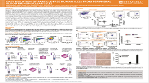 科学海报Easy Isolation of Particle-Free Human ILC2s from Peripheral Blood Mononuclear Cells
 沪公网安备31010102008431号