
 EasySep™小鼠TIL(CD45)正选试剂盒


搜索结果: 'methocult media formulations for human hematopoietic cells serum containing'
-

产品类型:
产品号#:
05850
05857
05870
05875
85850
85857
85870
85875
产品名:
mTeSR™1
mTeSR™1
-
产品类型:
产品号#:
19665
产品名:
EasySep™ Direct人NK细胞分选试剂盒
-
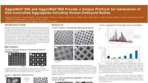 科学海报Aggrewell™400 and Aggrewell™800 Provide a Unique Platform for Generation of Size-Controlled Aggregates Including Human Embryoid Bodies
产品类型:
Conference:
AICHE 2010
产品号#:
产品名:
-
 新闻STEMCELL Technologies Launches mTeSR™ Plus, a Next-Generation Culture System for Human ES and iPS Cell Maintenance
产品类型:
产品号#:
产品名:
发布日期: 01/23/2019 -
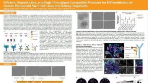 科学海报Efficient, Reproducible and High-Throughput-Compatible Protocols for Differentiation of Human Pluripotent Stem Cell Lines Into Kidney Organoids
产品类型:
Conference:
ISSCR 2019
产品号#:
产品名:
发布日期: 07/23/2019 -
产品类型:
产品号#:
05502
产品名:
-
产品类型:
产品号#:
73682
73684
产品名:
Fumonisin B1
Fumonisin B1
-
产品类型:
产品号#:
05700
05701
05702
产品名:
NeuroCult™ 基础培养基(小鼠和大鼠)
NeuroCult™ 扩增添加物(小鼠和大鼠)
NeuroCult™扩增试剂盒(小鼠和大鼠)
-
产品类型:
产品号#:
85850
85857
产品名:
mTeSR™1
mTeSR™1
-
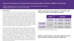 科学海报Immunomagnetic Cell Isolation of Human Gamma-Delta T Cells from PBMC
产品类型:
Conference:
ECI 2012
产品号#:
19255
19255RF
18000
产品名:
EasySep™人Gamma/Delta T细胞分选试剂盒
RoboSep™ 人Gamma/Delta T细胞分选试剂盒
EasySep™磁极
-
 56:31
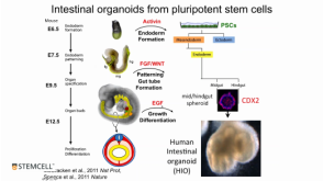
线上讲座Modeling Human Gastrointestinal Development and Disease Using Pluripotent Stem Cells发布日期: 03/06/2017
56:31
线上讲座Modeling Human Gastrointestinal Development and Disease Using Pluripotent Stem Cells发布日期: 03/06/2017 -
产品类型:
产品号#:
05850
05857
05870
05875
85850
85857
85870
85875
产品名:
mTeSR™1
mTeSR™1

 沪公网安备31010102008431号