Qu Q et al. (JUN 1999)
Journal of cellular biochemistry 73 4 500--7
Comparative effects of estrogen and antiestrogens on differentiation of osteoblasts in mouse bone marrow culture.
Estrogens as well as some antiestrogens have been shown to prevent bone loss in postmenopausal women. These compounds seem to inhibit bone resorption,but their anabolic effects have been less explored. In this study,bone marrow cultures were used to compare the effect of 17beta-estradiol (E2),and two triphenylethylene derivatives,tamoxifen (TAM),and FC1271a,and a benzothiophene derivative raloxifene (RAL) on differentiation of osteoblasts. All enhanced osteoblastic differentiation of 21-day cultures as indicated by increased mineralization and bone nodule formation. All,except RAL,stimulated cell proliferation during the first 6 days of the culture. However,in the presence of RAL the content of total protein was increased in 13-day cultures. SDS-PAGE and autoradiography of [14C]-proline labeled proteins revealed elevated level of the newly synthesized collagen type I. The pure antiestrogen ICI 182,780 abolished the increase of the specific activity of alkaline phosphatase by E2,TAM,and FC1271a but not the effect of RAL on protein synthesis. Our results show that E2 as well as TAM,FC1271a,and RAL stimulate bone formation in vitro but the mechanism of the anabolic action of RAL in bone clearly differs from that of E2,TAM,and FC1271a.
View Publication
产品类型:
产品号#:
72852
72854
产品名:
Fiorenzano A et al. (SEP 2016)
Nature communications 7 12589
Cripto is essential to capture mouse epiblast stem cell and human embryonic stem cell pluripotency.
Known molecular determinants of developmental plasticity are mainly transcription factors,while the extrinsic regulation of this process has been largely unexplored. Here we identify Cripto as one of the earliest epiblast markers and a key extracellular determinant of the naive and primed pluripotent states. We demonstrate that Cripto sustains mouse embryonic stem cell (ESC) self-renewal by modulating Wnt/β-catenin,whereas it maintains mouse epiblast stem cell (EpiSC) and human ESC pluripotency through Nodal/Smad2. Moreover,we provide unprecedented evidence that Cripto controls the metabolic reprogramming in ESCs to EpiSC transition. Remarkably,Cripto deficiency attenuates ESC lineage restriction in vitro and in vivo,and permits ESC transdifferentiation into trophectoderm lineage,suggesting that Cripto has earlier functions than previously recognized. All together,our studies provide novel insights into the current model of mammalian pluripotency and contribute to the understanding of the extrinsic regulation of the first cell lineage decision in the embryo.
View Publication
产品类型:
产品号#:
05850
05857
05870
05875
85850
85857
85870
85875
产品名:
mTeSR™1
mTeSR™1
Kim H et al. (JAN 2013)
Nature communications 4 2403
Modulation of β-catenin function maintains mouse epiblast stem cell and human embryonic stem cell self-renewal.
Wnt/β-catenin signalling has a variety of roles in regulating stem cell fates. Its specific role in mouse epiblast stem cell self-renewal,however,remains poorly understood. Here we show that Wnt/β-catenin functions in both self-renewal and differentiation in mouse epiblast stem cells. Stabilization and nuclear translocation of β-catenin and its subsequent binding to T-cell factors induces differentiation. Conversely,retention of stabilized β-catenin in the cytoplasm maintains self-renewal. Cytoplasmic retention of β-catenin is effected by stabilization of Axin2,a downstream target of β-catenin,or by genetic modifications to β-catenin that prevent its nuclear translocation. We also find that human embryonic stem cell and mouse epiblast stem cell fates are regulated by β-catenin through similar mechanisms. Our results elucidate a new role for β-catenin in stem cell self-renewal that is independent of its transcriptional activity and will have broad implications in understanding the molecular regulation of stem cell fate.
View Publication
产品类型:
产品号#:
72052
72054
72562
72564
100-1042
产品名:
CHIR99021
CHIR99021
IWR-1-endo
IWR-1-endo
CHIR99021
Sun SY et al. (SEP 2000)
Molecular pharmacology 58 3 508--14
Dual mechanisms of action of the retinoid CD437: nuclear retinoic acid receptor-mediated suppression of squamous differentiation and receptor-independent induction of apoptosis in UMSCC22B human head and neck squamous cell carcinoma cells.
The synthetic retinoid 6-[3-(adamantyl)-4-hydroxyphenyl]-2-naphthalene carboxylic acid (CD437),which can bind to and activate the nuclear retinoic acid receptors beta and gamma (RARbeta/gamma),is a potent inducer of apoptosis in various cancer cell lines. However,this effect was reported to be independent of RARs. In this study,we compared and contrasted the potencies and mechanisms of action of CD437 and several other receptor-selective retinoids in induction of apoptosis and modulation of squamous differentiation in UMSCC22B human head and neck squamous cell carcinoma cell line. CD437 and the structurally related retinoid CD2325 exhibited almost equal potency in inducing apoptosis,whereas several other retinoids failed to induce apoptosis. The RAR-specific pan antagonist AGN193109 failed to suppress CD437-induced apoptosis,indicating that the induction of apoptosis by CD437 was RAR-independent. c-Fos expression was induced by CD437 and CD2325 that induced apoptosis in the cell line but not by other retinoids that failed to induce apoptosis,suggesting a role for c-Fos in CD437-induced apoptosis. At low concentration (0.01 microM),CD437 shared with several other receptor-selective retinoids the ability to suppress the mRNA levels of the squamous differentiation markers Spr1,involucrin,and cytokeratin 1. This effect of CD437 could be blocked by AGN193109. We conclude that CD437 can exert its effects in UMSCC22B human human head and neck squamous cell carcinoma cells by at least two mechanisms: RAR-mediated suppression of squamous differentiation and RAR-independent induction of apoptosis.
View Publication
产品类型:
产品号#:
72722
72724
产品名:
CD437
CD437
Paul SR et al. (OCT 1990)
Proceedings of the National Academy of Sciences of the United States of America 87 19 7512--6
Molecular cloning of a cDNA encoding interleukin 11, a stromal cell-derived lymphopoietic and hematopoietic cytokine.
Hematopoiesis occurs in close association with a complex network of cells loosely termed the hematopoietic microenvironment. Analysis of the mechanisms of microenvironmental regulation of hematopoiesis has been hindered by the complexity of the microenvironment as well as the heterogeneity of hematopoietic stem cells and early progenitor cells. We have established immortalized primate bone marrow-derived stromal cell lines to facilitate analysis of the interactions of hematopoietic cells with the microenvironment in a large animal species. One such line,PU-34,was found to produce a variety of growth factors,including an activity that stimulates the proliferation of an interleukin 6-dependent murine plasmacytoma cell line. A cDNA encoding the plasmacytoma stimulatory activity was isolated through functional expression cloning in mammalian cells. The nucleotide sequence contained a single long reading frame of 597 nucleotides encoding a predicted 199-amino acid polypeptide. The amino acid sequence of this cytokine,designated interleukin 11 (IL-11),did not display significant similarity with any other sequence in the GenBank data base. Preliminary biological characterization indicates that in addition to stimulating plasmacytoma proliferation,IL-11 stimulates the T-cell-dependent development of immunoglobulin-producing B cells and synergizes with IL-3 in supporting murine megakaryocyte colony formation. These properties implicate IL-11 as an additional multifunctional regulator in the hematopoietic microenvironment.
View Publication
产品类型:
产品号#:
产品名:
J. Lam et al. (JUN 2018)
Nature communications 9 1 2418
miR-143/145 differentially regulate hematopoietic stem and progenitor activity through suppression of canonical TGFbeta$ signaling.
Expression of miR-143 and miR-145 is reduced in hematopoietic stem/progenitor cells (HSPCs) of myelodysplastic syndrome patients with a deletion in the long arm of chromosome 5. Here we show that mice lacking miR-143/145 have impaired HSPC activity with depletion of functional hematopoietic stem cells (HSCs),but activation of progenitor cells (HPCs). We identify components of the transforming growth factor beta$ (TGFbeta$) pathway as key targets of miR-143/145. Enforced expression of the TGFbeta$ adaptor protein and miR-145 target,Disabled-2 (DAB2),recapitulates the HSC defect seen in miR-143/145-/- mice. Despite reduced HSC activity,older miR-143/145-/- and DAB2-expressing mice show elevated leukocyte counts associated with increased HPC activity. A subset of mice develop a serially transplantable myeloid malignancy,associated with expansion of HPC. Thus,miR-143/145 play a cell context-dependent role in HSPC function through regulation of TGFbeta$/DAB2 activation,and loss of these miRNAs creates a preleukemic state.
View Publication
产品类型:
产品号#:
03434
03444
05350
产品名:
MethoCult™ GF M3434
MethoCult™ GF M3434
Farese AM et al. (JAN 1996)
Blood 87 2 581--91
Acceleration of hematopoietic reconstitution with a synthetic cytokine (SC-55494) after radiation-induced bone marrow aplasia.
The synthetic cytokine (Synthokine) SC-55494 is a high-affinity interleukin-3 (IL-3) receptor ligand that stimulates greater in vitro multilineage hematopoietic activity than native IL-3,while inducing no significant increase in inflammatory activity relative to native IL-3. The aim of this study was to investigate the in vivo hematopoietic response of rhesus monkeys receiving Synthokine after radiation-induced marrow aplasia. Administration schedule and dose of Synthokine were evaluated. All animals were total-body irradiated (TBI) with 700 cGy 60Co gamma radiation on day 0. Beginning on day 1,cohorts of animals (n = 5) received Synthokine subcutaneously (SC) twice daily with 25 micrograms/kg/d or 100 micrograms/kg/d for 23 days or 100 micrograms/kg/d for 14 days. Control animals (n = 9) received human serum albumin SC once daily at 15 micrograms/kg/d for 23 days. Complete blood counts were monitored for 60 days postirradiation and the durations of neutropenia (NEUT; absolute neutrophil count [ANC] textless 500/microL) and thrombocytopenia (THROM; platelet count textless 20,000/microL) were assessed. Synthokine significantly (P textless .05) reduced the duration of THROM versus the HSA-treated animals regardless of dose or protocol length. The most striking reduction was obtained in the animals receiving 100 micrograms/kg/d for 23 days (THROM = 3.5 v 12.5 days in HSA control animals). Although the duration of NEUT was not significantly altered,the depth of the nadir was significantly lessened in all animal cohorts treated with Synthokine regardless of dose versus schedule length. Bone marrow progenitor cell cultures indicated a beneficial effect of Synthokine on the recovery of granulocyte-macrophage colony-forming units that was significantly higher at day 24 post-TBI in both cohorts treated at 25 and 100 micrograms/kg/d for 23 days relative to the control animals. Plasma pharmacokinetic parameters were evaluated in both normal and irradiated animals. Pharmacokinetic analysis performed in irradiated animals after 1 week of treatment suggests an effect of repetitive Synthokine schedule and/or TBI on distribution and/or elimination of Synthokine. These data show that the Synthokine,SC55 94,administered therapeutically post-TBI,significantly enhanced platelet recovery and modulated neutrophil nadir and may be clinically useful in the treatment of the myeloablated host.
View Publication
Lewis C and Krieg PA (APR 2014)
Methods (San Diego,Calif.) 66 3 390--7
Reagents for developmental regulation of Hedgehog signaling.
We have examined a number of reagents for their ability to modulate activity of the Hh signaling pathway during embryonic development of Xenopus. In particular we have focused on regulation of events occurring during tailbud stages and later. Two inducible protein reagents based on the Gli1 and Gli3 transcription factors were generated and the activity of these proteins was compared to the Hh signaling pathway inhibitor,cyclopamine,and the activators,Smoothened agonist (SAG) and purmorphamine (PMA). Effectiveness of reagents was assayed using both molecular biological techniques and biological readouts. We found that the small molecule modulators of the Hh pathway were highly specific and effective and produced results generally superior to the more conventional protein reagents for examination of later stage developmental processes.
View Publication
产品类型:
产品号#:
73412
73414
产品名:
SAG
SAG
Chailangkarn T et al. (AUG 2016)
Nature 536 7616 338--343
A human neurodevelopmental model for Williams syndrome.
Williams syndrome is a genetic neurodevelopmental disorder characterized by an uncommon hypersociability and a mosaic of retained and compromised linguistic and cognitive abilities. Nearly all clinically diagnosed individuals with Williams syndrome lack precisely the same set of genes,with breakpoints in chromosome band 7q11.23 (refs 1-5). The contribution of specific genes to the neuroanatomical and functional alterations,leading to behavioural pathologies in humans,remains largely unexplored. Here we investigate neural progenitor cells and cortical neurons derived from Williams syndrome and typically developing induced pluripotent stem cells. Neural progenitor cells in Williams syndrome have an increased doubling time and apoptosis compared with typically developing neural progenitor cells. Using an individual with atypical Williams syndrome,we narrowed this cellular phenotype to a single gene candidate,frizzled 9 (FZD9). At the neuronal stage,layer V/VI cortical neurons derived from Williams syndrome were characterized by longer total dendrites,increased numbers of spines and synapses,aberrant calcium oscillation and altered network connectivity. Morphometric alterations observed in neurons from Williams syndrome were validated after Golgi staining of post-mortem layer V/VI cortical neurons. This model of human induced pluripotent stem cells fills the current knowledge gap in the cellular biology of Williams syndrome and could lead to further insights into the molecular mechanism underlying the disorder and the human social brain.
View Publication


 EasySep™小鼠TIL(CD45)正选试剂盒





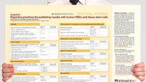 挂图Reporting Practices for Publishing Results with hPSCs Learn how to plan and conduct your human pluripotent stem cell (hPSC)-based research following the ISSCR’s Standards for Human Stem Cell Use in Research

 沪公网安备31010102008431号