
 EasySep™小鼠TIL(CD45)正选试剂盒


搜索结果: 'methocult media formulations for human hematopoietic cells serum containing'
-

产品类型:
产品号#:
05850
05857
05870
05875
85850
85857
85870
85875
产品名:
mTeSR™1
mTeSR™1
-
产品类型:
产品号#:
19059
19059RF
产品名:
EasySep™人单核细胞富集试剂盒
RoboSep™ 人单核细胞富集试剂盒含滤芯吸头
-
产品类型:
产品号#:
05850
05857
05870
05875
85850
85857
85870
85875
产品名:
mTeSR™1
mTeSR™1
-
产品类型:
产品号#:
15271HLA
产品名:
RosetteSep™ HLA 淋系细胞富集试剂盒
-
产品类型:
产品号#:
05910
05990
产品名:
用于hESC/hiPSC维持培养的TeSR™-E8™
-
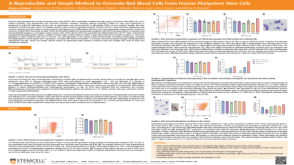 科学海报A Reproducible and Simple Method to Generate Red Blood Cells From Human Pluripotent Stem Cells
产品类型:
Conference:
ASH 2019
产品号#:
产品名:
-
产品类型:
产品号#:
17955
18000
19059
19059RF
17955RF
100-0960
产品名:
EasySep™人NK细胞分选试剂盒
EasySep™磁极
EasySep™人单核细胞富集试剂盒
RoboSep™ 人单核细胞富集试剂盒含滤芯吸头
RoboSep™ 人NK细胞分选试剂盒
EasySep™人NK细胞分离试剂盒
-
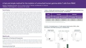 科学海报Cell Isolation of Untouched Human Gamma-Delta T Cells from PBMC
产品类型:
Conference:
AAI 2012
产品号#:
产品名:
-
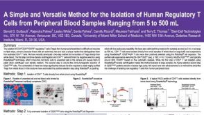 科学海报Isolation of Human Regulatory T Cells from Peripheral Blood Samples
产品类型:
Conference:
KEYSTONE 2007
产品号#:
21000
20155
20119
15862
15862RF
产品名:
RoboSep™- S
RoboSep™分选管套装(9个塑料管)
RoboSep™ 吸头组件抛光剂
-
产品类型:
产品号#:
72202
72204
100-1049
产品名:
Purmorphamine
Purmorphamine
Purmorphamine
-
产品类型:
产品号#:
05150
产品名:
MyeloCult™ H5100


 沪公网安备31010102008431号