Scale-down optimization of a robust, parallelizable human induced pluripotent stem cell bioprocess for high-throughput research
Highlights•Preformation of aggregates tuned by cell density enable cultivation of hiPSCs in scale-down shear environments.•Scale-down systems utilizing preformation protocols achieve comparable fold expansion with commercial systems.•Expression of pluripotency markers and functional differentiation capacity is maintained following passage in scale-down culture.•Successful application of hiPSC protocols at < 20 mL scales enable rapid and cost-effective research into cell phenotype under dynamic conditions. Human induced pluripotent stem cell (hiPSC) derived therapeutics require clinically relevant quantities of high-quality cell populations for applications in regenerative medicine. The lack of efficacy exhibited across clinical trials suggests deeper understanding of the networks governing phenotype is needed. Further,costs limit study throughput in characterizing the artificial niche relative to outcomes. We present herein an optimized strategy to enable high-throughput hiPSC expansion at <20 mL research scale. We assessed viability of single cell inoculation and aggregate preformation to facilitate proliferation. We modeled aggregate characteristics against agitation rate. Our results demonstrate tunable control with fold expansion comparable to commercial systems. Marker quantification and teratoma assay confirm functional pluripotency. This approach constitutes a scalable protocol to accelerate hiPSC research,and a significant step in advancing the rate of progress in elucidating links to derivative functionality. This work will enable statistically rigorous studies targeting hiPSC and downstream phenotype for clinical manufacturing. Graphical abstractImplementation of adapted protocols enable scale-down systems as a tool for high-throughput iPSC biomanufacturing research,in platforms conducive to scale-up for clinical manufacturing.Image,graphical abstract
View Publication
产品类型:
产品号#:
85850
85857
产品名:
mTeSR™1
mTeSR™1
S. Wendt et al. (Nov 2025)
Bio-protocol 15 21
Generation of 3D Human iPSC-Derived Multi-Cell Type Neurospheres for Studying Neuron, Astrocyte, and Microglia Crosstalk
Three-dimensional (3D) human brain tissue models derived from induced pluripotent stem cells (iPSCs) have transformed the study of neural development and disease in vitro. While cerebral organoids offer high structural complexity,their large size often leads to necrotic core formation,limiting reproducibility and challenging the integration of microglia. Here,we present a detailed,reproducible protocol for generating multi-cell type 3D neurospheres that incorporate neurons,astrocytes,and optionally microglia,all derived from the same iPSCs. While neurons and astrocytes differentiate spontaneously from neural precursor cells,generated by dual SMAD-inhibition (blocking BMP and TGF-b signaling),microglia are generated in parallel and can infiltrate the mature neurosphere tissue after plating neurospheres into 48-well plates. The system supports a range of downstream applications,including functional confocal live imaging of GCaMP6f after adeno-associated virus (AAV) transduction of neurospheres or immunofluorescence staining after fixation. Our approach has been successfully implemented across multiple laboratories,demonstrating its robustness and translational potential for studying neuron–glia interactions and modeling neurodegenerative processes.
View Publication
产品类型:
产品号#:
100-0483
100-0484
产品名:
Hausser Scientificᵀᴹ 明线血球计数板
ReLeSR™
C. B. Chhan et al. (Feb 2026)
Cell Reports Medicine 7 2
Transgenic mouse-derived human monoclonal antibodies targeting EBV gp350 and gp42 provide basis for therapeutic development
Epstein-Barr virus (EBV) causes infectious mononucleosis and contributes to neurodegenerative disorders and malignancies,particularly in immune-compromised hosts. Transplant patients face high risk of post-transplant lymphoproliferative disease,a life-threatening EBV-driven lymphoma. There are no EBV-specific vaccines or treatments; however,neutralizing antibodies against EBV glycoproteins may offer utility as therapeutic agents. EBV entry into B cells involves gp350,which binds complement receptors,and gp42,which engages HLA class II to trigger fusion. Most existing monoclonal antibodies (mAbs) against these antigens are non-human,limiting clinical use. Using a transgenic mouse model,we generate two gp350 and eight gp42 genetically human neutralizing mAbs that block receptor binding. Structural analyses reveal extended sites of vulnerability relevant to vaccine development. Delivery of a gp42 mAb protects humanized mice from EBV challenge,while a gp350 mAb provides partial protection. These mAbs highlight the utility of transgenic mice to produce therapeutic mAbs for preventing EBV-driven disease. Graphical abstract Highlights•Transgenic mice were used to make genetically human EBV mAbs against gp350 and gp42•mAbs potently neutralize EBV infection by blocking receptor-ligand interactions•mAbs prevent EBV infection following EBV challenge in humanized mice Epstein-Barr virus (EBV) can cause serious illness,including cancer,especially in immunocompromised patients. There are no EBV-specific treatments. Chhan et al. leverage a transgenic mouse model to develop human monoclonal antibodies that block EBV entry. These antibodies prevent EBV infection in a murine challenge model offering hope for new therapies.
View Publication
产品类型:
产品号#:
19054
19054RF
产品名:
EasySep™人B细胞富集试剂盒
RoboSep™ 人B细胞富集试剂盒含滤芯吸头
Ungrin MD et al. (APR 2012)
Biotechnology and bioengineering 109 4 853--66
Rational bioprocess design for human pluripotent stem cell expansion and endoderm differentiation based on cellular dynamics.
We present a predictive bioprocess design strategy employing cell- and molecular-level analysis of rate-limiting steps in human pluripotent stem cell (hPSC) expansion and differentiation,and apply it to produce definitive endoderm (DE) progenitors using a scalable directed-differentiation technology. We define a bioprocess optimization parameter (L; targeted cell Loss) and,with quantitative cell division tracking and fate monitoring,identify and overcome key suspension bioprocess bottlenecks. Adapting process operating conditions to pivotal parameters (single cell survival and growth rate) in a cell-line-specific manner enabled adherent-equivalent expansion of hPSCs in feeder- and matrix-free defined-medium suspension culture. Predominantly instructive differentiation mechanisms were found to underlie a subsequent 18-fold expansion,during directed differentiation,to high-purity DE competent for further commitment along pancreatic and hepatic lineages. This study demonstrates that iPSC expansion and differentiation conditions can be prospectively specified to guide the enhanced production of target cells in a scale-free directed differentiation system.
View Publication
产品类型:
产品号#:
27845
27945
27840
27865
27940
27965
产品名:
Ermakov A et al. (NOV 2012)
Stem Cell Research 9 3 171--184
A role for intracellular calcium downstream of G-protein signaling in undifferentiated human embryonic stem cell culture
Multiple signalling pathways maintain human embryonic stem cells (hESC) in an undifferentiated state. Here we sought to define the significance of G protein signal transduction in the preservation of this state distinct from other cellular processes. Continuous treatment with drugs targeting G(αs)-,G(α-i/o)- and G(α-q/11)-subunit signalling mediators were assessed in independent hESC lines after 7days to discern effects on normalised alkaline phosphatase positive colony frequency vs total cell content. This identified PLCβ,intracellular free calcium and CAMKII kinase activity downstream of G(α-q/11) as of particular importance to the former. To confirm the significance of this finding we generated an agonist-responsive hESC line transgenic for a G(α-q/11) subunit-coupled receptor and demonstrated that an undifferentiated state could be promoted in the presence of an agonist without exogenously supplied bFGF and that this correlated with elevated intracellular calcium. Similarly,treatment of unmodified hESCs with a range of intracellular free calcium-modulating drugs in biologically defined mTESR culture system lacking exogenous bFGF promoted an hESC phenotype after 1week of continuous culture as defined by co-expression of OCT4 and NANOG. At least one of these drugs,lysophosphatidic acid significantly elevates phosphorylation of calmodulin and STAT3 in this culture system (ptextless0.05). These findings substantiate a role for G-protein and calcium signalling in undifferentiated hESC culture.
View Publication
产品类型:
产品号#:
05850
05857
05870
05875
85850
85857
85870
85875
产品名:
mTeSR™1
mTeSR™1
(May 2024)
Molecular Therapy. Methods & Clinical Development 32 2
Preclinical specificity & activity of a fully human 41BB-expressing anti-CD19 CART- therapy for treatment-resistant autoimmune disease
Over 4% of the global population is estimated to live with autoimmune disease,necessitating immunosuppressive treatment that is often chronic,not curative,and carries associated risks. B cells have emerged as key players in disease pathogenesis,as evidenced by partial responsiveness to B cell depletion by antibody-based therapies. However,these treatments often have transient effects due to incomplete depletion of tissue-resident B cells. Chimeric antigen receptor (CAR) T cells targeting B cells have demonstrated efficacy in refractory systemic lupus erythematosus. To this end,we developed an anti-CD19 CAR T cell product candidate,CABA-201,containing a clinically evaluated fully human CD19 binder (IC78) with a 4-1BB costimulatory domain and CD3 zeta stimulation domain for treatment refractory autoimmune disease. Here,we demonstrate specific cytotoxic activity of CABA-201 against CD19+ Nalm6 cells with no off-target effects on primary human cells. Novel examination of CABA-201 generated from primary T cells from multiple patients with autoimmune disease displayed robust CAR surface expression and effective elimination of the intended target autologous CD19+ B cells in vitro. Together,these findings support the tolerability and activity of CABA-201 for clinical development in patients with autoimmune disease. Graphical abstract Basu and colleagues show CABA-201,a B cell-targeting CAR T cell product with a fully human CD19 binder and 4-1BB costimulatory domain,can precisely eliminate autoimmune patient B cells without off-target deleterious effects,demonstrating its ability as a robust therapeutic for B cell-driven autoimmune disorders.
View Publication
产品类型:
产品号#:
19554
17954
17951
100-0695
17951RF
19554RF
17954RF
100-0971
产品名:
EasySep™人Pan-B细胞富集试剂盒
EasySep™人B细胞分选试剂盒
EasySep™人T细胞分选试剂盒
EasySep™人T细胞分选试剂盒
RoboSep™ 人T细胞分选试剂盒
RoboSep™ 人Pan-B细胞富集试剂盒
RoboSep™ 人B细胞分选试剂盒
EasySep™人B细胞分离试剂盒
A. Wiegering et al. (Apr 2025)
Nature Communications 16
A differential requirement for ciliary transition zone proteins in human and mouse neural progenitor fate specification
Studying ciliary genes in the context of the human central nervous system is crucial for understanding the underlying causes of neurodevelopmental ciliopathies. Here,we use pluripotent stem cell-derived spinal organoids to reveal distinct functions of the ciliopathy gene RPGRIP1L in humans and mice,and uncover an unexplored role for cilia in human axial patterning. Previous research has emphasized Rpgrip1l critical functions in mouse brain and spinal cord development through the regulation of SHH/GLI pathway. Here,we show that RPGRIP1L is not required for SHH activation or motoneuron lineage commitment in human spinal progenitors and that this feature is shared by another ciliopathy gene,TMEM67 . Furthermore,human RPGRIP1L -mutant motoneurons adopt hindbrain and cervical identities instead of caudal brachial identity. Temporal transcriptome analysis reveals that this antero-posterior patterning defect originates in early axial progenitors and correlates with cilia loss. These findings provide important insights into the role of cilia in human neural development. Subject terms: Ciliogenesis,Pattern formation,Pluripotent stem cells,Neurogenesis
View Publication
产品类型:
产品号#:
100-0483
100-0484
产品名:
Hausser Scientificᵀᴹ 明线血球计数板
ReLeSR™
Son M-Y et al. (APR 2014)
Human molecular genetics 23 7 1802--1816
Comparative receptor tyrosine kinase profiling identifies a novel role for AXL in human stem cell pluripotency.
The extensive molecular characterization of human pluripotent stem cells (hPSCs),human embryonic stem cells (hESCs) and human-induced pluripotent stem cells (hiPSCs) is required before they can be applied in the future for personalized medicine and drug discovery. Despite the efforts that have been made with kinome analyses,we still lack in-depth insights into the molecular signatures of receptor tyrosine kinases (RTKs) that are related to pluripotency. Here,we present the first detailed and distinct repertoire of RTK characteristic for hPSC pluripotency by determining both the expression and phosphorylation profiles of RTKs in hESCs and hiPSCs using reverse transcriptase-polymerase chain reaction with degenerate primers that target conserved tyrosine kinase domains and phospho-RTK array,respectively. Among the RTKs tested,the up-regulation of EPHA1,ERBB2,FGFR4 and VEGFR2 and the down-regulation of AXL,EPHA4,PDGFRB and TYRO3 in terms of both their expression and phosphorylation levels were predominantly related to the maintenance of hPSC pluripotency. Notably,the specific inhibition of AXL was significantly advantageous in maintaining undifferentiated hESCs and hiPSCs and for the overall efficiency and kinetics of hiPSC generation. Additionally,a global phosphoproteomic analysis showed that ∼30% of the proteins (293 of 970 phosphoproteins) showed differential phosphorylation upon AXL inhibition in undifferentiated hPSCs,revealing the potential contribution of AXL-mediated phosphorylation dynamics to pluripotency-related signaling networks. Our findings provide a novel molecular signature of AXL in pluripotency control that will complement existing pluripotency-kinome networks.
View Publication
产品类型:
产品号#:
05850
05857
05870
05875
85850
85857
85870
85875
产品名:
mTeSR™1
mTeSR™1
Lei Y and Schaffer DV (DEC 2013)
Proceedings of the National Academy of Sciences of the United States of America 110 52 E5039----E5048
A fully defined and scalable 3D culture system for human pluripotent stem cell expansion and differentiation
Human pluripotent stem cells (hPSCs),including human embryonic stem cells and induced pluripotent stem cells,are promising for numerous biomedical applications,such as cell replacement therapies,tissue and whole-organ engineering,and high-throughput pharmacology and toxicology screening. Each of these applications requires large numbers of cells of high quality; however,the scalable expansion and differentiation of hPSCs,especially for clinical utilization,remains a challenge. We report a simple,defined,efficient,scalable,and good manufacturing practice-compatible 3D culture system for hPSC expansion and differentiation. It employs a thermoresponsive hydrogel that combines easy manipulation and completely defined conditions,free of any human- or animal-derived factors,and entailing only recombinant protein factors. Under an optimized protocol,the 3D system enables long-term,serial expansion of multiple hPSCs lines with a high expansion rate (∼20-fold per 5-d passage,for a 1072-fold expansion over 280 d),yield (∼2.0 × 10⁷ cells per mL of hydrogel),and purity (∼95% Oct4+),even with single-cell inoculation,all of which offer considerable advantages relative to current approaches. Moreover,the system enabled 3D directed differentiation of hPSCs into multiple lineages,including dopaminergic neuron progenitors with a yield of ∼8 × 10⁷ dopaminergic progenitors per mL of hydrogel and ∼80-fold expansion by the end of a 15-d derivation. This versatile system may be useful at numerous scales,from basic biological investigation to clinical development.
View Publication
产品类型:
产品号#:
05850
05857
05870
05875
85850
85857
85870
85875
产品名:
mTeSR™1
mTeSR™1
Zeng H et al. (SEP 2016)
Cell stem cell 19 3 326--340
An Isogenic Human ESC Platform for Functional Evaluation of Genome-wide-Association-Study-Identified Diabetes Genes and Drug Discovery.
Genome-wide association studies (GWASs) have increased our knowledge of loci associated with a range of human diseases. However,applying such findings to elucidate pathophysiology and promote drug discovery remains challenging. Here,we created isogenic human ESCs (hESCs) with mutations in GWAS-identified susceptibility genes for type 2 diabetes. In pancreatic beta-like cells differentiated from these lines,we found that mutations in CDKAL1,KCNQ1,and KCNJ11 led to impaired glucose secretion in vitro and in vivo,coinciding with defective glucose homeostasis. CDKAL1 mutant insulin+ cells were also hypersensitive to glucolipotoxicity. A high-content chemical screen identified a candidate drug that rescued CDKAL1-specific defects in vitro and in vivo by inhibiting the FOS/JUN pathway. Our approach of a proof-of-principle platform,which uses isogenic hESCs for functional evaluation of GWAS-identified loci and identification of a drug candidate that rescues gene-specific defects,paves the way for precision therapy of metabolic diseases.
View Publication
产品类型:
产品号#:
05850
05857
05870
05875
85850
85857
85870
85875
产品名:
mTeSR™1
mTeSR™1
McGrath PS et al. (JUL 2015)
Diabetes 64 7 2497--2505
The basic helix-loop-helix transcription factor neurog3 is required for development of the human endocrine pancreas
Neurogenin3 (NEUROG3) is a basic helix-loop-helix transcription factor required for development of the endocrine pancreas in mice. In contrast,humans with NEUROG3 mutations are born with endocrine pancreas function,calling into question whether NEUROG3 is required for human endocrine pancreas development. To test this directly,we generated human embryonic stem cell (hESC) lines where both alleles of NEUROG3 were disrupted using CRISPR/Cas9-mediated gene targeting. NEUROG3(-/-) hESC lines efficiently formed pancreatic progenitors but lacked detectible NEUROG3 protein and did not form endocrine cells in vitro. Moreover,NEUROG3(-/-) hESC lines were unable to form mature pancreatic endocrine cells after engraftment of PDX1(+)/NKX6.1(+) pancreatic progenitors into mice. In contrast,a 75-90% knockdown of NEUROG3 caused a reduction,but not a loss,of pancreatic endocrine cell development. We conclude that NEUROG3 is essential for endocrine pancreas development in humans and that as little as 10% NEUROG3 is sufficient for formation of pancreatic endocrine cells.
View Publication

 EasySep™小鼠TIL(CD45)正选试剂盒


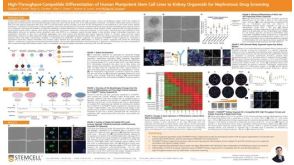 科学海报High-Throughput-Compatible Differentiation of Human Pluripotent Stem Cell Lines to Kidney Organoids for Nephrotoxic Drug Screening
 沪公网安备31010102008431号