Rasheed Z et al. (JAN 2010)
Journal of visualized experiments : JoVE 43
Isolation of stem cells from human pancreatic cancer xenografts.
Cancer stem cells (CSCs) have been identified in a growing number of malignancies and are functionally defined by their ability to undergo self-renewal and produce differentiated progeny. These properties allow CSCs to recapitulate the original tumor when injected into immunocompromised mice. CSCs within an epithelial malignancy were first described in breast cancer and found to display specific cell surface antigen expression (CD44+CD24(low/�?�)). Since then,CSCs have been identified in an increasing number of other human malignancies using CD44 and CD24 as well as a number of other surface antigens. Physiologic properties,including aldehyde dehydrogenase (ALDH) activity,have also been used to isolate CSCs from malignant tissues. Recently,we and others identified CSCs from pancreatic adenocarcinoma based on ALDH activity and the expression of the cell surface antigens CD44 and CD24,and CD133. These highly tumorigenic populations may or may not be overlapping and display other functions. We found that ALDH+ and CD44+CD24+ pancreatic CSCs are similarly tumorigenic,but ALDH+ cells are relatively more invasive. In this protocol we describe a method to isolate viable pancreatic CSCs from low-passage human xenografts. Xenografted tumors are harvested from mice and made into a single-cell suspension. Tissue debris and dead cells are separated from live cells and then stained using antibodies against CD44 and CD24 and using the ALDEFLUOR reagent,a fluorescent substrate of ALDH. CSCs are then isolated by fluorescence activated cell sorting. Isolated CSCs can then be used for analytical or functional assays requiring viable cells.
View Publication
Huang X et al. (DEC 2016)
Advanced materials (Deerfield Beach,Fla.) 28 48 10732--10737
Light-Patterned RNA Interference of 3D-Cultured Human Embryonic Stem Cells.
A new method of spatially controlled gene regulation in 3D-cultured human embryonic stem cells is developed using hollow gold nanoshells (HGNs) and near-infrared (NIR) light. Targeted cell(s) are discriminated from neighboring cell(s) by focusing NIR light emitted from a two-photon microscope. Irradiation of cells that have internalized HGNs releases surface attached siRNAs and leads to concomitant gene downregulation.
View Publication
产品类型:
产品号#:
05850
05857
05870
05875
85850
85857
85870
85875
产品名:
mTeSR™1
mTeSR™1
Butts JC et al. (APR 2017)
Proceedings of the National Academy of Sciences of the United States of America
Differentiation of V2a interneurons from human pluripotent stem cells.
The spinal cord consists of multiple neuronal cell types that are critical to motor control and arise from distinct progenitor domains in the developing neural tube. Excitatory V2a interneurons in particular are an integral component of central pattern generators that control respiration and locomotion; however,the lack of a robust source of human V2a interneurons limits the ability to molecularly profile these cells and examine their therapeutic potential to treat spinal cord injury (SCI). Here,we report the directed differentiation of CHX10(+) V2a interneurons from human pluripotent stem cells (hPSCs). Signaling pathways (retinoic acid,sonic hedgehog,and Notch) that pattern the neural tube were sequentially perturbed to identify an optimized combination of small molecules that yielded ∼25% CHX10(+) cells in four hPSC lines. Differentiated cultures expressed much higher levels of V2a phenotypic markers (CHX10 and SOX14) than other neural lineage markers. Over time,CHX10(+) cells expressed neuronal markers [neurofilament,NeuN,and vesicular glutamate transporter 2 (VGlut2)],and cultures exhibited increased action potential frequency. Single-cell RNAseq analysis confirmed CHX10(+) cells within the differentiated population,which consisted primarily of neurons with some glial and neural progenitor cells. At 2 wk after transplantation into the spinal cord of mice,hPSC-derived V2a cultures survived at the site of injection,coexpressed NeuN and VGlut2,extended neurites textgreater5 mm,and formed putative synapses with host neurons. These results provide a description of V2a interneurons differentiated from hPSCs that may be used to model central nervous system development and serve as a potential cell therapy for SCI.
View Publication
The Axl/Gas6 pathway is required for optimal cytokine signaling during human natural killer cell development.
Interleukin-15 (IL-15) is essential for natural killer (NK) cell differentiation. In this study,we assessed whether the receptor tyrosine kinase Axl and its ligand,Gas6,are involved in IL-15-mediated human NK differentiation from CD34(+) hematopoietic progenitor cells (HPCs). Blocking the Axl-Gas6 interaction with a soluble Axl fusion protein (Axl-Fc) or the vitamin K inhibitor warfarin significantly diminished the absolute number and percentage of CD3(-)CD56(+) NK cells derived from human CD34(+) HPCs cultured in the presence of IL-15,probably resulting in part from reduced phosphorylation of STAT5. In addition,CD3(-)CD56(+) NK cells derived from culture of CD34(+) HPCs with IL-15 and Axl-Fc had a significantly diminished capacity to express interferon-gamma or its master regulator,T-BET. Culture of CD34(+) HPCs in the presence of c-Kit ligand and Axl-Fc resulted in a significant decrease in the frequency of NK precursor cells responding to IL-15,probably the result of reduced c-Kit phosphorylation. Collectively,our data suggest that the Axl/Gas6 pathway contributes to normal human NK-cell development,at least in part via its regulatory effects on both the IL-15 and c-Kit signaling pathways in CD34(+) HPCs,and to functional NK-cell maturation via an effect on the master regulatory transcription factor T-BET.
View Publication
产品类型:
产品号#:
15026
15066
产品名:
RosetteSep™人造血祖细胞富集抗体混合物
RosetteSep™人造血祖细胞富集抗体混合物
Sakaki-Yumoto M et al. (JUN 2013)
Journal of Biological Chemistry 288 25 18546--18560
Smad2 Is essential for maintenance of the human and mouse primed pluripotent stem cell state
Human embryonic stem cells and mouse epiblast stem cells represent a primed pluripotent stem cell state that requires TGF-β/activin signaling. TGF-β and/or activin are commonly thought to regulate transcription through both Smad2 and Smad3. However,the different contributions of these two Smads to primed pluripotency and the downstream events that they may regulate remain poorly understood. We addressed the individual roles of Smad2 and Smad3 in the maintenance of primed pluripotency. We found that Smad2,but not Smad3,is required to maintain the undifferentiated pluripotent state. We defined a Smad2 regulatory circuit in human embryonic stem cells and mouse epiblast stem cells,in which Smad2 acts through binding to regulatory promoter sequences to activate Nanog expression while in parallel repressing autocrine bone morphogenetic protein signaling. Increased autocrine bone morphogenetic protein signaling caused by Smad2 down-regulation leads to cell differentiation toward the trophectoderm,mesoderm,and germ cell lineages. Additionally,induction of Cdx2 expression,as a result of decreased Smad2 expression,leads to repression of Oct4 expression,which,together with the decreased Nanog expression,accelerates the loss of pluripotency. These findings reveal that Smad2 is a unique integrator of transcription and signaling events and is essential for the maintenance of the mouse and human primed pluripotent stem cell state.
View Publication
产品类型:
产品号#:
05850
05857
05870
05875
85850
85857
85870
85875
产品名:
mTeSR™1
mTeSR™1
M. Doglio et al. (Jul 2025)
Frontiers in Immunology 16
CXCR5 engineered human and murine Tregs for targeted suppression in secondary and tertiary lymphoid organs
Secondary and tertiary lymphoid structures are a critical target of suppression in many autoimmune disorders,protein replacement therapies,and in transplantation. Although antigen-specific regulatory T cells (Tregs),such as chimeric antigen receptor (CAR) Tregs,generally persist longer and localize to target tissues more effectively than polyclonal Tregs in animal models,their numbers still progressively decline over time. A potential approach to maximize Treg activity in vivo is the expression of chemokine receptors such as CXCR5,which would enable localization of a greater number of engineered cells at sites of antigen presentation. Indeed,CXCR5 expression on follicular T helper cells and follicular Tregs enables migration toward lymph nodes,B cell zones,and tertiary lymphoid structures that appear in chronically inflamed non-lymphoid tissues. In this study,we generated human and murine CXCR5 co-expressing engineered receptor Tregs and tested them in preclinical mouse models of allo-immunity and hemophilia A,respectively. Additionally,we engineered a murine CXCR5 co-expressing clotting factor VIII (FVIII) specific T cell receptor fusion construct epsilon (FVIII TRuCe CXCR5) Treg to suppress anti-drug antibody development in a model of FVIII protein replacement therapy for hemophilia A. In vitro,anti-HLA-A2 CXCR5+ CAR-Tregs showed enhanced migratory and antigen-specific suppressive capacities compared to untransduced Tregs. When injected into an NSG mouse model of HLA-A2+ pancreatic islet transplantation,anti-HLA-A2 CXCR5+ CAR-Tregs maintained a good safety profile allowing for long-term graft survival in contrast to anti-HLA-A2 CXCR5+ conventional CAR-T (Tconv) cells that eliminated the graft. Similarly,FVIII TRuCe CXCR5 Treg demonstrated increased in vivo persistence and suppressive capacity in a murine model of hemophilia A. Collectively,our findings indicate that CXCR5 co-expression is safe and enhances in vivo localization and persistence in target tissues. This strategy can potentially promote targeted tolerance without the risk of off-target effects in multiple disease models.
View Publication
产品类型:
产品号#:
100-0956
10981
产品名:
ImmunoCult™ XF培养基
ImmunoCult™ XF 人T细胞扩增培养基,500 mL
L. Li et al. (nov 2019)
Proceedings of the National Academy of Sciences of the United States of America 116 46 23274--23283
Directing differentiation of human induced pluripotent stem cells toward androgen-producing Leydig cells rather than adrenal cells.
Reduced serum testosterone (T),or hypogonadism,affects millions of men and is associated with many pathologies,including infertility,cardiovascular diseases,metabolic syndrome,and decreased libido and sexual function. Administering T-replacement therapy (TRT) reverses many of the symptoms associated with low T levels. However,TRT is linked to side effects such as infertility and increased risk of prostate cancer and cardiovascular diseases. Thus,there is a need to obtain T-producing cells that could be used to treat hypogonadism via transplantation and reestablishment of T-producing cell lineages in the body. T is synthesized by Leydig cells (LCs),proposed to derive from mesenchymal cells of mesonephric origin. Although mesenchymal cells have been successfully induced into LCs,the limited source and possible trauma to donors hinders their application to clinical therapies. Alternatively,human induced pluripotent stem cells (hiPSCs),which are expandable in culture and have the potential to differentiate into all somatic cell types,have become the emerging source of autologous cell therapies. We have successfully induced the differentiation of hiPSCs into either human Leydig-like (hLLCs) or adrenal-like cells (hALCs) using chemically defined culture conditions. Factors critical for the development of LCs were added to both culture systems. hLLCs expressed all steroidogenic genes and proteins important for T biosynthesis,synthesized T rather than cortisol,secreted steroid hormones in response to dibutyryl-cAMP and 22(R)-hydroxycholesterol,and displayed ultrastructural features resembling LCs. By contrast,hALCs synthesized cortisol rather than T. The success in generating hiPSC-derived hLLCs with broad human LC (hLC) features supports the potential for hiPSC-based hLC regeneration.
View Publication
产品类型:
产品号#:
06005
产品名:
IntestiCult™ 类器官生长培养基 (小鼠)
Y. Tokumoto et al. (jan 2022)
Clinical and experimental immunology 207 1 95--103
Induction of memory-like CD8+ T cells and CD4+ T cells from human naive T cells in culture.
Memory T cells are crucial players in vertebrate adaptive immunity but their development is incompletely understood. Here,we describe a method to produce human memory-like T cells from naive human T cells in culture. Using commercially available human T-cell differentiation kits,both purified naive CD8+ T cells and purified naive CD4+ T cells were activated via T-cell receptor signaling and appropriate cytokines for several days in culture. All the T-cell activators were then removed from the medium and the cultures were continued in hypoxic condition (1% O2 atmosphere) for several more days; during this period,most of the cells died,but some survived in a quiescent state for a month. The survivors had small round cell bodies,expressed differentiation markers characteristic of memory T cells and restarted proliferation when the T-cell activators were added back. We could also induce memory-like T cells from naive human T cells without hypoxia,if we froze the activated T cells or prepared the naive T cells from chilled filter buffy coats.
View Publication
产品类型:
产品号#:
17968
19555
19555RF
17968RF
产品名:
EasySep™人Naïve CD8+ T细胞分选试剂盒 II
EasySep™人Naïve CD4+ T细胞分选试剂盒
RoboSep™ 人Naïve CD4+ T细胞分选试剂盒
RoboSep™ 人Naïve CD8+ T细胞分选试剂盒 II
Sikandar SS et al. (FEB 2010)
Cancer research 70 4 1469--78
NOTCH signaling is required for formation and self-renewal of tumor-initiating cells and for repression of secretory cell differentiation in colon cancer.
NOTCH signaling is critical for specifying the intestinal epithelial cell lineage and for initiating colorectal adenomas and colorectal cancers (CRC). Based on evidence that NOTCH is important for the maintenance and self-renewal of cancer-initiating cells in other malignancies,we studied the role of NOTCH signaling in colon cancer-initiating cells (CCIC). Tumors formed by CCICs maintain many properties of the primary CRCs from which they were derived,such as glandular organization,cell polarity,gap junctions,and expression of characteristic CRC molecular markers. Furthermore,CCICs have the property of self-renewal. In this study,we show that NOTCH signaling is 10- to 30-fold higher in CCIC compared with widely used colon cancer cell lines. Using small-molecule inhibition and short hairpin RNA knockdown,we show that NOTCH prevents CCIC apoptosis through repression of cell cycle kinase inhibitor p27 and transcription factor ATOH1. NOTCH is also critical to intrinsic maintenance of CCIC self-renewal and the repression of secretory cell lineage differentiation genes such as MUC2. Our findings describe a novel human cell system to study NOTCH signaling in CRC tumor initiation and suggest that inhibition of NOTCH signaling may improve CRC chemoprevention and chemotherapy.
View Publication
产品类型:
产品号#:
01700
01705
01702
产品名:
ALDEFLUOR™ 试剂盒
ALDEFLUOR™ DEAB试剂, 1.5 mM, 1 mL
ALDEFLUOR™检测缓冲液
Kim M et al. (JAN 2002)
Clinical cancer research : an official journal of the American Association for Cancer Research 8 1 22--8
The multidrug resistance transporter ABCG2 (breast cancer resistance protein 1) effluxes Hoechst 33342 and is overexpressed in hematopoietic stem cells.
The human ATP-binding cassette superfamily G (White) member 2 (ABCG2) gene and its murine homologue breast cancer resistance protein 1 (Bcrp1) are recently described ATP-binding cassette transporters associated with drug resistance in tumor cell lines,including the MCF-7 cell line,selected for its resistance to mitoxantrone (MCF-7/MitoR). Infection of MCF-7 cells with the retroviral vector containing ABCG2 cDNA (G1-ABCG2) resulted in cells (MCF-7/ABCG2) that were resistant to mitoxantrone at levels similar to those observed in MCF-7/MitoR cells. Previous studies have shown that pluripotent hematopoietic stem cells overexpress the multidrug-resistant transport (MDR1) gene and efflux rhodamine,a substrate for the MDR1 transporter. Other studies have identified a primitive hematopoietic stem cell population,or side population (SP) cells,which are identified by their efflux of the fluorescent dye,Hoechst 33342. In an attempt to identify the transport genes responsible for this phenotype,we examined the uptake of Hoechst 33342 into MCF-7,MCF-7/MitoR,and MCF-7 cells infected with a retroviral vector expressing the ABCG2 gene (MCF-7/ABCG2). MCF-7/MitoR cells as well as MCF-7/ABCG2 cells demonstrated lower levels of Hoechst 33342 uptake compared with the parental MCF-7 cells. We also examined the level of the mouse Bcrp1 RNA in SP cells and non-SP cells isolated from mouse hematopoietic cells. Mouse SP cells expressed relatively high levels of Bcrp1 mRNA relative to non-SP cells. These results suggest that Hoechst 33342 is a substrate for the ABCG2 transporter and that ABCG2/Bcrp1 expression may serve as a marker for hematopoietic stem cells in hematopoietic cells.
View Publication

 EasySep™小鼠TIL(CD45)正选试剂盒


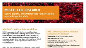 产品手册Derive, Expand, and Differentiate Human Skeletal Muscle Progenitor Cells

 沪公网安备31010102008431号