A Human Brain-Chip for Modeling Brain Pathologies and Screening Blood–Brain Barrier Crossing Therapeutic Strategies
Background/Objectives: The limited translatability of preclinical experimental findings to patients remains an obstacle for successful treatment of brain diseases. Relevant models to elucidate mechanisms behind brain pathogenesis,including cell-specific contributions and cell-cell interactions,and support successful targeting and prediction of drug responses in humans are urgently needed,given the species differences in brain and blood-brain barrier (BBB) functions. Human microphysiological systems (MPS),such as Organ-Chips,are emerging as a promising approach to address these challenges. Here,we examined and advanced a Brain-Chip that recapitulates aspects of the human cortical parenchyma and the BBB in one model. Methods: We utilized human primary astrocytes and pericytes,human induced pluripotent stem cell (hiPSC)-derived cortical neurons,and hiPSC-derived brain microvascular endothelial-like cells and included for the first time on-chip hiPSC-derived microglia. Results: Using Tumor necrosis factor alpha (TNF?) to emulate neuroinflammation,we demonstrate that our model recapitulates in vivo-relevant responses. Importantly,we show microglia-derived responses,highlighting the Brain-Chip’s sensitivity to capture cell-specific contributions in human disease-associated pathology. We then tested BBB crossing of human transferrin receptor antibodies and conjugated adeno-associated viruses. We demonstrate successful in vitro/in vivo correlation in identifying crossing differences,underscoring the model’s capacity as a screening platform for BBB crossing therapeutic strategies and ability to predict in vivo responses. Conclusions: These findings highlight the potential of the Brain-Chip as a reliable and time-efficient model to support therapeutic development and provide mechanistic insights into brain diseases,adding to the growing evidence supporting the value of MPS in translational research and drug discovery.
View Publication
产品类型:
产品号#:
100-0276
100-1130
产品名:
mTeSR™ Plus
mTeSR™ Plus
Kanninen LK et al. (FEB 2016)
Experimental cell research 341 2 207--217
Hepatic differentiation of human pluripotent stem cells on human liver progenitor HepaRG-derived acellular matrix.
Human hepatocytes are extensively needed in drug discovery and development. Stem cell-derived hepatocytes are expected to be an improved and continuous model of human liver to study drug candidates. Generation of endoderm-derived hepatocytes from human pluripotent stem cells (hPSCs),including human embryonic stem cells and induced pluripotent stem cells,is a complex,challenging process requiring specific signals from soluble factors and insoluble matrices at each developmental stage. In this study,we used human liver progenitor HepaRG-derived acellular matrix (ACM) as a hepatic progenitor-specific matrix to induce hepatic commitment of hPSC-derived definitive endoderm (DE) cells. The DE cells showed much better attachment to the HepaRG ACM than other matrices tested and then differentiated towards hepatic cells,which expressed hepatocyte-specific makers. We demonstrate that Matrigel overlay induced hepatocyte phenotype and inhibited biliary epithelial differentiation in two hPSC lines studied. In conclusion,our study demonstrates that the HepaRG ACM,a hepatic progenitor-specific matrix,plays an important role in the hepatic differentiation of hPSCs.
View Publication
产品类型:
产品号#:
05850
05857
05870
05875
07923
10215
85850
85857
85870
85875
产品名:
Dispase (1 U/mL)
mTeSR™1
mTeSR™1
Hervé et al. (JUL 2007)
The Journal of experimental medicine 204 7 1583--93
CD40 ligand and MHC class II expression are essential for human peripheral B cell tolerance.
Hyper-IgM (HIGM) syndromes are primary immunodeficiencies characterized by defects of class switch recombination and somatic hypermutation. HIGM patients who carry mutations in the CD40-ligand (CD40L) gene expressed by CD4(+) T cells suffer from recurrent infections and often develop autoimmune disorders. To investigate the impact of CD40L-CD40 interactions on human B cell tolerance,we tested by ELISA the reactivity of recombinant antibodies isolated from single B cells from three CD40L-deficient patients. Antibody characteristics and reactivity from CD40L-deficient new emigrant B cells were similar to those from healthy donors,suggesting that CD40L-CD40 interactions do not regulate central B cell tolerance. In contrast,mature naive B cells from CD40L-deficient patients expressed a high proportion of autoreactive antibodies,including antinuclear antibodies. Thus,CD40L-CD40 interactions are essential for peripheral B cell tolerance. In addition,a patient with the bare lymphocyte syndrome who could not express MHC class II molecules failed to counterselect autoreactive mature naive B cells,suggesting that peripheral B cell tolerance also depends on major histocompatibility complex (MHC) class II-T cell receptor (TCR) interactions. The decreased frequency of MHC class II-restricted CD4(+) regulatory T cells in CD40L-deficient patients suggests that these T cells may mediate peripheral B cell tolerance through CD40L-CD40 and MHC class II-TCR interactions.
View Publication
产品类型:
产品号#:
15024
15064
产品名:
RosetteSep™人B细胞富集抗体混合物
RosetteSep™人B细胞富集抗体混合物
Gibbs BF et al. (MAR 2008)
Clinical and experimental allergy : journal of the British Society for Allergy and Clinical Immunology 38 3 480--5
A rapid two-step procedure for the purification of human peripheral blood basophils to near homogeneity.
BACKGROUND: Basophils are increasingly utilized as indicators of allergic inflammation and as primary allergic effector cells to study signalling pathways. However,until the present,their enrichment has been time consuming,costly and limited to relatively few specialized laboratories. OBJECTIVE: We have therefore devised a reproducible and rapid method for the purification of human basophils from small quantities of peripheral blood within 1.5 h,which does not require the use of specialized equipment such as elutriators. METHODS: Human basophils were obtained from healthy volunteers undergoing venipuncture. Heparinized or K3-ethylenediaminetetraacetic acid blood samples were first subjected to centrifugation in Hetasep,directly followed by negative selection using immunomagnetic beads. Basophil morphology and purity were assessed by May-Grünwald staining of cytospins. IgE-mediated histamine release was analysed spectrofluorometrically and IL-4 and IL-13 production by quantitative RT-PCR. CD203c and CD63 surface expression was measured using flow cytometry before and after activation with anti-IgE. RESULTS: Using this protocol,basophils were enriched close to homogeneity in most cases with a mean purity of 99.34+/-0.88% (range 97-100%,n=18) and a mean recovery of 75.6 (range 39-100%,n=8). Basophil viability following purification was 99.6+/-0.89% using Trypan blue exclusion. The purification procedure gave rise to basophils with normal functional responses to anti-IgE regarding histamine release as well as IL-4 and IL-13 mRNA expression. Moreover,constitutive cell-surface CD203c/CD63 expressions were not elevated before anti-IgE stimulation. CONCLUSION: The rapidity,simplicity and reproducibility of this method will facilitate the employment of basophils in high-output ex vivo studies.
View Publication
产品类型:
产品号#:
19069
19069RF
产品名:
Daniels TR et al. ( 2011)
Journal of immunotherapy (Hagerstown,Md. : 1997) 34 6 500--8
An antibody-based multifaceted approach targeting the human transferrin receptor for the treatment of B-cell malignancies.
We previously developed an antibody-avidin fusion protein (ch128.1Av) targeting the human transferrin receptor 1 (TfR1,also known as CD71),which demonstrates direct in vitro cytotoxicity against malignant hematopoietic cells. This cytotoxicity is attributed to its ability to decrease the level of TfR1 leading to lethal iron deprivation. We now report that ch128.1Av shows the ability to bind the Fcγ receptors and the complement component C1q,suggesting that it is capable of eliciting Fc-mediated effector functions such as antibody-dependent cell-mediated cytotoxicity and complement-mediated cytotoxicity. In addition,in 2 disseminated multiple myeloma xenograft mouse models,we show that a single dose of ch128.1Av results in significant antitumor activity,including long-term survival. It is interesting to note that the parental antibody without avidin (ch128.1) also shows remarkable in vivo anticancer activity despite its limited in vitro cytotoxicity. Finally,we demonstrate that ch128.1Av is not toxic to pluripotent hematopoietic progenitor cells using the long-term cell-initiating culture assay suggesting that these important progenitors would be preserved in different therapeutic approaches,including the in vitro purging of cancer cells for autologous transplantation and in vivo passive immunotherapy. Our results suggest that ch128.1Av and ch128.1 may be effective in the therapy of human multiple myeloma and potentially other hematopoietic malignancies.
View Publication
Induction of cytotoxic T lymphocyte and antibody responses to enhanced green fluorescent protein following transplantation of transduced CD34(+) hematopoietic cells.
Genetic modification of hematopoietic stem cells often results in the expression of foreign proteins in pluripotent progenitor cells and their progeny. However,the potential for products of foreign genes introduced into hematopoietic stem cells to induce host immune responses is not well understood. Gene marking and induction of immune responses to enhanced green fluorescent protein (eGFP) were examined in rhesus macaques that underwent nonmyeloablative irradiation followed by infusions of CD34(+) bone marrow cells transduced with a retroviral vector expressing eGFP. CD34(+) cells were obtained from untreated animals or from animals treated with recombinant human granulocyte colony-stimulating factor (G-CSF) alone or G-CSF and recombinant human stem cell factor. Levels of eGFP-expressing cells detected by flow cytometry peaked at 0.1% to 0.5% of all leukocytes 1 to 4 weeks after transplantation. Proviral DNA was detected in 0% to 17% of bone marrow--derived colony-forming units at periods of 5 to 18 weeks after transplantation. However,5 of 6 animals studied demonstrated a vigorous eGFP-specific cytotoxic T lymphocyte (CTL) response that was associated with a loss of genetically modified cells in peripheral blood,as demonstrated by both flow cytometry and polymerase chain reaction. The eGFP-specific CTL responses were MHC-restricted,mediated by CD8(+) lymphocytes,and directed against multiple epitopes. eGFP-specific CTLs were able to efficiently lyse autologous CD34(+) cells expressing eGFP. Antibody responses to eGFP were detected in 3 of 6 animals. These data document the potential for foreign proteins expressed in CD34(+) hematopoietic cells and their progeny to induce antibody and CTL responses in the setting of a clinically applicable transplantation protocol. (Blood. 2001;97:1951-1959)
View Publication
产品类型:
产品号#:
09600
09650
产品名:
StemSpan™ SFEM
StemSpan™ SFEM
S. Bari et al. ( 2018)
Stem cells translational medicine 7 5 376--393
Ex Vivo Expansion of CD34+ CD90+ CD49f+ Hematopoietic Stem and Progenitor Cells from Non-Enriched Umbilical Cord Blood with Azole Compounds.
Umbilical cord blood (UCB) transplants in adults have slower hematopoietic recovery compared to bone marrow (BM) or peripheral blood (PB) stem cells mainly due to low number of total nucleated cells and hematopoietic stem and progenitor cells (HSPC). As such in this study,we aimed to perform ex vivo expansion of UCB HSPC from non-enriched mononucleated cells (MNC) using novel azole-based small molecules. Freshly-thawed UCB-MNC were cultured in expansion medium supplemented with small molecules and basal cytokine cocktail. The effects of the expansion protocol were measured based on in vitro and in vivo assays. The proprietary library of {\textgreater}50 small molecules were developed using structure-activity-relationship studies of SB203580,a known p38-MAPK inhibitor. A particular analog,C7,resulted in 1,554.1 ± 27.8-fold increase of absolute viable CD45+ CD34+ CD38- CD45RA- progenitors which was at least 3.7-fold higher than control cultures (p {\textless} .001). In depth phenotypic analysis revealed {\textgreater}600-fold expansion of CD34+ /CD90+ /CD49f+ rare HSPCs coupled with significant (p {\textless} .01) increase of functional colonies from C7 treated cells. Transplantation of C7 expanded UCB grafts to immunodeficient mice resulted in significantly (p {\textless} .001) higher engraftment of human CD45+ and CD45+ CD34+ cells in the PB and BM by day 21 compared to non-expanded and cytokine expanded grafts. The C7 expanded grafts maintained long-term human multilineage chimerism in the BM of primary recipients with sustained human CD45 cell engraftment in secondary recipients. In conclusion,a small molecule,C7,could allow for clinical development of expanded UCB grafts without pre-culture stem cell enrichment that maintains in vitro and in vivo functionality. Stem Cells Translational Medicine 2018;7:376-393.
View Publication
产品类型:
产品号#:
05010
05240
产品名:
STEMdiff™ 心室肌细胞分化试剂盒
STEMdiff™ 间充质祖细胞试剂盒
Bielawska-Pohl A et al. (MAY 2005)
Journal of immunology (Baltimore,Md. : 1950) 174 9 5573--82
Human NK cells lyse organ-specific endothelial cells: analysis of adhesion and cytotoxic mechanisms.
Human organ-specific microvascular endothelial cells (ECs) were established and used in the present study to investigate their susceptibility to natural killer cell line (NKL)-induced lysis. Our data indicate that although IL-2-stimulated NKL (NKL2) cells adhered to the human peripheral (HPLNEC.B3),mesenteric lymph node (HMLNEC),brain (HBrMEC),and lung (HLMEC) and skin (HSkMEC.2) ECs,they significantly killed these cells quite differently. A more pronounced lysis of OSECs was also observed when IL-2-stimulated,purified peripheral blood NK cells were used as effector cells. In line with the correlation observed between adhesion pattern and the susceptibility to NKL2-mediated killing,we demonstrated using different chelators that the necessary adhesion step was governed by an Mg(2+)-dependent,but Ca(2+)-independent,mechanism as opposed to the subsequent Ca(2+)-dependent killing. To identify the cytotoxic pathway used by NKL2 cells,the involvement of the classical and alternate pathways was examined. Blocking of the Ca(2+)-dependent cytotoxicity pathway by EGTA/MgCl(2) significantly inhibited endothelial target cell killing,suggesting a predominant role for the perforin/granzyme pathway. Furthermore,using confocal microscopy,we demonstrated that the interaction between NKL2 effectors and ECs induced cytochrome c release and Bid translocation in target cells,indicating an involvement of the mitochondrial pathway in NKL2-induced EC death. In addition,although all tested cells were sensitive to the cytotoxic action of TNF,no susceptibility to TRAIL or anti-Fas mAb was observed. The present studies emphasize that human NK cell cytotoxicity toward ECs may be a potential target to block vascular injury.
View Publication

 EasySep™小鼠TIL(CD45)正选试剂盒


 新闻Applied Cells Enters into a Supply Agreement with STEMCELL Technologies on Reagent Kits for Use in a Next-Generation Solution for Cell Separation
 实验方案How to Dissociate and Plate Human Pluripotent Stem Cell-Derived Cardiomyocytes for Microelectrode Array (MEA) Assay
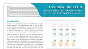 产品手册ClonaCell™-CHO ACF Supplement for Robust Growth of CHO Cells at Low Cell Density

 沪公网安备31010102008431号