S. Murty et al. (nov 2020)
Cancer research 80 21 4731--4740
PET Reporter Gene Imaging and Ganciclovir-Mediated Ablation of Chimeric Antigen Receptor T Cells in Solid Tumors.
Imaging strategies to monitor chimeric antigen receptor (CAR) T-cell biodistribution and proliferation harbor the potential to facilitate clinical translation for the treatment of both liquid and solid tumors. In addition,the potential adverse effects of CAR T cells highlight the need for mechanisms to modulate CAR T-cell activity. The herpes simplex virus type 1 thymidine kinase (HSV1-tk) gene has previously been translated as a PET reporter gene for imaging of T-cell trafficking in patients with brain tumor. The HSV1-TK enzyme can act as a suicide gene of transduced cells through treatment with the prodrug ganciclovir. Here we report the molecular engineering,imaging,and ganciclovir-mediated destruction of B7H3 CAR T cells incorporating a mutated version of the HSV1-tk gene (sr39tk) with improved enzymatic activity for ganciclovir. The sr39tk gene did not affect B7H3 CAR T-cell functionality and in vitro and in vivo studies in osteosarcoma models showed no significant effect on B7H3 CAR T-cell antitumor activity. PET/CT imaging with 9-(4-[18F]-fluoro-3-[hydroxymethyl]butyl)guanine ([18F]FHBG) of B7H3-sr39tk CAR T cells in an orthotopic model of osteosarcoma revealed tumor homing and systemic immune expansion. Bioluminescence and PET imaging of B7H3-sr39tk CAR T cells confirmed complete tumor ablation with intraperitoneal ganciclovir administration. This imaging and suicide ablation system can provide insight into CAR T-cell migration and proliferation during clinical trials while serving as a suicide switch to limit potential toxicities. SIGNIFICANCE: This study showcases the only genetically engineered system capable of serving the dual role both as an effective PET imaging reporter and as a suicide switch for CAR T cells.
View Publication
产品类型:
产品号#:
15021
15061
产品名:
RosetteSep™人T细胞富集抗体混合物
RosetteSep™人T细胞富集抗体混合物
(Feb 2024)
Cancer Immunology Research 12 4
High-Specificity CRISPR-Mediated Genome Engineering in Anti-BCMA Allogeneic CAR T Cells Suppresses Allograft Rejection in Preclinical Models
Allogeneic CAR T–cell therapies are being developed for hematologic malignancies. The authors implement a Cas12a chRDNA platform to generate allogeneic immune-cloaked BCMA-specific CAR T cells with resistance to host response–mediated rejection for evaluation in multiple myeloma. AbstractAllogeneic chimeric antigen receptor (CAR) T cell therapies hold the potential to overcome many of the challenges associated with patient-derived (autologous) CAR T cells. Key considerations in the development of allogeneic CAR T cell therapies include prevention of graft-vs-host disease (GvHD) and suppression of allograft rejection. Here,we describe preclinical data supporting the ongoing first-in-human clinical study,the CaMMouflage trial (NCT05722418),evaluating CB-011 in patients with relapsed/refractory multiple myeloma. CB-011 is a hypoimmunogenic,allogeneic anti–B-cell maturation antigen (BCMA) CAR T cell therapy candidate. CB-011 cells feature 4 genomic alterations and were engineered from healthy donor–derived T cells using a Cas12a CRISPR hybrid RNA–DNA (chRDNA) genome-editing technology platform. To address allograft rejection,CAR T cells were engineered to prevent endogenous HLA class I complex expression and overexpress a single-chain polyprotein complex composed of beta-2 microglobulin (B2M) tethered to HLA-E. In addition,T-cell receptor (TCR) expression was disrupted at the TCR alpha constant locus in combination with the site-specific insertion of a humanized BCMA-specific CAR. CB-011 cells exhibited robust plasmablast cytotoxicity in vitro in a mixed lymphocyte reaction in cell cocultures derived from patients with multiple myeloma. In addition,CB-011 cells demonstrated suppressed recognition by and cytotoxicity from HLA-mismatched T cells. CB-011 cells were protected from natural killer cell–mediated cytotoxicity in vitro and in vivo due to endogenous promoter-driven expression of B2M–HLA-E. Potent antitumor efficacy,when combined with an immune-cloaking armoring strategy to dampen allograft rejection,offers optimized therapeutic potential in multiple myeloma. See related Spotlight by Caimi and Melenhorst,p. 385
View Publication
产品类型:
产品号#:
100-0956
10981
17951
100-0695
17951RF
产品名:
ImmunoCult™ XF培养基
ImmunoCult™ XF 人T细胞扩增培养基,500 mL
EasySep™人T细胞分选试剂盒
EasySep™人T细胞分选试剂盒
RoboSep™ 人T细胞分选试剂盒
Yamamizu K et al. (MAY 2016)
Scientific reports 6 1 25667
Generation and gene expression profiling of 48 transcription-factor-inducible mouse embryonic stem cell lines.
Mouse embryonic stem cells (ESCs) can differentiate into a wide range - and possibly all cell types in vitro,and thus provide an ideal platform to study systematically the action of transcription factors (TFs) in cell differentiation. Previously,we have generated and analyzed 137 TF-inducible mouse ESC lines. As an extension of this NIA Mouse ESC Bank we generated and characterized 48 additional mouse ESC lines,in which single TFs in each line could be induced in a doxycycline-controllable manner. Together,with the previous ESC lines,the bank now comprises 185 TF-manipulable ESC lines (>10% of all mouse TFs). Global gene expression (transcriptome) profiling revealed that the induction of individual TFs in mouse ESCs for 48 hours shifts their transcriptomes toward specific differentiation fates (e.g.,neural lineages by Myt1 Isl1,and St18; mesodermal lineages by Pitx1,Pitx2,Barhl2,and Lmx1a; white blood cells by Myb,Etv2,and Tbx6,and ovary by Pitx1,Pitx2,and Dmrtc2). These data also provide and lists of inferred target genes of each TF and possible functions of these TFs. The results demonstrate the utility of mouse ESC lines and their transcriptome data for understanding the mechanism of cell differentiation and the function of TFs.
View Publication
产品类型:
产品号#:
05700
05703
05704
产品名:
NeuroCult™ 基础培养基(小鼠和大鼠)
NeuroCult™ 分化添加物(小鼠和大鼠)
NeuroCult™ 分化试剂盒(小鼠和大鼠)
Y. Kuwano et al. (MAY 2016)
Journal of Immunology 196 9 3828--33
G$\alpha$i2 and G$\alpha$i3 Differentially Regulate Arrest from Flow and Chemotaxis in Mouse Neutrophils.
Leukocyte recruitment to inflammation sites progresses in a multistep cascade. Chemokines regulate multiple steps of the cascade,including arrest,transmigration,and chemotaxis. The most important chemokine receptor in mouse neutrophils is CXCR2,which couples through G$\alpha$i2- and G$\alpha$i3-containing heterotrimeric G proteins. Neutrophils arrest in response to CXCR2 stimulation. This is defective in G$\alpha$i2-deficient neutrophils. In this study,we show that G$\alpha$i3-deficient neutrophils showed reduced transmigration but normal arrest in mice. We also tested G$\alpha$i2- or G$\alpha$i3-deficient neutrophils in a CXCL1 gradient generated by a microfluidic device. G$\alpha$i3-,but not G$\alpha$i2-,deficient neutrophils showed significantly reduced migration and directionality. This was confirmed in a model of sterile inflammation in vivo. G$\alpha$i2-,but not G$\alpha$i3-,deficient neutrophils showed decreased Ca(2+) flux in response to CXCR2 stimulation. Conversely,G$\alpha$i3-,but not G$\alpha$i2-,deficient neutrophils exhibited reduced AKT phosphorylation upon CXCR2 stimulation. We conclude that G$\alpha$i2 controls arrest and G$\alpha$i3 controls transmigration and chemotaxis in response to chemokine stimulation of neutrophils.
View Publication
产品类型:
产品号#:
19762
19762RF
20155
产品名:
EasySep™小鼠中性粒细胞富集试剂盒
RoboSep™ 小鼠中性粒细胞富集试剂盒含滤芯吸头
RoboSep™分选管套装(9个塑料管)
(Mar 2025)
Molecular Neurodegeneration 20 2
A versatile mouse model to advance human microglia transplantation research in neurodegenerative diseases
BackgroundRecent studies highlight the critical role of microglia in neurodegenerative disorders,and emphasize the need for humanized models to accurately study microglial responses. Human-mouse microglia xenotransplantation models are a valuable platform for functional studies and for testing therapeutic approaches,yet currently those models are only available for academic research. This hampers their implementation for the development and testing of medication that targets human microglia.MethodsWe developed the hCSF1Bdes mouse line,which is suitable as a new transplantation model and available to be crossed to any disease model of interest. The hCSF1Bdes model created by CRISPR gene editing is RAG2 deficient and expresses human CSF1. Additionally,we crossed this model with two humanized App KI mice,the AppHu and the AppSAA. Flow cytometry,immunohistochemistry and bulk sequencing was used to study the response of microglia in the context of Alzheimer’s disease.ResultsOur results demonstrate the successful transplantation of iPSC-derived human microglia into the brains of hCSF1Bdes mice without triggering a NK-driven immune response. Furthermore,we confirmed the multipronged response of microglia in the context of Alzheimer’s disease. The hCSF1Bdes and the crosses with the Alzheimer’s disease knock-in model AppSAA and the humanized App knock-in control mice,AppHu are deposited with EMMA and fully accessible to the research community.ConclusionThe hCSF1Bdes mouse is available for both non-profit and for-profit organisations,facilitating the use of the xenotransplantation paradigm for human microglia to study complex human disease.Supplementary InformationThe online version contains supplementary material available at 10.1186/s13024-025-00823-2.
View Publication
Lai W-H et al. (DEC 2010)
Cellular reprogramming 12 6 641--653
ROCK inhibition facilitates the generation of human-induced pluripotent stem cells in a defined, feeder-, and serum-free system.
Human-induced pluripotent stem cells (iPSCs) generated from human adult somatic cells through reprogramming hold great promises for future regenerative medicine. However,exposure of human iPSCs to animal feeder and serum in the process of their generation and maintenance imposes risk of transmitting animal pathogens to human subjects,thus hindering the potential therapeutic applications. Here,we report the successful generation of human iPSCs in a feeder-independent culture system with defined factors. Two stable human iPSC lines were established from primary human dermal fibroblasts of two healthy volunteers. These human iPSCs expressed a panel of pluripotency markers including stage-specific embryonic antigen (SSEA)-4,tumor-rejection antigen (TRA)-1-60,TRA-1-81,and alkaline phosphatase,while maintaining normal karyotypes and the exogenous reprogramming factors being silenced. In addition,these human iPSCs can differentiate along lineages representative of the three embryonic germ layers upon formation of embryoid bodies,indicating their pluripotency. Furthermore,subcutaneous transplantation of these cells into immunodeficient mice resulted in teratoma formation in 6 to 8 weeks. Our findings are an important step toward generating patient-specific iPSCs in a more clinically compliant manner by eliminating the need of animal feeder cells and animal serum.
View Publication
产品类型:
产品号#:
05850
05857
05870
05875
85850
85857
85870
85875
产品名:
mTeSR™1
mTeSR™1
Kabanova A et al. (APR 2016)
Cell Reports 15 1 9--18
Human Cytotoxic T Lymphocytes Form Dysfunctional Immune Synapses with B Cells Characterized by Non-Polarized Lytic Granule Release.
Suppression of the cytotoxic T cell (CTL) immune response has been proposed as one mechanism for immune evasion in cancer. In this study,we have explored the underlying basis for CTL suppression in the context of B cell malignancies. We document that human B cells have an intrinsic ability to resist killing by freshly isolated cytotoxic T cells (CTLs),but are susceptible to lysis by IL-2 activated CTL blasts and CTLs isolated from immunotherapy-treated patients with chronic lymphocytic leukemia (CLL). Impaired killing was associated with the formation of dysfunctional non-lytic immune synapses characterized by the presence of defective linker for activation of T cells (LAT) signaling and non-polarized release of the lytic granules transported by ADP-ribosylation factor-like protein 8 (Arl8). We propose that non-lytic degranulation of CTLs are a key regulatory mechanism of evasion through which B cells may interfere with the formation of functional immune synapses by CTLs.
View Publication
产品类型:
产品号#:
15024
15064
15023
15063
产品名:
RosetteSep™人B细胞富集抗体混合物
RosetteSep™人B细胞富集抗体混合物
RosetteSep™人CD8+ T细胞富集抗体混合物
RosetteSep™人CD8+ T细胞富集抗体混合物
(Apr 2024)
Cell Communication and Signaling : CCS 22 9274
Gut microbiota-derived butyrate restores impaired regulatory T cells in patients with AChR myasthenia gravis via mTOR-mediated autophagy
More than 80% of patients with myasthenia gravis (MG) are positive for anti-acetylcholine receptor (AChR) antibodies. Regulatory T cells (Tregs) suppress overproduction of these antibodies,and patients with AChR antibody-positive MG (AChR MG) exhibit impaired Treg function and reduced Treg numbers. The gut microbiota and their metabolites play a crucial role in maintaining Treg differentiation and function. However,whether impaired Tregs correlate with gut microbiota activity in patients with AChR MG remains unknown. Here,we demonstrate that butyric acid-producing gut bacteria and serum butyric acid level are reduced in patients with AChR MG. Butyrate supplementation effectively enhanced Treg differentiation and their suppressive function of AChR MG. Mechanistically,butyrate activates autophagy of Treg cells by inhibiting the mammalian target of rapamycin. Activation of autophagy increased oxidative phosphorylation and surface expression of cytotoxic T-lymphocyte-associated protein 4 on Treg cells,thereby promoting Treg differentiation and their suppressive function in AChR MG. This observed effect of butyrate was blocked using chloroquine,an autophagy inhibitor,suggesting the vital role of butyrate-activated autophagy in Tregs of patients with AChR MG. We propose that gut bacteria derived butyrate has potential therapeutic efficacy against AChR MG by restoring impaired Tregs.Supplementary InformationThe online version contains supplementary material available at 10.1186/s12964-024-01588-9.
View Publication
产品类型:
产品号#:
18063
18063RF
100-1136
产品名:
EasySep™人CD4+CD127low CD25+调节性T细胞分选试剂盒
EasySep™人CD4+CD127lowCD25+调节性T细胞分离试剂盒
EasySep™人CD4+CD127low CD25+调节性T细胞分选试剂盒
Kortylewski M et al. (DEC 2005)
Nature medicine 11 12 1314--21
Inhibiting Stat3 signaling in the hematopoietic system elicits multicomponent antitumor immunity.
The immune system can act as an extrinsic suppressor of tumors. Therefore,tumor progression depends in part on mechanisms that downmodulate intrinsic immune surveillance. Identifying these inhibitory pathways may provide promising targets to enhance antitumor immunity. Here,we show that Stat3 is constitutively activated in diverse tumor-infiltrating immune cells,and ablating Stat3 in hematopoietic cells triggers an intrinsic immune-surveillance system that inhibits tumor growth and metastasis. We observed a markedly enhanced function of dendritic cells,T cells,natural killer (NK) cells and neutrophils in tumor-bearing mice with Stat3(-/-) hematopoietic cells,and showed that tumor regression requires immune cells. Targeting Stat3 with a small-molecule drug induces T cell- and NK cell-dependent growth inhibition of established tumors otherwise resistant to direct killing by the inhibitor. Our findings show that Stat3 signaling restrains natural tumor immune surveillance and that inhibiting hematopoietic Stat3 in tumor-bearing hosts elicits multicomponent therapeutic antitumor immunity.
View Publication
The endothelial antigen ESAM marks primitive hematopoietic progenitors throughout life in mice.
Although recent advances have enabled hematopoietic stem cells (HSCs) to be enriched to near purity,more information about their characteristics will improve our understanding of their development and stage-related functions. Here,using microarray technology,we identified endothelial cell-selective adhesion molecule (ESAM) as a novel marker for murine HSCs in fetal liver. Esam was expressed at high levels within a Rag1(-) c-kit(Hi) Sca1(+) HSC-enriched fraction,but sharply down-regulated with activation of the Rag1 locus,a valid marker for the most primitive lymphoid progenitors in E14.5 liver. The HSC-enriched fraction could be subdivided into 2 on the basis of ESAM levels. Among endothelial antigens on hematopoietic progenitors,ESAM expression showed intimate correlation with HSC activity. The ESAM(Hi) population was highly enriched for multipotent myeloid-erythroid progenitors and primitive progenitors with lymphopoietic activity,and exclusively reconstituted long-term lymphohematopoiesis in lethally irradiated recipients. Tie2(+) c-kit(+) lymphohematopoietic cells in the E9.5-10.5 aorta-gonad-mesonephros region also expressed high levels of ESAM. Furthermore,ESAM was detected on primitive hematopoietic progenitors in adult bone marrow. Interestingly,ESAM expression in the HSC-enriched fraction was up-regulated in aged mice. We conclude that ESAM marks HSC in murine fetal liver and will facilitate studies of hematopoiesis throughout life.
View Publication

 EasySep™小鼠TIL(CD45)正选试剂盒


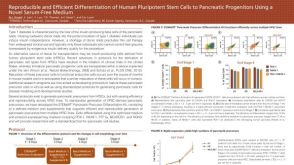 科学海报Reproducible and Efficient Differentiation of Human Pluripotent Stem Cells to Pancreatic Progenitors Using a Novel Serum-Free Medium
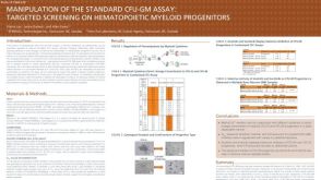 科学海报Manipulation of the Standard CFU-GM Assay Targeted Screening of Hematopoietic Myeloid Progenitors
 沪公网安备31010102008431号