
 EasySep™小鼠TIL(CD45)正选试剂盒


搜索结果: 'methocult media formulations for human hematopoietic cells serum containing'
-

产品类型:
产品号#:
05850
05857
05870
05875
05940
27310
85850
85857
85870
85875
产品名:
缺氧小室
mTeSR™1
mTeSR™1
-
产品类型:
产品号#:
04330
产品名:
MethoCult™ H4330
-
产品类型:
产品号#:
17954
17954RF
100-0971
产品名:
EasySep™人B细胞分选试剂盒
RoboSep™ 人B细胞分选试剂盒
EasySep™人B细胞分离试剂盒
-
产品类型:
产品号#:
09605
09655
04034
04044
产品名:
StemSpan™ SFEM II
StemSpan™ SFEM II
MethoCult™ H4034 Optimum
MethoCult™ H4034 Optimum
-
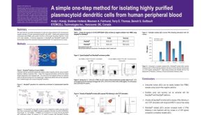 科学海报Isolation of Plasmacytoid Dendritic Cells from Human Peripheral Blood
产品类型:
Conference:
AAI 2009
产品号#:
19062
19062RF
21000
20155
20119
产品名:
EasySep™人浆细胞样DC富集试剂盒
RoboSep™ 人浆细胞样DC富集试剂盒含滤芯吸头
RoboSep™- S
RoboSep™分选管套装(9个塑料管)
RoboSep™ 吸头组件抛光剂
-
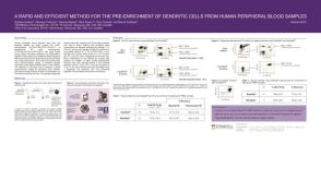 科学海报Pre-Enrichment of Dendritic Cells from Human Peripheral Blood Samples
产品类型:
Conference:
ISCT 2008
产品号#:
19251
19251RF
21000
20155
20119
产品名:
EasySep™人Pan-DC预富集试剂盒
RoboSep™ 人Pan-DC预富集试剂盒含滤芯吸头
RoboSep™- S
RoboSep™分选管套装(9个塑料管)
RoboSep™ 吸头组件抛光剂
-
产品类型:
产品号#:
05850
05857
05870
05875
85850
85857
85870
85875
产品名:
mTeSR™1
mTeSR™1
-
产品类型:
产品号#:
05850
05857
05870
05875
85850
85857
85870
85875
产品名:
mTeSR™1
mTeSR™1
-
产品类型:
产品号#:
05850
05857
05870
05875
85850
85857
85870
85875
产品名:
mTeSR™1
mTeSR™1
-
产品类型:
产品号#:
05850
05857
05870
05875
85850
85857
85870
85875
产品名:
mTeSR™1
mTeSR™1
-
产品类型:
产品号#:
09600
09605
09650
09655
产品名:
StemSpan™ SFEM
StemSpan™ SFEM II
StemSpan™ SFEM
StemSpan™ SFEM II
-
技术手册Maintenance of Human Pluripotent Stem Cells in mTeSR™ Plus
产品类型:
产品号#:
100-0276
100-1130
产品名:
mTeSR™ Plus
mTeSR™ Plus
发布日期: 08/19/2020

 沪公网安备31010102008431号