A Micropatterned Human Pluripotent Stem Cell-Based Ventricular Cardiac Anisotropic Sheet for Visualizing Drug-Induced Arrhythmogenicity.
A novel cardiomimetic biohybrid material,termed as the human ventricular cardiac anisotropic sheet (hvCAS) is reported. Well-characterized human pluripotent stem-cell-derived ventricular cardiomyocytes are strategically aligned to reproduce key electrophysiological features of native human ventricle,which,along with specific selection criteria,allows for a direct visualization of arrhythmic spiral re-entry and represents a revolutionary tool to assess preclinical drug-induced arrhythmogenicity.
View Publication
Ishizawa K et al. (SEP 2010)
Cell stem cell 7 3 279--82
Tumor-initiating cells are rare in many human tumors.
Tumor-initiating cells (TICs) are defined by their ability to form tumors after xenotransplantation in immunodeficient mice and appear to be relatively rare in most human cancers. Recent data in melanoma indicate that the frequency of TICs increases dramatically via more permissive xenotransplantation conditions,raising the possibility that the true frequency of TICs has been greatly underestimated in most human tumors. We compared the growth of human pancreatic,non-small cell lung,and head and neck carcinomas in NOD/SCID and NSG mice. Although TIC frequency was detected up to 10-fold higher in NSG mice,it remained low (textless1 in 2500 cells) in all cases. Moreover,aldehyde dehydrogenase-positive (ALDH(+)) and CD44(+)CD24(+) cells,phenotypically distinct cells enriched in TICs,were equally tumorigenic in NOD/SCID and NSG mice. Our findings demonstrate that TICs are rare in these cancers and that the identification of TICs and their frequency in other human malignancies should be validated via primary tumors and highly permissive xenotransplantation conditions.
View Publication
产品类型:
产品号#:
01700
01705
01701
01702
产品名:
ALDEFLUOR™ 试剂盒
ALDEFLUOR™ DEAB试剂, 1.5 mM, 1 mL
ALDEFLUOR™检测缓冲液
Ben-David U and Benvenisty N (MAR 2014)
Nature protocols 9 3 729--740
Chemical ablation of tumor-initiating human pluripotent stem cells.
The tumorigenicity of human pluripotent stem cells (hPSCs) is widely acknowledged as a major obstacle that withholds their application in regenerative medicine. This protocol describes two efficient and robust ways to chemically eliminate the tumor-initiating hPSCs from monolayer culture. The protocol details how to maintain and differentiate hPSCs,how to apply chemical inhibitors to cultures of hPSCs and their differentiated progeny,and how to assess the purity of the resultant cell cultures using in vitro and in vivo assays. It also describes how to rescue the cytotoxic effect. The elimination and the rescue assay can be completed within 3-5 d,the in vitro assessment requires another day,and the in vivo assessment requires up to 12 additional weeks.
View Publication
产品类型:
产品号#:
05850
05857
05870
05875
85850
85857
85870
85875
产品名:
mTeSR™1
mTeSR™1
Kurian L et al. (JAN 2013)
Nature methods 10 1 77--83
Conversion of human fibroblasts to angioblast-like progenitor cells.
Lineage conversion of one somatic cell type to another is an attractive approach for generating specific human cell types. Lineage conversion can be direct,in the absence of proliferation and multipotent progenitor generation,or indirect,by the generation of expandable multipotent progenitor states. We report the development of a reprogramming methodology in which cells transition through a plastic intermediate state,induced by brief exposure to reprogramming factors,followed by differentiation. We use this approach to convert human fibroblasts to mesodermal progenitor cells,including by non-integrative approaches. These progenitor cells demonstrated bipotent differentiation potential and could generate endothelial and smooth muscle lineages. Differentiated endothelial cells exhibited neo-angiogenesis and anastomosis in vivo. This methodology for indirect lineage conversion to angioblast-like cells adds to the armamentarium of reprogramming approaches aimed at the study and treatment of ischemic pathologies.
View Publication
产品类型:
产品号#:
05850
05857
05870
05875
85850
85857
85870
85875
产品名:
mTeSR™1
mTeSR™1
Ward E et al. (MAY 2017)
Stem cells and development
Feeder-Free Derivation of Naïve Human Pluripotent Stem Cells.
Human pluripotent stem cells (HPSCs) cultured in conditions that maintain pluripotency via FGF and TGFβ signaling have been described as being in a primed state. These cells have been shown to exhibit characteristics more closely related to mouse epiblast-derived stem cells than to so called naïve mouse PSCs said to possess a more ground state pluripotency that mimics the early mouse embryo inner cell mass. Initial attempts to create culture conditions favorable for generation of naïve HPSCs from primed HPSCs has required the use of mouse embryonic fibroblasts as a feeder layer to support this transition. A protocol for the routine derivation and maintenance of naïve HPSCs in completely defined conditions is highly desirable for stem cell researchers to enhance the study and clinical translation of naïve HPSCs. Here we describe a standard protocol for transitioning primed HPSCs to a naïve state using commercial RSet media and xeno-free recombinant vitronectin.
View Publication
Characterization of mouse lymphohematopoietic stem cells lacking spleen colony-forming activity.
The classical definition of lymphohematopoietic stem cells (LHSC),the most primitive progenitors of all blood cells,requires that they have the capacity for self-renewal and for the long-term production of all blood cell lineages. However,other characteristics of LHSC have been debated. Our previous data suggested that mouse LHSC are very slowly proliferating cells that generate delayed multilineage engraftment,while radioprotection" (rapid engraftment that will prevent early death from radiation-induced marrow aplasia) results from more committed progenitors. Alternatively�
View Publication
产品类型:
产品号#:
01700
01705
01701
01702
产品名:
ALDEFLUOR™ 试剂盒
ALDEFLUOR™ DEAB试剂, 1.5 mM, 1 mL
ALDEFLUOR™检测缓冲液
Pei Y et al. (MAR 2015)
Scientific reports 5 9205
A platform for rapid generation of single and multiplexed reporters in human iPSC lines.
Induced pluripotent stem cells (iPSC) are important tools for drug discovery assays and toxicology screens. In this manuscript,we design high efficiency TALEN and ZFN to target two safe harbor sites on chromosome 13 and 19 in a widely available and well-characterized integration-free iPSC line. We show that these sites can be targeted in multiple iPSC lines to generate reporter systems while retaining pluripotent characteristics. We extend this concept to making lineage reporters using a C-terminal targeting strategy to endogenous genes that express in a lineage-specific fashion. Furthermore,we demonstrate that we can develop a master cell line strategy and then use a Cre-recombinase induced cassette exchange strategy to rapidly exchange reporter cassettes to develop new reporter lines in the same isogenic background at high efficiency. Equally important we show that this recombination strategy allows targeting at progenitor cell stages,further increasing the utility of the platform system. The results in concert provide a novel platform for rapidly developing custom single or dual reporter systems for screening assays.
View Publication
Time-integrated BMP signaling determines fate in a stem cell model for early human development
How paracrine signals are interpreted to yield multiple cell fate decisions in a dynamic context during human development in vivo and in vitro remains poorly understood. Here we report an automated tracking method to follow signaling histories linked to cell fate in large numbers of human pluripotent stem cells (hPSCs). Using an unbiased statistical approach,we discover that measured BMP signaling history correlates strongly with fate in individual cells. We find that BMP response in hPSCs varies more strongly in the duration of signaling than the level. However,both the level and duration of signaling activity control cell fate choices only by changing the time integral. Therefore,signaling duration and level are interchangeable in this context. In a stem cell model for patterning of the human embryo,we show that signaling histories predict the fate pattern and that the integral model correctly predicts changes in cell fate domains when signaling is perturbed. Our data suggest that mechanistically,BMP signaling is integrated by SOX2. The interpretation of the key developmental signal BMP remains poorly understood. Here,the authors show that the total time-integrated signaling controls differentiation in a stem cell embryo model and provide a possible mechanism.
View Publication

 EasySep™小鼠TIL(CD45)正选试剂盒


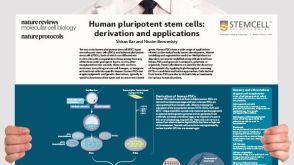 挂图Derivation and Applications of Human Pluripotent Stem Cells Overview of the derivation of human embryonic stem cells (hESCs) and induced pluripotent stem cells (iPSCs)发布日期: 11/26/2020


 沪公网安备31010102008431号