HELLS is required for maintaining proper DNA modification at human satellite repeats
DNA methylation regulation involves multi-layered chromatin interactions that require remodeling proteins like the helicase,lymphoid-specific (HELLS). Here,we generate HELLS and DNA methyltransferase 3A and B (DNMT3A/B) knockout human pluripotent stem cells and report telomere-to-telomere maps of whole genome bisulfite sequencing data combined with ATAC-sequencing. Disrupting HELLS induces a global loss of DNA methylation that is distinct from the DNMTs,in particular over peri/centromeric satellite repeats as defined in the telomere-to-telomere genome assembly. However,HELLS appears dispensable for local enhancer remodeling and the potential to differentiate into the three embryonic germ layers. Taken together,our results further clarify the genomic targets and role of HELLS in human cells.Supplementary InformationThe online version contains supplementary material available at 10.1186/s13059-025-03681-9.
View Publication
产品类型:
产品号#:
05120
100-0276
100-1130
产品名:
STEMdiff™胰腺祖细胞试剂盒
mTeSR™ Plus
mTeSR™ Plus
R. Lorenzetti et al. (jul 2019)
Journal of autoimmunity 101 145--152
Abatacept modulates CD80 and CD86 expression and memory formation in human B-cells.
BACKGROUND Cytotoxic T lymphocyte antigen-4 (CTLA-4) limits T-cell activation and is expressed on T-regulatory cells. Human CTLA-4 deficiency results in severe immune dysregulation. Abatacept (CTLA-4 Ig) is approved for the treatment of rheumatoid arthritis (RA) and its mechanism of action is attributed to effects on T-cells. It is known that CTLA-4 modulates the expression of its ligands CD80 and CD86 on antigen presenting cells (APC) by transendocytosis. As B-cells express CD80/CD86 and function as APC,we hypothesize that B-cells are a direct target of abatacept. OBJECTIVES To investigate direct effects of abatacept on human B-lymphocytes in vitro and in RA patients. METHODS The effect of abatacept on healthy donor B-cells' phenotype,activation and CD80/CD86 expression was studied in vitro. Nine abatacept-treated RA patients were studied. Seven of these were followed up to 24 months,and two up to 12 months only and treatment response,immunoglobulins,ACPA,RF concentrations,B-cell phenotype and ACPA-specific switched memory B-cell frequency were assessed. RESULTS B-cell development was unaffected by abatacept. Abatacept treatment resulted in a dose-dependent decrease of CD80/CD86 expression on B-cells in vitro,which was due to dynamin-dependent internalization. RA patients treated with abatacept showed a progressive decrease in plasmablasts and serum IgG. While ACPA-titers only moderately declined,the frequency of ACPA-specific switched memory B-cells significantly decreased. CONCLUSIONS Abatacept directly targets B-cells by reducing CD80/CD86 expression. Impairment of antigen presentation and T-cell activation may result in altered B-cell selection,providing a new therapeutic mechanism and a base for abatacept use in B-cell mediated autoimmunity.
View Publication
产品类型:
产品号#:
17954
17954RF
100-0971
产品名:
EasySep™人B细胞分选试剂盒
RoboSep™ 人B细胞分选试剂盒
EasySep™人B细胞分离试剂盒
Safi R et al. (FEB 2009)
Molecular endocrinology (Baltimore,Md.) 23 2 188--201
Pharmacological manipulation of the RAR/RXR signaling pathway maintains the repopulating capacity of hematopoietic stem cells in culture.
The retinoid X receptor (RXR) contributes to the regulation of diverse biological pathways via its role as a heterodimeric partner of several nuclear receptors. However,RXR has no established role in the regulation of hematopoietic stem cell (HSC) fate. In this study,we sought to determine whether direct modulation of RXR signaling could impact human HSC self-renewal or differentiation. Treatment of human CD34(+)CD38(-)lin(-) cells with LG1506,a selective RXR modulator,inhibited the differentiation of HSCs in culture and maintained long-term repopulating HSCs in culture that were otherwise lost in response to cytokine treatment. Further studies revealed that LG1506 had a distinct mechanism of action in that it facilitated the recruitment of corepressors to the retinoic acid receptor (RAR)/RXR complex at target gene promoters,suggesting that this molecule was functioning as an inverse agonist in the context of this heterodimer. Interestingly,using combinatorial peptide phage display,we identified unique surfaces presented on RXR when occupied by LG1506 and demonstrated that other modulators that exhibited these properties functioned similarly at both a mechanistic and biological level. These data indicate that the RAR/RXR heterodimer is a critical regulator of human HSC differentiation,and pharmacological modulation of RXR signaling prevents the loss of human HSCs that otherwise occurs in short-term culture.
View Publication
产品类型:
产品号#:
04434
04444
产品名:
MethoCult™ H4434 Classic
MethoCult™ H4434 Classic
Eggimann L et al. (MAY 2015)
Bone marrow transplantation 50 5 743--5
Kinetics of peripheral blood chimerism for surveillance of patients with leukemia and chronic myeloid malignancies after reduced-intensity conditioning allogeneic hematopoietic SCT.
Prowse A et al. (JUL 2009)
BioTechniques 47 1 599--606
A rapid, cost-effective method for counting human embryonic stem cell numbers as clumps.
Enumeration of human embryonic stem cell (hESC) numbers through single cell digestion can be time consuming especially in high-throughput or multi-factorial analysis containing 50+ samples. We have developed a reproducible,cost-effective method of counting hESCs in clumps circumventing the need to manually dissociate each sample to single cells. The method is based on the DNA binding capacity of propidium iodide (PI) and subsequent fluorescent signal detection. Standard curves generated for cell numbers versus PI fluorescence as single cells or clumps showed an almost identical relationship in the lines of best fit. The reproducibility of the assay was first demonstrated by seeding hESC clumps at specific cell densities ranging 0.05[x02013]2x10⁵ cells/well and then secondly by using the assay to count cell numbers after different growth conditions. Validation tests showed that consistent seeding densities are important in maintaining undifferentiated hESC culture and that the assay can be used to estimate relative cell numbers and growth curves with high accuracy.
View Publication
产品类型:
产品号#:
05850
05857
05870
05875
85850
85857
85870
85875
产品名:
mTeSR™1
mTeSR™1
Naylor RW et al. ( 2016)
PloS one 11 10 e0165464
Derivation of Corneal Keratocyte-Like Cells from Human Induced Pluripotent Stem Cells.
Corneal diseases such as keratoconus represent a relatively common disorder in the human population. However,treatment is restricted to corneal transplantation,which only occurs in the most advanced cases. Cell based therapies may offer an alternative approach given that the eye is amenable to such treatments and corneal diseases like keratoconus have been associated specifically with the death of corneal keratocytes. The ability to generate corneal keratocytes in vitro may enable a cell-based therapy to treat patients with keratoconus. Human induced pluripotent stem cells (hiPSCs) offer an abundant supply of cells from which any cell in the body can be derived. In the present study,hiPSCs were successfully differentiated into neural crest cells (NCCs),the embryonic precursor to keratocytes,and then cultured on cadaveric corneal tissue to promote keratocyte differentiation. The hiPSC-derived NCCs were found to migrate into the corneal stroma where they acquired a keratocyte-like morphology and an expression profile similar to corneal keratocytes in vivo. These results indicate that hiPSCs can be used to generate corneal keratocytes in vitro and lay the foundation for using these cells in cornea cell-based therapies.
View Publication
Gronski P et al. (AUG 1988)
Behring Institute Mitteilungen 7 83 246--9
E. coli derived human granulocyte-macrophage colony-stimulating factor (rh GM-CSF) available for clinical trials.
Recombinant human GM-CSF has been expressed as a fusion protein in E. coli in the form of inclusion bodies. Using denaturing agents,acid cleavage and sulfitolysis,the biologically inactive GM-CSF protein could be highly purified and additionally renaturated under suitable reoxidizing conditions. The thorough repair of the two disulfide bridges could be confirmed by sequencing fragments obtained by tryptic digestion. Refolding of the molecule has been studied by CD spectrometry and identity by Western blotting and SDS-PAGE analysis. As could be demonstrated,full biological activity (colony-forming assay with fresh human bone marrow cells) was restored during renaturation of the GM-CSF protein. Further proof of biological equivalence of the E. coli-derived protein with a yeast-derived biologically active rh GM-CSF has been published elsewhere.
View Publication
Santoni de Sio FR et al. (JUN 2006)
Blood 107 11 4257--65
Proteasome activity restricts lentiviral gene transfer into hematopoietic stem cells and is down-regulated by cytokines that enhance transduction.
The therapeutic potential of hematopoietic stem cell (HSC) gene therapy can be fully exploited only by reaching efficient gene transfer into HSCs without compromising their biologic properties. Although HSCs can be transduced by HIV-derived lentiviral vectors (LVs) in short ex vivo culture,they display low permissivity to the vector,requiring cytokine stimulation to reach high-frequency transduction. Using stringent assays of competitive xenograft repopulation,we show that early-acting cytokines synergistically enhanced human HSC gene transfer by LVs without impairing engraftment and repopulation capacity. Using S-phase suicide assays,we show that transduction enhancement by cytokines was not dependent on cell cycle progression and that LVs can transduce quiescent HSCs. Pharmacologic inhibition of the proteasome during transduction dramatically enhanced HSC gene transfer,allowing the reach of very high levels of vector integration in their progeny in vivo. Thus,LVs are effectively restricted at a postentry step by the activity of this proteolytic complex. Unexpectedly,cytokine stimulation rapidly and substantially down-regulated proteasome activity in hematopoietic progenitors,highlighting one mechanism by which cytokines may enhance permissiveness to LV gene transfer. These findings demonstrate that antiviral responses ultimately mediated by proteasomes strongly limit the efficiency of HSC transduction by LVs and establish improved conditions for HSC-based gene therapy.
View Publication
产品类型:
产品号#:
09600
09650
产品名:
StemSpan™ SFEM
StemSpan™ SFEM
Kandasamy M et al. (MAR 2017)
Cell and Tissue Research 368 3 531--549
Glycoconjugates reveal diversity of human neural stem cells (hNSCs) derived from human induced pluripotent stem cells (hiPSCs)
Neural stem cells (NSCs) have the ability to self-renew and to differentiate into various cell types of the central nervous system. This potential can be recapitulated by human induced pluripotent stem cells (hiPSCs) in vitro. The differentiation capacity of hiPSCs is characterized by several stages with distinct morphologies and the expression of various marker molecules. We used the monoclonal antibodies (mAbs) 487(LeX),5750(LeX) and 473HD to analyze the expression pattern of particular carbohydrate motifs as potential markers at six differentiation stages of hiPSCs. Mouse ESCs were used as a comparison. At the pluripotent stage,487(LeX)-,5750(LeX)- and 473HD-related glycans were differently expressed. Later,cells of the three germ layers in embryoid bodies (hEBs) and,even after neuralization of hEBs,subpopulations of cells were labeled with these surface antibodies. At the human rosette-stage of NSCs (hR-NSC),LeX- and 473HD-related epitopes showed antibody-specific expression patterns. We also found evidence that these surface antibodies could be used to distinguish the hR-NSCs from the hSR-NSCs stages. Characterization of hNSCs(FGF-2/EGF) derived from hSR-NSCs revealed that both LeX antibodies and the 473HD antibody labeled subpopulations of hNSCs(FGF-2/EGF). Finally,we identified potential LeX carrier molecules that were spatiotemporally regulated in early and late stages of differentiation. Our study provides new insights into the regulation of glycoconjugates during early human stem cell development. The mAbs 487(LeX),5750(LeX) and 473HD are promising tools for identifying distinct stages during neural differentiation.
View Publication
产品类型:
产品号#:
05832
05850
05857
05870
05875
85850
85857
85870
85875
产品名:
STEMdiff™ 神经花环选择试剂
mTeSR™1
mTeSR™1
Xi J et al. (JAN 2010)
PLoS ONE 5 12 e14457
Human fetal liver stromal cells that overexpress bFGF support growth and maintenance of human embryonic stem cells
In guiding hES cell technology toward the clinic,one key issue to be addressed is to culture and maintain hES cells much more safely and economically in large scale. In order to avoid using mouse embryonic fibroblasts (MEFs) we isolated human fetal liver stromal cells (hFLSCs) from 14 weeks human fetal liver as new human feeder cells. hFLSCs feeders could maintain hES cells for 15 passages (about 100 days). Basic fibroblast growth factor (bFGF) is known to play an important role in promoting self-renewal of human embryonic stem (hES) cells. So,we established transgenic hFLSCs that stably express bFGF by lentiviral vectors. These transgenic human feeder cells--bFGF-hFLSCs maintained the properties of H9 hES cells without supplementing with any exogenous growth factors. H9 hES cells culturing under these conditions maintained all hES cell features after prolonged culture,including the developmental potential to differentiate into representative tissues of all three embryonic germ layers,unlimited and undifferentiated proliferative ability,and maintenance of normal karyotype. Our results demonstrated that bFGF-hFLSCs feeder cells were central to establishing the signaling network among bFGF,insulin-like growth factor 2 (IGF-2),and transforming growth factor β (TGF-β),thereby providing the framework in which hES cells were instructed to self-renew or to differentiate. We also found that the conditioned medium of bFGF-hFLSCs could maintain the H9 hES cells under feeder-free conditions without supplementing with bFGF. Taken together,bFGF-hFLSCs had great potential as feeders for maintaining pluripotent hES cell lines more safely and economically.
View Publication

 EasySep™小鼠TIL(CD45)正选试剂盒


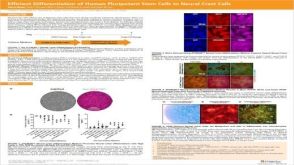 科学海报Efficient Generation of Human Pluripotent Stem Cells to Neural Crest Cells

 沪公网安备31010102008431号