
 EasySep™小鼠TIL(CD45)正选试剂盒


搜索结果: 'methocult media formulations for human hematopoietic cells serum containing'
-
-

产品类型:
产品号#:
05401
05402
05411
产品名:
MesenCult™ MSC 基础培养基(人)
MesenCult™ MSC刺激添加物(人)
MesenCult™ 增殖试剂盒(人)
-
产品类型:
产品号#:
05150
05350
产品名:
MyeloCult™ H5100
-
产品类型:
产品号#:
05850
05857
05870
05875
85850
85857
85870
85875
产品名:
mTeSR™1
mTeSR™1
-
产品类型:
产品号#:
05850
05857
05870
05875
85850
85857
85870
85875
产品名:
mTeSR™1
mTeSR™1
-
产品类型:
产品号#:
03231
09600
09650
产品名:
MethoCult™ M3231
StemSpan™ SFEM
StemSpan™ SFEM
-
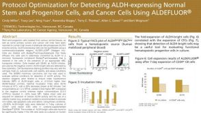 科学海报Protocol Optimization for Detecting ALDH-Expressing Normal Stem and Progenitor Cells and Cancer Cells Using ALDEFLUOR™
产品类型:
Conference:
LORNE 2010
产品号#:
产品名:
-
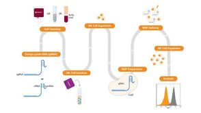
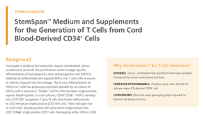 技术公告StemSpan™ Medium and Supplements for the Generation of T Cells from Cord Blood-Derived CD34+ Cells
产品类型:
细胞类型:
CD4+ T细胞,CD8+ T细胞,T细胞,造血干祖细胞
产品号#:
09605
09655
09900
09910
09920
09915
09925
09930
09940
产品名:
StemSpan™ SFEM II
StemSpan™ SFEM II
StemSpan™淋系祖细胞扩增添加物(10X)
StemSpan™淋系祖细胞分化包板材料(100X)
StemSpan™ T细胞祖细胞成熟添加物(10X)
StemSpan™ T细胞生成试剂盒
发布日期: 08/24/2020 -
产品类型:
产品号#:
01700
01705
01702
产品名:
ALDEFLUOR™ 试剂盒
ALDEFLUOR™ DEAB试剂, 1.5 mM, 1 mL
ALDEFLUOR™检测缓冲液
-
产品类型:
产品号#:
01700
01705
01702
产品名:
ALDEFLUOR™ 试剂盒
ALDEFLUOR™ DEAB试剂, 1.5 mM, 1 mL
ALDEFLUOR™检测缓冲液
-
产品类型:
产品号#:
05850
05857
05870
05875
85850
85857
85870
85875
产品名:
mTeSR™1
mTeSR™1
-
产品类型:
产品号#:
05850
05857
05870
05875
85850
85857
85870
85875
产品名:
mTeSR™1
mTeSR™1

 沪公网安备31010102008431号