
 EasySep™小鼠TIL(CD45)正选试剂盒


搜索结果: 'methocult media formulations for mouse hematopoietic cells serum containing'
-

产品类型:
产品号#:
18000
17951
100-0695
17951RF
产品名:
EasySep™磁极
EasySep™人T细胞分选试剂盒
EasySep™人T细胞分选试剂盒
RoboSep™ 人T细胞分选试剂盒
-
-
产品类型:
产品号#:
产品名:
-
产品类型:
产品号#:
05790
产品名:
BrainPhys™神经元培养基
-
产品类型:
产品号#:
72852
72854
产品名:
-
产品类型:
产品号#:
100-0276
100-1130
05946
产品名:
mTeSR™ Plus
mTeSR™ Plus
TeSR™-E6
-
产品类型:
产品号#:
72412
产品名:
骨化三醇(Calcitriol)
-
产品类型:
产品号#:
04434
28600
04444
产品名:
MethoCult™ H4434 Classic
L-Calc™有限稀释软件
MethoCult™ H4434 Classic
-
产品类型:
产品号#:
05150
产品名:
MyeloCult™ H5100
-
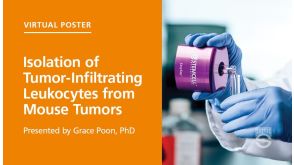
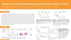 科学海报Isolation of Tumor-Infiltrating Leukocytes from Mouse Tumors
产品类型:
Conference:
AAI 2020
产品号#:
100-0350
产品名:
EasySep™小鼠TIL(CD45)正选试剂盒
发布日期: 10/22/2020 -


 沪公网安备31010102008431号