Chen X et al. (SEP 2015)
Stem Cell Research 15 2 395--402
OP9-Lhx2 stromal cells facilitate derivation of hematopoietic progenitors both in vitro and in vivo
Generating engraftable hematopoietic stem cells (HSCs) from pluripotent stem cells (PSCs) is an ideal approach for obtaining induced HSCs for cell therapy. However,the path from PSCs to robustly induced HSCs (iHSCs) in vitro remains elusive. We hypothesize that the modification of hematopoietic niche cells by transcription factors facilitates the derivation of induced HSCs from PSCs. The Lhx2 transcription factor is expressed in fetal liver stromal cells but not in fetal blood cells. Knocking out Lhx2 leads to a fetal hematopoietic defect in a cell non-autonomous role. In this study,we demonstrate that the ectopic expression of Lhx2 in OP9 cells (OP9-Lhx2) accelerates the hematopoietic differentiation of PSCs. OP9-Lhx2 significantly increased the yields of hematopoietic progenitor cells via co-culture with PSCs in vitro. Interestingly,the co-injection of OP9-Lhx2 and PSCs into immune deficient mice also increased the proportion of hematopoietic progenitors via the formation of teratomas. The transplantation of phenotypic HSCs from OP9-Lhx2 teratomas but not from the OP9 control supported a transient repopulating capability. The upregulation of Apln gene by Lhx2 is correlated to the hematopoietic commitment property of OP9-Lhx2. Furthermore,the enforced expression of Apln in OP9 cells significantly increased the hematopoietic differentiation of PSCs. These results indicate that OP9-Lhx2 is a good cell line for regeneration of hematopoietic progenitors both in vitro and in vivo.
View Publication
产品类型:
产品号#:
05850
05857
05870
05875
85850
85857
85870
85875
产品名:
mTeSR™1
mTeSR™1
Vetter ML and D'Aquila RT (SEP 2009)
Journal of virology 83 17 8646--54
Cytoplasmic APOBEC3G restricts incoming Vif-positive human immunodeficiency virus type 1 and increases two-long terminal repeat circle formation in activated T-helper-subtype cells.
Cytoplasmic APOBEC3G has been reported to block wild-type human immunodeficiency virus type 1 (HIV-1) infection in some primary cells. It is not known whether cytoplasmic APOBEC3G has residual activity in activated T cells,even though virion-packaged APOBEC3G does restrict HIV-1 in activated T cells. Because we found that APOBEC3G expression is greater in activated CD4(+) T-helper type 1 (Th1) lymphocytes than in T-helper type 2 (Th2) lymphocytes,we hypothesized that residual target cell restriction of incoming Vif-positive virions that lack APOBEC3G,if present,would be greater in Th1 than Th2 lymphocytes. Infection of activated Th1 cells with APOBEC3-negative virions did result in decreased amounts of early and late reverse transcription products and integrated virus relative to infection of activated Th2 cells. Two-long terminal repeat (2-LTR) circles,which are formed in the nucleus when reverse transcripts do not integrate,were increased after APOBEC3-negative virus infection of activated Th1 cells relative to infection of activated Th2 cells. In contrast,2-LTR circle forms were decreased after infection of APOBEC3G-negative cells with APOBEC3G-containing virions relative to APOBEC3G-negative virions and with Th1 cell-produced virions relative to Th2 cell-produced virions. Increasing APOBEC3G in Th2 cells and decreasing APOBEC3G in Th1 cells modulated the target cell phenotypes,indicating causation by APOBEC3G. The comparison between activated Th1 and Th2 cells indicates that cytoplasmic APOBEC3G in activated Th1 cells partially restricts reverse transcription and integration of incoming Vif-positive,APOBEC3G-negative HIV-1. The differing effects of cytoplasmic and virion-packaged APOBEC3G on 2-LTR circle formation indicate a difference in their antiviral mechanisms.
View Publication
产品类型:
产品号#:
19052
19052RF
21000
20119
20155
产品名:
EasySep™人CD4+ T细胞富集试剂盒
RoboSep™ 人CD4+ T细胞富集试剂盒含滤芯吸头
RoboSep™- S
RoboSep™ 吸头组件抛光剂
RoboSep™分选管套装(9个塑料管)
L. L. M. Derks et al. (Dec 2025)
HemaSphere 9 12
Posttransplantation clonal dynamics of hematopoietic stem cells carrying prenatal and early‐life DNMT3A mutations
Clonal hematopoiesis (CH),a prevalent and premalignant state in the elderly,has been detected in young individuals under selective pressures such as hematopoietic cell transplantation (HCT). However,the origin of CH and mutational processes underlying CH driver mutations in young blood systems remain unclear. Here,we used genome‐wide somatic mutation profiles to retrospectively trace the origin of DNMT3A‐mutant CH in three individuals,14–41 years after childhood HCT. Both the rate and spectrum of somatic mutations in individuals with posttransplant CH were consistent with normal age‐associated mutagenesis. Phylogenetic analysis revealed that DNMT3A‐mutant HSPCs were present in the donor before 6.8 years of age,including during fetal development,despite being undetectable with a limit of detection of variant allele frequency of 0.001 at the time of transplantation. These findings were validated by comparing the observed mutations to expected age‐dependent mutational signatures. Our results reveal that undetectable DNMT3A‐mutant clones in young donors can expand into significant CH clones within decades upon transplantation. The rapid expansion of these clones in this context indicates that specific environmental pressures,rather than solely mutation acquisition,drive the development of CH.
View Publication
产品类型:
产品号#:
09600
09605
09650
09655
产品名:
StemSpan™ SFEM
StemSpan™ SFEM II
StemSpan™ SFEM
StemSpan™ SFEM II
Booth L et al. (JUL 2015)
Journal of cellular physiology 230 7 1661--76
GRP78/BiP/HSPA5/Dna K is a universal therapeutic target for human disease.
The chaperone GRP78/Dna K is conserved throughout evolution down to prokaryotes. The GRP78 inhibitor OSU-03012 (AR-12) interacted with sildenafil (Viagra) or tadalafil (Cialis) to rapidly reduce GRP78 levels in eukaryotes and as a single agent reduce Dna K levels in prokaryotes. Similar data with the drug combination were obtained for: HSP70,HSP90,GRP94,GRP58,HSP27,HSP40 and HSP60. OSU-03012/sildenafil treatment killed brain cancer stem cells and decreased the expression of: NPC1 and TIM1; LAMP1; and NTCP1,receptors for Ebola/Marburg/Hepatitis A,Lassa fever,and Hepatitis B viruses,respectively. Pre-treatment with OSU-03012/sildenafil reduced expression of the coxsakie and adenovirus receptor in parallel with it also reducing the ability of a serotype 5 adenovirus or coxsakie virus B4 to infect and to reproduce. Similar data were obtained using Chikungunya,Mumps,Measles,Rubella,RSV,CMV,and Influenza viruses. OSU-03012 as a single agent at clinically relevant concentrations killed laboratory generated antibiotic resistant E. coli and clinical isolate multi-drug resistant N. gonorrhoeae and MRSE which was in bacteria associated with reduced Dna K and Rec A expression. The PDE5 inhibitors sildenafil or tadalafil enhanced OSU-03012 killing in N. gonorrhoeae and MRSE and low marginally toxic doses of OSU-03012 could restore bacterial sensitivity in N. gonorrhoeae to multiple antibiotics. Thus,Dna K and bacterial phosphodiesterases are novel antibiotic targets,and inhibition of GRP78 is of therapeutic utility for cancer and also for bacterial and viral infections.
View Publication
产品类型:
产品号#:
05750
05751
产品名:
NeuroCult™ NS-A 基础培养基(人)
NeuroCult™ NS-A 扩增试剂盒(人)
Gallego MJ et al. (JUN 2009)
Stem cells and development 18 5 737--740
Opioid and progesterone signaling is obligatory for early human embryogenesis.
The growth factors that drive the division and differentiation of stem cells during early human embryogenesis are unknown. The secretion of endorphins,progesterone (P(4)),human chorionic gonadotropin,17beta-estradiol,and gonadotropin-releasing hormone by trophoblasts that lie adjacent to the embryoblast in the blastocyst suggests that these pregnancy-associated factors may directly signal the growth and development of the embryoblast. To test this hypothesis,we treated embryoblast-derived human embryonic stem cells (hESCs) with ICI 174,864,a delta-opioid receptor antagonist,and RU-486 (mifepristone),a P(4) receptor competitive antagonist. Both antagonists potently inhibited the differentiation of hESC into embryoid bodies,an in vitro structure akin to the blastocyst containing all three germ layers. Furthermore,these agents prevented the differentiation of hESC aggregates into columnar neuroectodermal cells and their organization into neural tube-like rosettes as determined morphologically. Immunoblot analyses confirmed the obligatory role of these hormones; both antagonists inhibited nestin expression,an early marker of neural precursor cells normally detected during rosette formation. Conversely,addition of P(4) to hESC aggregates induced nestin expression and the formation of neuroectodermal rosettes. These results demonstrate that trophoblast-associated hormones induce blastulation and neurulation during early human embryogenesis.
View Publication
产品类型:
产品号#:
05850
05857
05870
05875
85850
85857
85870
85875
产品名:
mTeSR™1
mTeSR™1
Lin M et al. (AUG 2012)
PLoS ONE 7 8 e44017
Allele-biased expression in differentiating human neurons: implications for neuropsychiatric disorders.
Stochastic processes and imprinting,along with genetic factors,lead to monoallelic or allele-biased gene expression. Stochastic monoallelic expression fine-tunes information processing in immune cells and the olfactory system,and imprinting plays an important role in development. Recent studies suggest that both stochastic events and imprinting may be more widespread than previously considered. We are interested in allele-biased gene expression occurring in the brain because parent-of-origin effects suggestive of imprinting appear to play a role in the transmission of schizophrenia (SZ) and autism spectrum disorders (ASD) in some families. In addition,allele-biased expression could help explain monozygotic (MZ) twin discordance and reduced penetrance. The ability to study allele-biased expression in human neurons has been transformed with the advent of induced pluripotent stem cell (iPSC) technology and next generation sequencing. Using transcriptome sequencing (RNA-Seq) we identified 801 genes in differentiating neurons that were expressed in an allele-biased manner. These included a number of putative SZ and ASD candidates,such as A2BP1 (RBFOX1),ERBB4,NLGN4X,NRG1,NRG3,NRXN1,and NLGN1. Overall,there was a modest enrichment for SZ and ASD candidate genes among those that showed evidence for allele-biased expression (chi-square,p = 0.02). In addition to helping explain MZ twin discordance and reduced penetrance,the capacity to group many candidate genes affecting a variety of molecular and cellular pathways under a common regulatory process - allele-biased expression - could have therapeutic implications.
View Publication
Yang H et al. (MAY 2005)
Bone marrow transplantation 35 9 881--7
Association of post-thaw viable CD34+ cells and CFU-GM with time to hematopoietic engraftment.
In all,78 peripheral hematopoietic progenitor cell collections from 52 patients were evaluated using our previously published validated post-thaw assays at the time of collection and following transplantation by assessment of viable CD34(+) cells,and granulocyte-macrophage colony-forming units (CFU-GM) cryopreserved in quality control vials. The median (range) post-thaw recovery of viable CD34(+) cells and CFU-GM was 66.4% (36.1-93.6%) and 63.0% (28.6-85.7%),respectively,which did not show significant correlation with the engraftment of either neutrophils (P=0.136 and 0.417,respectively) or platelets (P=0.88 and 0.126,respectively). However,the reinfused viable CD34(+) cells/kg of patient weight pre- or post-cryopreservation showed significant correlation to engraftment of neutrophils (P=0.0001 and 0.001,respectively) and platelets (P=0.023 and 0.010,respectively),whereas CFU-GM pre- or post-cryopreservation was significantly correlated to neutrophils (P=0.011 and 0.007,respectively) but not to platelets (P=0.112 and 0.100,respectively). The results show that post-cryopreservation assessment of viable CD34(+) cells or CFU-GM is as reliable a predictor of rapid engraftment as that of pre-cryopreservation measures. Therefore,the post-cryopreservation number of viable CD34(+) cells or CFU-GM should be used to eliminate the risks of unforeseen cell loss that could occur during cryopreservation or long-term storage.
View Publication
产品类型:
产品号#:
04437
04447
产品名:
MethoCult™ Express
MethoCult™ Express
Johansson BM and Wiles MV (JAN 1995)
Molecular and cellular biology 15 1 141--51
Evidence for involvement of activin A and bone morphogenetic protein 4 in mammalian mesoderm and hematopoietic development.
Xenopus in vitro studies have implicated both transforming growth factor beta (TGF-beta) and fibroblast growth factor (FGF) families in mesoderm induction. Although members of both families are present during mouse mesoderm formation,there is little evidence for their functional role in mesoderm induction. We show that mouse embryonic stem cells,which resemble primitive ectoderm,can differentiate to mesoderm in vitro in a chemically defined medium (CDM) in the absence of fetal bovine serum. In CDM,this differentiation is responsive to TGF-beta family members in a concentration-dependent manner,with activin A mediating the formation of dorsoanterior-like mesoderm and bone morphogenetic protein 4 mediating the formation of ventral mesoderm,including hematopoietic precursors. These effects are not observed in CDM alone or when TGF-beta 1,-beta 2,or -beta 3,acid FGF,or basic FGF is added individually to CDM. In vivo,at day 6.5 of mouse development,activin beta A RNA is detectable in the decidua and bone morphogenetic protein 4 RNA is detectable in the egg cylinder. Together,our data strongly implicate the TGF-beta family in mammalian mesoderm development and hematopoietic cell formation.
View Publication
产品类型:
产品号#:
06902
06952
00321
00322
00323
00324
00325
产品名:
Dobo I et al. (AUG 1995)
Journal of hematotherapy 4 4 281--7
Collagen matrix: an attractive alternative to agar and methylcellulose for the culture of hematopoietic progenitors in autologous transplantation products.
Autografts using untreated or in vitro manipulated bone marrow and peripheral blood stem cells represent promising approaches to the treatment of malignant diseases. In this work,the collagen gel culture technique was compared with agar and methylcellulose for its capacity to permit the growth of human granulomonocytic (day 14 CFU-GM; collagen vs agar or MTC) or erythroblastic (day 7 CFU-E and day 14 BFU-E; collagen versus methylcellulose) colonies in autologous transplantation products. Our results show that the collagen culture system always gave as many or more colonies than the other techniques. It also allowed harvesting of gels onto glass slides and subsequent May-Grünwald-Giemsa,cytochemical or immunocytochemical staining. We suggest that the collagen assay represents an interesting alternative to the widely used agar or methylcellulose systems for the culture of hematopoietic progenitors because of the equal or higher number of colonies detected,the easy phenotypical identification of colonies in stained gels,and the ability to store high-quality documentation. This technique is particularly attractive for use in the quality control of autologous bone marrow transplantation procedures.
View Publication
产品类型:
产品号#:
04961
04965
04962
04915
04807
04809
04906
04913
04803
04804
04905
04850
04974
04902
04960
04900
04901
04963
04970
04971
产品名:
MegaCult™-C胶原和含细胞因子培养基
MegaCult™-C CFU-Mk染色试剂盒
MegaCult-C 10% BSA, 6mL
MegaCult-C Human Serum, 6mL
Alkaline Phosphatase Substrate Tabs, pk
Biotin/Conjugate Goat Anti-Mu lgG, 125uL
MegaCult-C Evans Blue Stain, 5mL
Primary Ab, Anti-HuAnti-GPIIb/IIIa 360uL
MegaCult-C Control Antibody, 100 µL
Avidin-Alk Phosphatase Conjugate, 200 uL
MegaCult™-C含脂质培养基
MegaCult™-C胶原和含脂质培养基
胶原蛋白溶液
MegaCult™-C胶原和无细胞因子培养基
MegaCult™-C无细胞因子培养基
MegaCult™-C含细胞因子培养基
双室载玻片套件
MegaCult™-C无细胞因子全套试剂盒
MegaCult™-C含细胞因子全套试剂盒
H. C. Lee et al. (Nov 2015)
Biology of blood and marrow transplantation : journal of the American Society for Blood and Marrow Transplantation 21 1948-54
Mixed T Lymphocyte Chimerism after Allogeneic Hematopoietic Transplantation Is Predictive for Relapse of Acute Myeloid Leukemia and Myelodysplastic Syndromes.
Chimerism testing after allogeneic hematopoietic stem cell transplantation (allo-HSCT) in patients with acute myeloid leukemia (AML) and myelodysplastic syndromes (MDS) represents a promising tool for predicting disease relapse,although its precise role in this setting remains unclear. We investigated the predictive value of T lymphocyte chimerism analysis at 90 to 120 days after allo-HSCT in 378 patients with AML/MDS who underwent busulfan/fludarabine-based myeloablative preparative regimens. Of 265 (70%) patients with available T lymphocyte chimerism data,43% of patients in first or second complete remission (CR1/CR2) at the time of transplantation had complete (100%) donor T lymphocytes at day +90 to +120 compared with 60% of patients in the non-CR1/CR2 cohort (P = .005). In CR1/CR2 patients,donor T lymphocyte chimerism ?85% at day +90 to +120 was associated with a higher frequency of 3-year disease progression (29%; 95% confidence interval [CI],18% to 46% versus 15%; 95% CI,9% to 23%; hazard ratio [HR],2.1; P = .04). However,in the more advanced,non-CR1/CR2 cohort,mixed T lymphocyte chimerism was not associated with relapse (37%; 95% CI,20% to 66% versus 34%; 95% CI,25% to 47%; HR,1.3; P = .60). These findings demonstrate that early T lymphocyte chimerism testing at day +90 to +120 is a useful approach for predicting AML/MDS disease recurrence in patients in CR1/CR2 at the time of transplantation.
View Publication
产品类型:
产品号#:
21000
产品名:
RoboSep™- S
Miller CL and Eaves CJ (DEC 1997)
Proceedings of the National Academy of Sciences of the United States of America 94 25 13648--53
Expansion in vitro of adult murine hematopoietic stem cells with transplantable lympho-myeloid reconstituting ability.
Elucidation of mechanisms that regulate hematopoietic stem cell self-renewal and differentiation would be facilitated by the identification of defined culture conditions that allow these cells to be amplified. We now demonstrate a significant net increase (3-fold,P textless 0.001) in vitro of cells that are individually able to permanently and competitively reconstitute the lymphoid and myeloid systems of syngeneic recipient mice when Sca-1(+)lin- adult marrow cells are incubated for 10 days in serum-free medium with interleukin 11,flt3-ligand,and Steel factor. Moreover,the culture-derived repopulating cells continued to expand their numbers in the primary hosts at the same rate seen in recipients of noncultured stem cells. In the expansion cultures,long-term culture-initiating cells increased 7- +/- 2-fold,myeloid colony-forming cells increased 140- +/- 36-fold,and total nucleated cells increased 230- +/- 62-fold. Twenty-seven of 100 cultures initiated with 15 Sca-1(+)lin- marrow cells were found to contain transplantable stem cells 10 days later. This frequency of positive cultures is the same as the frequency of transplantable stem cells in the original input suspension,suggesting that most had undergone at least one self-renewal division in vitro. No expansion of stem cells was seen when Sca-1+TER119- CD34+ day 14.5 fetal liver cells were cultured under the same conditions. These findings set the stage for further investigations of the mechanisms by which cytokine stimulation may elicit different outcomes in mitotically activated hematopoietic stem cells during ontogeny and in the adult.
View Publication

 EasySep™小鼠TIL(CD45)正选试剂盒


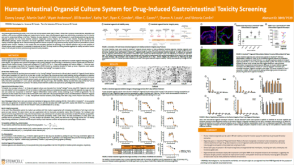 科学海报Human Intestinal Organoid Culture System for Drug-Induced Gastrointestinal Toxicity Screening
 沪公网安备31010102008431号