Fourier transform infrared microspectroscopy reveals that tissue culture conditions affect the macromolecular phenotype of human embryonic stem cells
We employed Fourier transform infrared (FTIR) microspectroscopy to investigate the effects of different tissue culture environments on the FTIR spectra of undifferentiated human embryonic stem cells (hESCs) and their differentiated progeny. First we tested whether there were any possible spectral artifacts resulting from the use of transflectance measurements by comparing them with transmission measurements and found no evidence of these concluding that the lack of any differences resulted from the homogeneity of the dried cytospun cellular monolayers. We found that hESCs that were enzymatically passaged onto mouse embryonic fibroblasts (MEFs) in KOSR based hESC medium,hESCs enzymatically passaged onto Matrigel in mTESR medium and hESCs mechanically passaged onto MEFs in KOSR-based hESC medium,possessed unique FTIR spectroscopic signatures that reflect differences in their macromolecular chemistry. Further,these spectroscopic differences persisted even upon differentiation towards mesendodermal lineages. Our results suggest that FTIR microspectroscopy is a powerful,objective,measurement modality that complements existing methods for studying the phenotype of hESCs and their progeny,particularly changes induced by the cellular environment.
View Publication
C. Alcaino et al. (JUL 2018)
Proceedings of the National Academy of Sciences of the United States of America
A population of gut epithelial enterochromaffin cells is mechanosensitive and requires Piezo2 to convert force into serotonin release.
Enterochromaffin (EC) cells constitute the largest population of intestinal epithelial enteroendocrine (EE) cells. EC cells are proposed to be specialized mechanosensory cells that release serotonin in response to epithelial forces,and thereby regulate intestinal fluid secretion. However,it is unknown whether EE and EC cells are directly mechanosensitive,and if so,what the molecular mechanism of their mechanosensitivity is. Consequently,the role of EE and EC cells in gastrointestinal mechanobiology is unclear. Piezo2 mechanosensitive ion channels are important for some specialized epithelial mechanosensors,and they are expressed in mouse and human EC cells. Here,we use EC and EE cell lineage tracing in multiple mouse models to show that Piezo2 is expressed in a subset of murine EE and EC cells,and it is distributed near serotonin vesicles by superresolution microscopy. Mechanical stimulation of a subset of isolated EE cells leads to a rapid inward ionic current,which is diminished by Piezo2 knockdown and channel inhibitors. In these mechanosensitive EE cells force leads to Piezo2-dependent intracellular Ca2+ increase in isolated cells as well as in EE cells within intestinal organoids,and Piezo2-dependent mechanosensitive serotonin release in EC cells. Conditional knockout of intestinal epithelial Piezo2 results in a significant decrease in mechanically stimulated epithelial secretion. This study shows that a subset of primary EE and EC cells is mechanosensitive,uncovers Piezo2 as their primary mechanotransducer,defines the molecular mechanism of their mechanotransduction and mechanosensitive serotonin release,and establishes the role of epithelial Piezo2 mechanosensitive ion channels in regulation of intestinal physiology.
View Publication
产品类型:
产品号#:
06005
产品名:
IntestiCult™ 类器官生长培养基 (小鼠)
文献
Wang D et al. (OCT 2013)
Transfusion 53 10 2134--40
Antibody-mediated glycophorin C coligation on K562 cells induces phosphatidylserine exposure and cell death in an atypical apoptotic process.
BACKGROUND Glycophorin C (GPC) is necessary in the maintenance of red blood cell structure. Severe autoimmune hemolytic anemia and hemolytic disease of the fetus and newborn (HDFN) have been associated with Gerbich (Ge) blood group system antigens expressed on GPC. Previous in vitro studies with cord blood progenitor cells have shown that anti-Ge suppresses erythropoiesis. STUDY DESIGN AND METHODS Here,we evaluated the K562 erythroleukemic cell line to study the cellular effects of a murine anti-GPC. Cell proliferation was evaluated after treatment with anti-GPC. Flow cytometry was used to evaluate exofacial phosphatidylserine (PS) expression and cell viability (propidium iodide binding). Cell morphology was evaluated under light microscopy with cytospin preparations stained with May-Grünwald Giemsa. RESULTS Anti-GPC dramatically inhibited K562 proliferation and increased PS expression,consistent with cytoplasmic blebbing,suggesting evidence of apoptosis. Z-VAD-FMK,an inhibitor of classical apoptosis,was unable to reverse the suppressive effect of anti-GPC. However,hemin was able to attenuate growth suppression. CONCLUSION Together,the data suggest that anti-GPC suppresses erythroid proliferation through the induction of nonclassical apoptosis.
View Publication
产品类型:
产品号#:
70008
70008.1
70008.2
70008.3
70008.4
70008.5
产品名:
冻存的人脐带血CD34+细胞
冻存的人脐带血CD34+细胞
冻存的人脐带血CD34+细胞
冻存的人脐带血CD34+细胞
冻存的人脐带血CD34+细胞
冻存的人脐带血CD34+细胞
文献
S. Mikawa et al. (sep 2019)
FASEB journal : official publication of the Federation of American Societies for Experimental Biology 33 2 fj201701200RR
Serotonin 3 receptor signaling regulates 5-fluorouracil-mediated apoptosis indirectly via TNF-alpha production by enhancing serotonin release from enterochromaffin cells.
Antagonists of the 5-hydroxytryptamine (serotonin) 3 receptor (5-HT3R) have anti-inflammatory and anti-apoptotic activities,but the detailed,underlying mechanisms are not well understood. We focused on anti-apoptotic activities via 5-HT3R signaling to clarify the underlying mechanisms. Mice were administered 5-fluorouracil (5-FU),which induced apoptosis in intestinal epithelial cells. Coadministration with 5-HT3R antagonists or agonists tended to decrease or increase the number of apoptotic cells,respectively. In serotonin 3A receptor (5-HT3AR) null (HTR3A-/-) mice,the number of apoptotic cells induced by 5-FU was decreased compared with that in wild-type (WT) mice. Bone marrow (BM) transplantation was performed to determine if BM-derived immune cells regulated 5-FU-induced apoptosis,but they were found to be unrelated to this process. Data from 5-HT3AR/enhanced green fluorescent protein reporter mice revealed that 50{\%} of enterochromaffin (EC) cells expressed 5-HT3AR,but the number of apoptotic cells induced by 5-FU in the intestinal crypt organoids of HTR3A-/- mice was not altered compared with WT mice. In contrast,plasma 5-HT concentrations in WT mice but not in HTR3A-/- mice administered 5-FU were increased significantly. In conclusion,5-HT3R signaling may enhance 5-HT release,possibly from EC cells intravascularly,or paracrine,resulting in increases in plasma 5-HT concentration,which in turn,enhances apoptotic activities induced by 5-FU.-Mikawa,S.,Kondo,M.,Kaji,N.,Mihara,T.,Yoshitake,R.,Nakagawa,T.,Takamoto,M.,Nishimura,R.,Shimada,S.,Ozaki,H.,Hori,M. Serotonin 3 receptor signaling regulates 5-fluorouracil-mediated apoptosis indirectly via TNF-alpha production by enhancing serotonin release from enterochromaffin cells.
View Publication
产品类型:
产品号#:
06005
产品名:
IntestiCult™ 类器官生长培养基 (小鼠)
文献
R. A. Gardner et al. ( 2017)
Blood 129 25 3322--3331
Intent-to-treat leukemia remission by CD19 CAR T cells of defined formulation and dose in children and young adults.
Transitioning CD19-directed chimeric antigen receptor (CAR) T cells from early-phase trials in relapsed patients to a viable therapeutic approach with predictable efficacy and low toxicity for broad application among patients with high unmet need is currently complicated by product heterogeneity resulting from transduction of undefined T-cell mixtures,variability of transgene expression,and terminal differentiation of cells at the end of culture. A phase 1 trial of 45 children and young adults with relapsed or refractory B-lineage acute lymphoblastic leukemia was conducted using a CD19 CAR product of defined CD4/CD8 composition,uniform CAR expression,and limited effector differentiation. Products meeting all defined specifications occurred in 93{\%} of enrolled patients. The maximum tolerated dose was 106 CAR T cells per kg,and there were no deaths or instances of cerebral edema attributable to product toxicity. The overall intent-to-treat minimal residual disease-negative (MRD-) remission rate for this phase 1 study was 89{\%}. The MRD- remission rate was 93{\%} in patients who received a CAR T-cell product and 100{\%} in the subset of patients who received fludarabine and cyclophosphamide lymphodepletion. Twenty-three percent of patients developed reversible severe cytokine release syndrome and/or reversible severe neurotoxicity. These data demonstrate that manufacturing a defined-composition CD19 CAR T cell identifies an optimal cell dose with highly potent antitumor activity and a tolerable adverse effect profile in a cohort of patients with an otherwise poor prognosis. This trial was registered at www.clinicaltrials.gov as {\#}NCT02028455.
View Publication
Yu C-HC-C et al. (JUN 2013)
Cancer research 73 11 3425--3440
miR145 targets the SOX9/ADAM17 axis to inhibit tumor-initiating cells and IL-6-mediated paracrine effects in head and neck cancer.
ALDH1(+)CD44(+) cells are putative tumor-initiating cells (TIC) in head and neck squamous cell carcinomas (HNC). miR-145 regulates tumorigenicity in various cancers but the breadth of its mechanistic contributions and potential therapeutic applications are not completely known. Here,we report that ALDH1(+)CD44(+)-HNC cells express reduced levels of miR145. SPONGE-mediated inhibition of miR-145 (Spg-miR145) was sufficient to drive tumor-initiating characteristics in non-TICs/ALDH1(-)CD44-negative HNC cells. Mechanistic analyses identified SOX9 and ADAM17 as two novel miR145 targets relevant to this process. miR-145 expression repressed TICs in HNC in a manner associated with SOX9 interaction with the ADAM17 promoter,thereby activating ADAM17 expression. Notably,the SOX9/ADAM17 axis dominated the TIC-inducing activity of miR-145. Either miR-145 suppression or ADAM17 overexpression in non-TICs/ALDH1(-)CD44(-)-HNC cells increased expression and secretion of interleukin (IL)-6 and soluble-IL-6 receptor (sIL-6R). Conversely,conditioned medium from Spg-miR145-transfected non-TICs/ALDH1(-)CD44(-)-HNC cells was sufficient to confer tumor-initiating properties in non-TICs/ALDH1(-)CD44(-)-HNC and this effect could be abrogated by an IL-6-neutralizing antibody. We found that curcumin administration increased miR-145 promoter activity,thereby decreasing SOX9/ADAM17 expression and eliminating TICs in HNC cell populations. Delivery of lentivral-miR145 or orally administered curcumin blocked tumor progression in HNC-TICs in murine xenotransplant assays. Finally,immunohistochemical analyses of patient specimens confirmed that an miR-145(low)/SOX9(high)/ADAM17(high) phenotype correlated with poor survival. Collectively,our results show how miR-145 targets the SOX9/ADAM17 axis to regulate TIC properties in HNC,and how altering this pathway may partly explain the anticancer effects of curcumin. By inhibiting IL-6 and sIL-6R as downstream effector cytokines in this pathway,miR-145 seems to suppress a paracrine signaling pathway in the tumor microenvironment that is vital to maintain TICs in HNC.
View Publication
产品类型:
产品号#:
01700
01705
产品名:
ALDEFLUOR™工具
ALDEFLUOR™DEAB试剂
文献
Lagarkova MA et al. (NOV 2008)
Cell Cycle 7 22 3610--3612
CD 30 is a marker of undifferentiated human embryonic stem cells rather than a biomarker of transformed hESCs
Recently it has been demonstrated that CD30 expression was rather specific for transformed than for normal human ES cells and therefore CD30 maybe suggested as a potential marker for human ES cells bearing chromosomal abnormalities. Using immunohistochemistry and RT-PCR analysis we examined �?¡D30 expression in 10 hESCs lines with normal and abberant karyotypes. All hESC lines expressed CD30 antigen and RNA in undifferentiated state whether cell line beared chromosomal abnormalities or not. In contrast to previous notions our data demonstrate that CD30 could be considered as marker of undifferentiated hESCs without respect to karyotype changes.
View Publication
产品类型:
产品号#:
85850
85857
产品名:
mTeSR™1
mTeSR™1
文献
Doehle BP et al. (OCT 2009)
Journal of virology 83 20 10395--405
Human immunodeficiency virus type 1 mediates global disruption of innate antiviral signaling and immune defenses within infected cells.
Interferon regulatory factor 3 (IRF-3) is essential for innate intracellular immune defenses that limit virus replication,but these defenses fail to suppress human immunodeficiency virus (HIV) infection,which can ultimately associate with opportunistic coinfections and the progression to AIDS. Here,we examined antiviral defenses in CD4+ cells during virus infection and coinfection,revealing that HIV type 1 (HIV-1) directs a global disruption of innate immune signaling and supports a coinfection model through suppression of IRF-3. T cells responded to paramyxovirus infection to activate IRF-3 and interferon-stimulated gene expression,but they failed to mount a response against HIV-1. The lack of response associated with a marked depletion of IRF-3 but not IRF-7 in HIV-1-infected cells,which supported robust viral replication,whereas ectopic expression of active IRF-3 suppressed HIV-1 infection. IRF-3 depletion was dependent on a productive HIV-1 replication cycle and caused the specific disruption of Toll-like receptor and RIG-I-like receptor innate immune signaling that rendered cells permissive to secondary virus infection. IRF-3 levels were reduced in vivo within CD4+ T cells from patients with acute HIV-1 infection but not from long-term nonprogressors. Our results indicate that viral suppression of IRF-3 promotes HIV-1 infection by disrupting IRF-3-dependent signaling pathways and innate antiviral defenses of the host cell. IRF-3 may direct an innate antiviral response that regulates HIV-1 replication and viral set point while governing susceptibility to opportunistic virus coinfections.
View Publication
产品类型:
产品号#:
19052
19052RF
21000
20119
20155
产品名:
EasySep™人CD4+ T细胞富集试剂盒
RoboSep™ 人CD4+ T细胞富集试剂盒含滤芯吸头
RoboSep™- S
RoboSep™ 吸头组件抛光剂
RoboSep™分选试管套装(9个塑料管+吸头保护器)
文献
A. Srinivasan et al. (JUN 2018)
Biomaterials 167 153--167
Substrate stiffness modulates the multipotency of human neural crest derived ectomesenchymal stem cells via CD44 mediated PDGFR signaling.
Mesenchymal stem cells (MSCs) have been isolated from various mesodermal and ectodermal tissues. While the phenotypic and functional heterogeneity of MSCs stemming from their developmental origins has been acknowledged,the genetic and environmental factors underpinning these differences are not well-understood. Here,we investigated whether substrate stiffness mediated mechanical cues can directly modulate the development of ectodermal MSCs (eMSCs) from a precursor human neural crest stem cell (NCSC) population. We showed that NCSC-derived eMSCs were transcriptionally and functionally distinct from mesodermal bone marrow MSCs. eMSCs derived on lower substrate stiffness specifically increased their expression of the MSC marker,CD44 in a Rho-ROCK signaling dependent manner,which resulted in a concomitant increase in the eMSCs' adipogenic and chondrogenic differentiation potential. This mechanically-induced effect can only be maintained for short-term upon switching back to a stiff substrate but can be sustained for longer-term when the eMSCs were exclusively maintained on soft substrates. We also discovered that CD44 expression modulated eMSC self-renewal and multipotency via the downregulation of downstream platelet-derived growth factor receptor beta (PDGFRbeta$) signaling. This is the first instance demonstrating that substrate stiffness not only influences the differentiation trajectories of MSCs but also their derivation from upstream progenitors,such as NCSCs.
View Publication

 EasySep™小鼠TIL(CD45)正选试剂盒


 文献
文献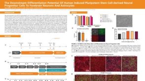 科学海报The Downstream Differentiation Potential of Human Induced Pluripotent Stem Cell-Derived Neural Progenitor Cells to Forebrain Neurons and Astrocytes