Singbrant S et al. (JUN 2010)
Blood 115 23 4689--98
Canonical BMP signaling is dispensable for hematopoietic stem cell function in both adult and fetal liver hematopoiesis, but essential to preserve colon architecture.
Numerous publications have described the importance of bone morphogenetic protein (BMP) signaling in the specification of hematopoietic tissue in developing embryos. Here we investigate the full role of canonical BMP signaling in both adult and fetal liver hematopoiesis using conditional knockout strategies because conventional disruption of components of the BMP signaling pathway result in early death of the embryo. By targeting both Smad1 and Smad5,we have generated a double-knockout mouse with complete disruption of canonical BMP signaling. Interestingly,concurrent deletion of Smad1 and Smad5 results in death because of extrahematopoietic pathologic changes in the colon. However,Smad1/Smad5-deficient bone marrow cells can compete normally with wild-type cells and display unaffected self-renewal and differentiation capacity when transplanted into lethally irradiated recipients. Moreover,although BMP receptor expression is increased in fetal liver,fetal liver cells deficient in both Smad1 and Smad5 remain competent to long-term reconstitute lethally irradiated recipients in a multilineage manner. In conclusion,canonical BMP signaling is not required to maintain either adult or fetal liver hematopoiesis,despite its crucial role in the initial patterning of hematopoiesis in early embryonic development.
View Publication
Nicolini FE et al. (AUG 2002)
Blood 100 4 1257--64
Expression of a human beta-globin transgene in erythroid cells derived from retrovirally transduced transplantable human fetal liver and cord blood cells.
Transfer of therapeutic genes to human hematopoietic stem cells (HSCs) using complex vectors at clinically relevant efficiencies remains a major challenge. Recently we described a stable retroviral vector that sustains long-term expression of green fluorescent protein (GFP) and a human beta-globin gene in the erythroid progeny of transduced murine HSCs. We now report the efficient transduction of primitive human CD34(+) fetal liver or cord blood cells with this vector and expression of the beta-globin transgene in the erythroid progeny of these human cells for at least 2 months. After growth factor prestimulation and then a 2- to 3-day exposure to the virus,35% to 55% GFP(+) progeny were seen in assays of transduced colony-forming cells,primitive erythroid precursors that generate large numbers of glycophorin A(+) cells in 3-week suspension cultures,and 6-week long-term culture-initiating cells. In immunodeficient mice injected with unselected infected cells,5% to 15% of the human cells regenerated in the marrow (including the erythroid cells) were GFP(+) 3 and 6 weeks after transplantation. Importantly,the numbers of GFP(+) human lymphoid and either granulopoietic or erythroid cells in individual mice 6 weeks after transplantation were significantly correlated,indicative of the initial transduction of human multipotent cells with in vivo repopulating activity. Expression of the transduced beta-globin gene in human cells obtained directly from the mice or after their differentiation into erythroid cells in vitro was demonstrated by reverse transcriptase-polymerase chain reaction using specific primers. These experiments represent a significant step toward the realization of a gene therapy approach for human beta-globin gene disorders.
View Publication
产品类型:
产品号#:
04330
产品名:
MethoCult™H4330
文献
Santoni de Sio FR et al. (JUN 2006)
Blood 107 11 4257--65
Proteasome activity restricts lentiviral gene transfer into hematopoietic stem cells and is down-regulated by cytokines that enhance transduction.
The therapeutic potential of hematopoietic stem cell (HSC) gene therapy can be fully exploited only by reaching efficient gene transfer into HSCs without compromising their biologic properties. Although HSCs can be transduced by HIV-derived lentiviral vectors (LVs) in short ex vivo culture,they display low permissivity to the vector,requiring cytokine stimulation to reach high-frequency transduction. Using stringent assays of competitive xenograft repopulation,we show that early-acting cytokines synergistically enhanced human HSC gene transfer by LVs without impairing engraftment and repopulation capacity. Using S-phase suicide assays,we show that transduction enhancement by cytokines was not dependent on cell cycle progression and that LVs can transduce quiescent HSCs. Pharmacologic inhibition of the proteasome during transduction dramatically enhanced HSC gene transfer,allowing the reach of very high levels of vector integration in their progeny in vivo. Thus,LVs are effectively restricted at a postentry step by the activity of this proteolytic complex. Unexpectedly,cytokine stimulation rapidly and substantially down-regulated proteasome activity in hematopoietic progenitors,highlighting one mechanism by which cytokines may enhance permissiveness to LV gene transfer. These findings demonstrate that antiviral responses ultimately mediated by proteasomes strongly limit the efficiency of HSC transduction by LVs and establish improved conditions for HSC-based gene therapy.
View Publication
产品类型:
产品号#:
09600
09650
产品名:
StemSpan™ SFEM
StemSpan™ SFEM
文献
Zhang J et al. (NOV 2011)
Stem Cell Reviews and Reports 7 4 987--996
Electrically Guiding Migration of Human Induced Pluripotent Stem Cells
A major road-block in stem cell therapy is the poor homing and integration of transplanted stem cells with the targeted host tissue. Human induced pluripotent stem (hiPS) cells are considered an excellent alternative to embryonic stem (ES) cells and we tested the feasibility of using small,physiological electric fields (EFs) to guide hiPS cells to their target. Applied EFs stimulated and guided migration of cultured hiPS cells toward the anode,with a stimulation threshold of textless30 mV/mm; in three-dimensional (3D) culture hiPS cells remained stationary,whereas in an applied EF they migrated directionally. This is of significance as the therapeutic use of hiPS cells occurs in a 3D environment. EF exposure did not alter expression of the pluripotency markers SSEA-4 and Oct-4 in hiPS cells. We compared EF-directed migration (galvanotaxis) of hiPS cells and hES cells and found that hiPS cells showed greater sensitivity and directedness than those of hES cells in an EF,while hES cells migrated toward cathode. Rho-kinase (ROCK) inhibition,a method to aid expansion and survival of stem cells,significantly increased the motility,but reduced directionality of iPS cells in an EF by 70-80%. Thus,our study has revealed that physiological EF is an effective guidance cue for the migration of hiPS cells in either 2D or 3D environments and that will occur in a ROCK-dependent manner. Our current finding may lead to techniques for applying EFs in vivo to guide migration of transplanted stem cells.
View Publication
产品类型:
产品号#:
85850
85857
产品名:
mTeSR™1
mTeSR™1
文献
Lu Y et al. (FEB 2012)
Stem cells and development 21 3 394--403
Avian-Induced Pluripotent Stem Cells Derived Using Human Reprogramming Factors
Avian species are important model animals for developmental biology and disease research. However,unlike in mice,where clonal lines of pluripotent stem cells have enabled researchers to study mammalian gene function,clonal and highly proliferative pluripotent avian cell lines have been an elusive goal. Here we demonstrate the generation of avian induced pluripotent stem cells (iPSCs),the first nonmammalian iPSCs,which were clonally isolated and propagated,important attributes not attained in embryo-sourced avian cells. This was accomplished using human pluripotency genes rather than avian genes,indicating that the process in which mammalian and nonmammalian cells are reprogrammed is a conserved process. Quail iPSCs (qiPSCs) were capable of forming all 3 germ layers in vitro and were directly differentiated in culture into astrocytes,oligodendrocytes,and neurons. Ultimately,qiPSCs were capable of generating live chimeric birds and incorporated into tissues from all 3 germ layers,extraembryonic tissues,and potentially the germline. These chimera competent qiPSCs and in vitro differentiated cells offer insight into the conserved nature of reprogramming and genetic tools that were only previously available in mammals.
View Publication
产品类型:
产品号#:
85850
85857
产品名:
mTeSR™1
mTeSR™1
文献
Krawetz R and Rancourt DE (JAN 2012)
Methods in molecular biology (Clifton,N.J.) 873 227--235
Suspension bioreactor expansion of undifferentiated human embryonic stem cells
Embryonic stem cells (ESCs) are unique cells,which have the ability to differentiate into all cell types that comprise the adult organism. Furthermore,ESCs can infinitely self-renew under optimized conditions. These features place human ESCs (hESCs) in a position where these cells can be exploited for tissue engineering and regenerative medicine approaches in treating human degenerative disorders. However,cell therapy approaches will require large amounts of clinically useable cells,not typically achievable using standard static cell culture methods. Here,we describe a method wherein clinically relevant numbers of hESCs can be generated in a cost and time effective manner.
View Publication

 EasySep™小鼠TIL(CD45)正选试剂盒


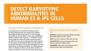 产品手册Detect Karyotypic Abnormalities in Human ES & iPS Cells
 技术手册Maintenance of Human Pluripotent Stem Cells in mTeSR™ Plus
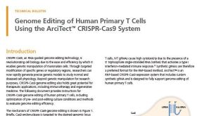 技术公告Genome Editing of Human Primary T Cells Using CRISPR-Cas9
 产品手册Derive, Expand, and Differentiate Human Skeletal Muscle Progenitor Cells