Stumpf M et al. (DEC 2010)
Proceedings of the National Academy of Sciences of the United States of America 107 50 21541--6
Specific erythroid-lineage defect in mice conditionally deficient for Mediator subunit Med1.
The Mediator complex forms the bridge between transcriptional activators and the RNA polymerase II. Med1 (also known as PBP or TRAP220) is a key component of Mediator that interacts with nuclear hormone receptors and GATA transcription factors. Here,we show dynamic recruitment of GATA-1,TFIIB,Mediator,and RNA polymerase II to the β-globin locus in induced mouse erythroid leukemia cells and in an erythropoietin-inducible hematopoietic progenitor cell line. Using Med1 conditional knockout mice,we demonstrate a specific block in erythroid development but not in myeloid or lymphoid development,highlighted by the complete absence of β-globin gene expression. Thus,Mediator subunit Med1 plays a pivotal role in erythroid development and in β-globin gene activation.
View Publication
产品类型:
产品号#:
03334
产品名:
MethoCult™M3334
文献
Wood ER et al. ( 2004)
Cancer research 64 18 6652--6659
A unique structure for epidermal growth factor receptor bound to GW572016 (Lapatinib): relationships among protein conformation, inhibitor off-rate, and receptor activity in tumor cells.
GW572016 (Lapatinib) is a tyrosine kinase inhibitor in clinical development for cancer that is a potent dual inhibitor of epidermal growth factor receptor (EGFR,ErbB-1) and ErbB-2. We determined the crystal structure of EGFR bound to GW572016. The compound is bound to an inactive-like conformation of EGFR that is very different from the active-like structure bound by the selective EGFR inhibitor OSI-774 (Tarceva) described previously. Surprisingly,we found that GW572016 has a very slow off-rate from the purified intracellular domains of EGFR and ErbB-2 compared with OSI-774 and another EGFR selective inhibitor,ZD-1839 (Iressa). Treatment of tumor cells with these inhibitors results in down-regulation of receptor tyrosine phosphorylation. We evaluated the duration of the drug effect after washing away free compound and found that the rate of recovery of receptor phosphorylation in the tumor cells reflected the inhibitor off-rate from the purified intracellular domain. The slow off-rate of GW572016 correlates with a prolonged down-regulation of receptor tyrosine phosphorylation in tumor cells. The differences in the off-rates of these drugs and the ability of GW572016 to inhibit ErbB-2 can be explained by the enzyme-inhibitor structures.
View Publication
产品类型:
产品号#:
73242
产品名:
拉帕替尼
文献
Lambert AA et al. (AUG 2008)
Blood 112 4 1299--307
The C-type lectin surface receptor DCIR acts as a new attachment factor for HIV-1 in dendritic cells and contributes to trans- and cis-infection pathways.
The dynamic interplay between dendritic cells (DCs) and human immunodeficiency virus type-1 (HIV-1) is thought to result in viral dissemination and evasion of antiviral immunity. Although initial observations suggested that the C-type lectin receptor (CLR) DC-SIGN was responsible for the trans-infection function of the virus,subsequent studies demonstrated that trans-infection of CD4(+) T cells with HIV-1 can also occur through DC-SIGN-independent mechanisms. We demonstrate that a cell surface molecule designated DCIR (for DC immunoreceptor),a member of a recently described family of DC-expressing CLRs,can participate in the capture of HIV-1 and promote infection in trans and in cis of autologous CD4(+) T cells from human immature monocyte-derived DCs. The contribution of DCIR to these processes was revealed using DCIR-specific siRNAs and a polyclonal antibody specific for the carbohydrate recognition domain of DCIR. Data from transfection experiments indicated that DCIR acts as a ligand for HIV-1 and is involved in events leading to productive virus infection. Finally,we show that the neck domain of DCIR is important for the DCIR-mediated effect on virus binding and infection. These results point to a possible role for DCIR in HIV-1 pathogenesis by supporting the productive infection of DCs and promoting virus propagation.
View Publication
Kwok CTD et al. (MAR 2016)
Stem Cell Research 16 3 651--661
The Forkhead box transcription factor FOXM1 is required for the maintenance of cell proliferation and protection against oxidative stress in human embryonic stem cells
Human embryonic stem cells (hESCs) exhibit unique cell cycle structure,self-renewal and pluripotency. The Forkhead box transcription factor M1 (FOXM1) is critically required for the maintenance of pluripotency in mouse embryonic stem cells and mouse embryonal carcinoma cells,but its role in hESCs remains unclear. Here,we show that FOXM1 expression was enriched in undifferentiated hESCs and was regulated in a cell cycle-dependent manner with peak levels detected at the G2/M phase. Expression of FOXM1 did not correlate with OCT4 and NANOG during in vitro differentiation of hESCs. Importantly,knockdown of FOXM1 expression led to aberrant cell cycle distribution with impairment in mitotic progression but showed no profound effect on the undifferentiated state. Interestingly,FOXM1 depletion sensitized hESCs to oxidative stress. Moreover,genome-wide analysis of FOXM1 targets by ChIP-seq identified genes important for M phase including CCNB1 and CDK1,which were subsequently confirmed by ChIP and RNA interference analyses. Further peak set comparison against a differentiating hESC line and a cancer cell line revealed a substantial difference in the genomic binding profile of FOXM1 in hESCs. Taken together,our findings provide the first evidence to support FOXM1 as an important regulator of cell cycle progression and defense against oxidative stress in hESCs.
View Publication
产品类型:
产品号#:
05110
85850
85857
产品名:
STEMdiff™权威内胚层检测试剂盒
mTeSR™1
mTeSR™1
文献
Beeton C et al. (NOV 2006)
Proceedings of the National Academy of Sciences of the United States of America 103 46 17414--9
Kv1.3 channels are a therapeutic target for T cell-mediated autoimmune diseases.
Autoreactive memory T lymphocytes are implicated in the pathogenesis of autoimmune diseases. Here we demonstrate that disease-associated autoreactive T cells from patients with type-1 diabetes mellitus or rheumatoid arthritis (RA) are mainly CD4+ CCR7- CD45RA- effector memory T cells (T(EM) cells) with elevated Kv1.3 potassium channel expression. In contrast,T cells with other antigen specificities from these patients,or autoreactive T cells from healthy individuals and disease controls,express low levels of Kv1.3 and are predominantly naïve or central-memory (T(CM)) cells. In T(EM) cells,Kv1.3 traffics to the immunological synapse during antigen presentation where it colocalizes with Kvbeta2,SAP97,ZIP,p56(lck),and CD4. Although Kv1.3 inhibitors [ShK(L5)-amide (SL5) and PAP1] do not prevent immunological synapse formation,they suppress Ca2+-signaling,cytokine production,and proliferation of autoantigen-specific T(EM) cells at pharmacologically relevant concentrations while sparing other classes of T cells. Kv1.3 inhibitors ameliorate pristane-induced arthritis in rats and reduce the incidence of experimental autoimmune diabetes in diabetes-prone (DP-BB/W) rats. Repeated dosing with Kv1.3 inhibitors in rats has not revealed systemic toxicity. Further development of Kv1.3 blockers for autoimmune disease therapy is warranted.
View Publication
产品类型:
产品号#:
19051
19051RF
产品名:
EasySep™人T细胞富集试剂盒
RoboSep™ 人T细胞富集试剂盒含滤芯吸头
文献
Liu C et al. (SEP 2013)
Biochemical and Biophysical Research Communications 439 1 154--159
Neural differentiation of human embryonic stem cells as an in vitro tool for the study of the expression patterns of the neuronal cytoskeleton during neurogenesis
The neural differentiation of human embryonic stem cells (ESCs) is a potential tool for elucidating the key mechanisms involved in human neurogenesis. Nestin and ??-III-tubulin,which are cytoskeleton proteins,are marker proteins of neural stem cells (NSCs) and neurons,respectively. However,the expression patterns of nestin and ??-III-tubulin in neural derivatives from human ESCs remain unclear. In this study,we found that neural progenitor cells (NPCs) derived from H9 cells express high levels of nestin and musashi-1. In contrast,??-III-tubulin was weakly expressed in a few NPCs. Moreover,in these cells,nestin formed filament networks,whereas ??-III-tubulin was distributed randomly as small particles. As the differentiation proceeded,the nestin filament networks and the ??-III-tubulin particles were found in both the cell soma and the cellular processes. Moreover,the colocalization of nestin and ??-III-tubulin was found mainly in the cell processes and neurite-like structures and not in the cell soma. These results may aid our understanding of the expression patterns of nestin and ??-III-tubulin during the neural differentiation of H9 cells. ?? 2013 Elsevier Inc.
View Publication
产品类型:
产品号#:
85850
85857
产品名:
mTeSR™1
mTeSR™1
文献
Cashman JD et al. (JAN 1990)
Blood 75 1 96--101
Mechanisms that regulate the cell cycle status of very primitive hematopoietic cells in long-term human marrow cultures. I. Stimulatory role of a variety of mesenchymal cell activators and inhibitory role of TGF-beta.
Long-term marrow cultures (LTMC) allow the proliferation and differentiation of primitive human hematopoietic progenitor cells to be maintained for many weeks in the absence of exogenously provided hematopoietic growth factors. Previous investigations focused on defining various types of cells that are present in this culture system and on measuring the cycling behavior of the different subpopulations of colony-forming cells maintained within it. These studies suggested that mesenchymal stromal elements derived from the input marrow play a key role in regulating the turnover of the most primitive,high-proliferative potential erythroid and granulopoietic colony-forming cells that are found almost exclusively in the adherent layer of LTMC. In this study we show that the re-entry into S-phase of these primitive hematopoietic progenitors that occurs after each weekly medium change is due to an as yet undefined constituent of horse serum,which is absent from fetal calf serum. However,this effect is not unique to the factor present in horse serum. It is also elicited by the addition to LTMC of several well-defined growth regulatory molecules,ie,platelet-derived growth factor (PDGF),interleukin-1 (IL-1),transforming growth factor alpha (TGF-alpha),and IL-2. None of these was able to stimulate hematopoietic colony-forming cells in methylcellulose assays,although all have known actions on mesenchymal cells including,in some cases,the ability to increase production of growth factors that can stimulate primitive high-proliferative potential hematopoietic progenitors in clonogenic assays. Interestingly,a stimulating effect was not obtained after addition of endotoxin to LTMC. TGF-beta,a direct-acting negative regulator that acts selectively on primitive hematopoietic progenitor cells if added to LTMC simultaneously with new medium or IL-1,blocked their stimulating activity. These results suggest a model in which indirect,local modulation of both positive and negative regulatory factors via effects on mesenchymal elements determines the rate of turnover of adjacent populations of very primitive hematopoietic cells that are normally maintained in a quiescent state in vivo.
View Publication

 EasySep™小鼠TIL(CD45)正选试剂盒


 文献
文献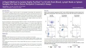 科学海报A Rapid Method to Isolate Highly Purified T or B Cells from Blood, Lymph Node or Spleen Samples For Use in Donor-Recipient Crossmatch Assays
 科学海报Dendritic Cell Isolation from Mouse Spleen Made Simple
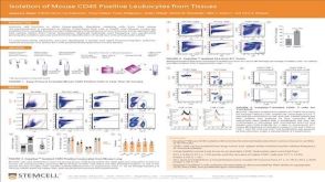 科学海报Isolation of Mouse CD45 Positive Leukocytes from Tissues