Grievink HW et al. (OCT 2016)
Biopreservation and biobanking 14 5 410--415
Comparison of Three Isolation Techniques for Human Peripheral Blood Mononuclear Cells: Cell Recovery and Viability, Population Composition, and Cell Functionality.
Routine techniques for the isolation of human peripheral blood mononuclear cells (PBMCs) include density centrifugation with Ficoll-Paque and isolation by cell preparation tubes (CPTs) and SepMate tubes with Lymphoprep. In a series of experiments,these three PBMC isolation techniques were compared for cell recovery and viability,PBMC population composition,and cell functionality,aiming to provide a starting basis for the selection of the most appropriate method of PBMC isolation for a specific downstream application. PBMCs were freshly isolated from venous blood of healthy male donors,applying the different techniques in parallel. Cell recovery and viability were assessed using a hemacytometer and trypan blue. Immunophenotyping was performed by flow cytometry. Cell functionality was assessed in stimulated (100 ng/mL staphylococcal enterotoxin B [SEB]) and unstimulated 24 hours PBMC cultures,with cytokine production and lactate dehydrogenase (LDH) release as readout measures. PBMC isolation by SepMate and CPT resulted in a 70% higher recovery than Ficoll isolation. CPT-isolated populations contained more erythrocyte contamination. Cell viability,assessed by trypan blue exclusion,was 100% for all three isolation techniques. SepMate and CPT isolation gave higher SEB-induced cytokine responses in cell cultures,for IFNγ and for secondary cytokines. IL-6 and IL-8 release in unstimulated cultures was higher for CPT-isolated PBMCs compared to Ficoll- and SepMate-isolated PBMCs. LDH release did not differ between cell isolation techniques. In addition to criteria such as cost and application practicalities,these data may support selection of a specific PBMC isolation technique for downstream analysis.
View Publication
产品类型:
产品号#:
07811
07861
85450
85460
86450
86460
产品名:
Lymphoprep™
Lymphoprep™
SepMate™-50 (IVD)
SepMate™-50 (IVD)
SepMate™-50 (RUO)
SepMate™-50 (RUO)
文献
Wu X et al. (DEC 2008)
Blood 112 12 4675--82
Alternative splicing regulates activation-induced cytidine deaminase (AID): implications for suppression of AID mutagenic activity in normal and malignant B cells.
The mutagenic enzyme activation-induced cytidine deaminase (AID) is required for immunoglobulin class switch recombination (CSR) and somatic hypermutation (SHM) in germinal center (GC) B cells. Deregulated expression of AID is associated with various B-cell malignancies and,currently,it remains unclear how AID activity is extinguished to avoid illegitimate mutations. AID has also been shown to be alternatively spliced in malignant B cells,and there is limited evidence that this also occurs in normal blood B cells. The functional significance of these splice variants remains unknown. Here we show that normal GC human B cells and blood memory B cells similarly express AID splice variants and show for the first time that AID splicing variants are singly expressed in individual normal B cells as well as malignant B cells from chronic lymphocytic leukemia patients. We further demonstrate that the alternative AID splice variants display different activities ranging from inactivation of CSR to inactivation or heightened SHM activity. Our data therefore suggest that CSR and SHM are differentially switched off by varying the expression of splicing products of AID at the individual cell level. Most importantly,our findings suggest a novel tumor suppression mechanism by which unnecessary AID mutagenic activities are promptly contained for GC B cells.
View Publication
R. Ravichandran et al. (sep 2022)
American journal of transplantation : official journal of the American Society of Transplantation and the American Society of Transplant Surgeons 22 9 2180--2194
Low-dose IL-2 prevents murine chronic cardiac allograft rejection: Role for IL-2-induced T regulatory cells and exosomes with PD-L1 and CD73.
To determine the effects and immunological mechanisms of low-dose interleukin-2 (IL-2) in a murine model of chronic cardiac allograft rejection (BALB/c to C57BL/6) after costimulatory blockade consisting of MR1 (250??$\mu$g/ip day 0) and CTLA4-Ig (200??$\mu$g/ip day 2),we administered low-dose IL-2 (2000??IU/day) starting on posttransplant day 14 for 3??weeks. T regulatory (Treg) cell infiltration of the grafts was determined by immunohistochemistry; circulating exosomes by western blot and aldehyde bead flow cytometry; antibodies to donor MHC by immunofluorescent staining of donor cells; and antibodies to cardiac self-antigens (myosin,vimentin) by ELISA. We demonstrated that costimulation blockade after allogeneic heart transplantation induced circulating exosomes containing cardiac self-antigens and antibodies to both donor MHC and self-antigens,leading to chronic rejection by day 45. Treatment with low-dose IL-2 prolonged allograft survival (>100??days),prevented chronic rejection,and induced splenic and graft-infiltrating CD4+ CD25+ Foxp3 Treg cells by day 45 and circulating exosomes (Foxp3+) with PD-L1 and CD73. MicroRNA 142,associated with the TGF$\beta$ pathway,was significantly downregulated in exosomes from IL-2-treated mice. In conclusion,low-dose IL-2 delays rejection in a murine model of chronic cardiac allograft rejection and also induces graft-infiltrating Tregs and circulating exosomes with immunoregulatory molecules.
View Publication
Taubert I et al. (APR 2011)
Cytotherapy 13 4 459--66
Characterization of hematopoietic stem cell subsets from patients with multiple myeloma after mobilization with plerixafor.
BACKGROUND AIMS: Previous studies have demonstrated that the combination of granulocyte-colony-stimulating factor (G-CSF) + plerixafor is more efficient in mobilizing CD34(+) hematopoietic stem cells (HSC) into the peripheral blood than G-CSF alone. In this study we analyzed the impact of adding plerixafor to G-CSF upon the mobilization of different HSC subsets. METHODS: We characterized the immunophenotype of HSC subsets isolated from the peripheral blood of eight patients with multiple myeloma (MM) before and after treatment with plerixafor. All patients were supposed to collect stem cells prior to high-dose chemotherapy and consecutive autologous stem cell transplantation,and therefore received front-line mobilization with 4 days of G-CSF followed by a single dose of plerixafor. Samples of peripheral blood were analyzed comparatively by flow cytometry directly before and 12 h after administration of plerixafor. RESULTS: The number of aldehyde dehydrogenase (ALDH)(bright) and CD34(+) cells was significantly higher after plerixafor treatment (1.2-5.0 and 1.5-6.0 times; both P textless 0.01) and an enrichment of the very primitive CD34(+) CD38(-) and ALDH(bright) CD34(+) CD38(-) HSC subsets was detectable. Additionally,two distinct ALDH(+) subsets could be clearly distinguished. The small ALDH(high) subset showed a higher number of CD34(+) CD38(-) cells in contrast to the total ALDH(bright) subpopulation and probably represented a very primitive subpopulation of HSC. CONCLUSIONS: A combined staining of ALDH,CD34 and CD38 might represent a powerful tool for the identification of a very rare and primitive hematopoietic stem cell subset. The addition of plerixafor mobilized not only more CD34(+) cells but was also able to increase the proportion of more primitive stem cell subsets.
View Publication
产品类型:
产品号#:
01700
01705
产品名:
ALDEFLUOR™工具
ALDEFLUOR™DEAB试剂
文献
Croker AK et al. (AUG 2009)
Journal of cellular and molecular medicine 13 8B 2236--52
High aldehyde dehydrogenase and expression of cancer stem cell markers selects for breast cancer cells with enhanced malignant and metastatic ability.
Cancer stem cells (CSCs) have recently been identified in leukaemia and solid tumours; however,the role of CSCs in metastasis remains poorly understood. This dearth of knowledge about CSCs and metastasis is due largely to technical challenges associated with the use of primary human cancer cells in pre-clinical models of metastasis. Therefore,the objective of this study was to develop suitable pre-clinical model systems for studying stem-like cells in breast cancer metastasis,and to test the hypothesis that stem-like cells play a key role in metastatic behaviour. We assessed four different human breast cancer cell lines (MDA-MB-435,MDA-MB-231,MDA-MB-468,MCF-7) for expression of prospective CSC markers CD44/CD24 and CD133,and for functional activity of aldehyde dehydrogenase (ALDH),an enzyme involved in stem cell self-protection. We then used fluorescence-activated cell sorting and functional assays to characterize differences in malignant/metastatic behaviour in vitro (proliferation,colony-forming ability,adhesion,migration,invasion) and in vivo (tumorigenicity and metastasis). Sub-populations of cells demonstrating stem-cell-like characteristics (high expression of CSC markers and/or high ALDH) were identified in all cell lines except MCF-7. When isolated and compared to ALDH(low)CD44(low/-) cells,ALDH(hi)CD44(+)CD24(-) (MDA-MB-231) and ALDH(hi)CD44(+)CD133(+) (MDA-MB-468) cells demonstrated increased growth (P textless 0.05),colony formation (P textless 0.05),adhesion (P textless 0.001),migration (P textless 0.001) and invasion (P textless 0.001). Furthermore,following tail vein or mammary fat pad injection of NOD/SCID/IL2gamma receptor null mice,ALDH(hi)CD44(+)CD24(-) and ALDH(hi)CD44(+)CD133(+) cells showed enhanced tumorigenicity and metastasis relative to ALDH(low)CD44(low/-) cells (P textless 0.05). These novel results suggest that stem-like ALDH(hi)CD44(+)CD24(-) and ALDH(hi)CD44(+)CD133(+) cells may be important mediators of breast cancer metastasis.
View Publication
产品类型:
产品号#:
01700
01705
产品名:
ALDEFLUOR™工具
ALDEFLUOR™DEAB试剂
文献
Carvalho JL et al. (NOV 2012)
Journal of tissue science & engineering Suppl 11 002
Characterization of Decellularized Heart Matrices as Biomaterials for Regular and Whole Organ Tissue Engineering and Initial In-vitro Recellularization with Ips Cells.
Tissue engineering strategies,based on solid/porous scaffolds,suffer from several limitations,such as ineffective vascularization,poor cell distribution and organization within scaffold,in addition to low final cell density,among others. Therefore,the search for other tissue engineering approaches constitutes an active area of investigation. Decellularized matrices (DM) present major advantages compared to solid scaffolds,such as ideal chemical composition,the preservation of vascularization structure and perfect three-dimensional structure. In the present study,we aimed to characterize and investigate murine heart decellularized matrices as biomaterials for regular and whole organ tissue engineering. Heart decellularized matrices were characterized according to: 1. DNA content,through DNA quantificationo and PCR of isolated genomic DNA; 2. Histological structure,assessed after Hematoxylin and Eosin,as well as Masson's Trichrome stainings; 3. Surface nanostructure analysis,performed,using SEM. Those essays allowed us to conclude that DM was indeed decellularized,with preserved extracellular matrix structure. Following characterization,decellularized heart slices were seeded with induced Pluripotent Stem cells (iPS). As expected,but - to the best of our knowledge - never shown before,decellularization of murine heart matrices maintained matrix biocompatibility,as iPS cells rapidly attached to the surface of the material and proliferated. Strikingly though,heart DM presented a differentiation induction effect over those cells,which lost their pluripotency markers after 7 days of culture in the DM. Such loss of differentiation markers was observed,even though bFGF containing media mTSR was used during such period. Gene expression of iPS cells cultured on DM will be further analyzed,in order to assess the effects of culturing pluripotent stem cells in decellularized heart matrices.
View Publication
产品类型:
产品号#:
85850
85857
产品名:
mTeSR™1
mTeSR™1
文献
Bone HK et al. (JUN 2011)
Journal of cell science 124 Pt 12 1992--2000
A novel chemically directed route for the generation of definitive endoderm from human embryonic stem cells based on inhibition of GSK-3.
The use of small molecules to 'chemically direct' differentiation represents a powerful approach to promote specification of embryonic stem cells (ESCs) towards particular functional cell types for use in regenerative medicine and pharmaceutical applications. Here,we demonstrate a novel route for chemically directed differentiation of human ESCs (hESCs) into definitive endoderm (DE) exploiting a selective small-molecule inhibitor of glycogen synthase kinase 3 (GSK-3). This GSK-3 inhibitor,termed 1m,when used as the only supplement to a chemically defined feeder-free culture system,effectively promoted differentiation of ESC lines towards primitive streak (PS),mesoderm and DE. This contrasts with the role of GSK-3 in murine ESCs,where GSK-3 inhibition promotes pluripotency. Interestingly,1m-mediated induction of differentiation involved transient NODAL expression and Nodal signalling. Prolonged treatment of hESCs with 1m resulted in the generation of a population of cells displaying hepatoblast characteristics,that is expressing α-fetoprotein and HNF4α. Furthermore,1m-induced DE had the capacity to mature and generate hepatocyte-like cells capable of producing albumin. These findings describe,for the first time,the utility of GSK-3 inhibition,in a chemically directed approach,to a method of DE generation that is robust,potentially scalable and applicable to different hESC lines.
View Publication
产品类型:
产品号#:
85850
85857
产品名:
mTeSR™1
mTeSR™1
文献
Baksh D et al. (NOV 2005)
Blood 106 9 3012--9
Soluble factor cross-talk between human bone marrow-derived hematopoietic and mesenchymal cells enhances in vitro CFU-F and CFU-O growth and reveals heterogeneity in the mesenchymal progenitor cell compartment.
The homeostatic adult bone marrow (BM) is a complex tissue wherein physical and biochemical interactions serve to maintain a balance between the hematopoietic and nonhematopoietic compartments. To focus on soluble factor interactions occurring between mesenchymal and hematopoietic cells,a serum-free adhesion-independent culture system was developed that allows manipulation of the growth of both mesenchymal and hematopoietic human BM-derived progenitors and the balance between these compartments. Factorial experiments demonstrated a role for stem cell factor (SCF) and interleukin 3 (IL-3) in the concomitant growth of hematopoietic (CD45+) and nonhematopoietic (CD45-) cells,as well as their derivatives. Kinetic tracking of IL-3alpha receptor (CD123) and SCF receptor (CD117) expression on a sorted CD45- cell population revealed the emergence of CD45-CD123+ cells capable of osteogenesis. Of the total fibroblast colony-forming units (CFU-Fs) and osteoblast colony-forming units (CFU-O),approximately 24% of CFU-Fs and about 22% of CFU-Os were recovered from this population. Cell-sorting experiments demonstrated that the CD45+ cell population secreted soluble factors that positively affect the survival and proliferation of CFU-Fs and CFU-Os generated from the CD45- cells. Together,our results provide insight into the intercellular cytokine network between hematopoietic and mesenchymal cells and provide a strategy to mutually culture both mesenchymal and hematopoietic cells in a defined scalable bioprocess.
View Publication
产品类型:
产品号#:
产品名:
文献
Nguyen AT et al. (JUN 2011)
Blood 117 25 6912--22
DOT1L, the H3K79 methyltransferase, is required for MLL-AF9-mediated leukemogenesis.
Chromosomal translocations of the mixed lineage leukemia (MLL) gene are a common cause of acute leukemias. The oncogenic function of MLL fusion proteins is,in part,mediated through aberrant activation of Hoxa genes and Meis1,among others. Here we demonstrate using a tamoxifen-inducible Cre-mediated loss of function mouse model that DOT1L,an H3K79 methyltransferase,is required for both initiation and maintenance of MLL-AF9-induced leukemogenesis in vitro and in vivo. Through gene expression and chromatin immunoprecipitation analysis we demonstrate that mistargeting of DOT1L,subsequent H3K79 methylation,and up-regulation of Hoxa and Meis1 genes underlie the molecular mechanism of how DOT1L contributes to MLL-AF9-mediated leukemogenesis. Our study not only provides the first in vivo evidence for the function of DOT1L in leukemia,but also reveals the molecular mechanism for DOT1L in MLL-AF9 mediated leukemia. Thus,DOT1L may serve as a potential therapeutic target for the treatment of leukemia caused by MLL translocations.
View Publication

 EasySep™小鼠TIL(CD45)正选试剂盒


 文献
文献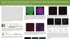 科学海报Neurofluor CDr3 a Fluorescent Probe for the Detection of Live CNS and Human Pluripotent Stem Cell Derived Neural Progenitor Cells
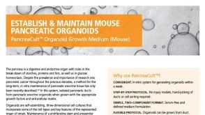 产品手册PancreaCult™ Organoid Growth Medium (Mouse)