Woods EJ et al. (OCT 2009)
Cryobiology 59 2 150--7
Optimized cryopreservation method for human dental pulp-derived stem cells and their tissues of origin for banking and clinical use.
Dental pulp is a promising source of mesenchymal stem cells with the potential for cell-mediated therapies and tissue engineering applications. We recently reported that isolation of dental pulp-derived stem cells (DPSC) is feasible for at least 120h after tooth extraction,and that cryopreservation of early passage cultured DPSC leads to high-efficiency recovery post-thaw. This study investigated additional processing and cryobiological characteristics of DPSC,ending with development of procedures for banking. First,we aimed to optimize cryopreservation of established DPSC cultures,with regards to optimizing the cryoprotective agent (CPA),the CPA concentration,the concentration of cells frozen,and storage temperatures. Secondly,we focused on determining cryopreservation characteristics of enzymatically digested tissue as a cell suspension. Lastly,we evaluated the growth,surface markers and differentiation properties of DPSC obtained from intact teeth and undigested,whole dental tissue frozen and thawed using the optimized procedures. In these experiments it was determined that Me(2)SO at a concentration between 1 and 1.5M was the ideal cryopreservative of the three studied. It was also determined that DPSC viability after cryopreservation is not limited by the concentration of cells frozen,at least up to 2x10(6) cells/mL. It was further established that DPSC can be stored at -85 degrees C or -196 degrees C for at least six months without loss of functionality. The optimal results with the least manipulation were achieved by isolating and cryopreserving the tooth pulp tissues,with digestion and culture performed post-thaw. A recovery of cells from textgreater85% of the tissues frozen was achieved and cells isolated post-thaw from tissue processed and frozen with a serum free,defined cryopreservation medium maintained morphological and developmental competence and demonstrated MSC-hallmark trilineage differentiation under the appropriate culture conditions.
View Publication
产品类型:
产品号#:
05401
05402
05411
产品名:
MesenCult™ MSC 基础培养基(人)
MesenCult™ MSC 刺激补充剂(人)
MesenCult™ 增殖试剂盒(人)
文献
Yu F et al. (MAY 2011)
Oncogene 30 18 2161--72
Kruppel-like factor 4 (KLF4) is required for maintenance of breast cancer stem cells and for cell migration and invasion.
Kruppel-like factor 4 (KLF4) is highly expressed in more than 70% of breast cancers and functions as an oncogene. However,an exact mechanism by which KLF4 enhances tumorigenesis of breast cancer remains unknown. In this study,we show that KLF4 was highly expressed in cancer stem cell (CSC)-enriched populations in mouse primary mammary tumor and breast cancer cell lines. Knockdown of KLF4 in breast cancer cells (MCF-7 and MDA-MB-231) decreased the proportion of stem/progenitor cells as demonstrated by expression of stem cell surface markers such as aldehyde dehydrogenase 1,side population and by in vitro mammosphere assay. Consistently KLF4 overexpression led to an increase of the cancer stem cell population. KLF4 knockdown also suppressed cell migration and invasion in MCF-7 and MDA-MB-231 cells. Furthermore,knockdown of KLF4 reduced colony formation in vitro and inhibited tumorigenesis in immunocompromised non-obese diabetic/severe combined immunodeficiency mice,supporting an oncogenic role for KLF4 in breast cancer development. Further mechanistic studies revealed that the Notch signaling pathway was required for KLF4-mediated cell migration and invasion,but not for CSC maintenance. Taken together,our study provides evidence that KLF4 has a potent oncogenic role in mammary tumorigenesis likely by maintaining stem cell-like features and by promoting cell migration and invasion. Thus,targeting KLF4 may provide an effective therapeutic approach to suppress tumorigenicity in breast cancer.
View Publication
产品类型:
产品号#:
72782
产品名:
Kenpaullone
文献
Kuroki MM et al. ( 2005)
Anticancer Research 25 6A 3733--9
Preparation of human IgG and IgM monoclonal antibodies for MK-1/Ep-CAM by using human immunoglobulin gene-transferred mouse and gene cloning of their variable regions.
For antibody-based therapy of cancer,monoclonal antibodies (mAbs) of human origin are superior to mouse,mouse/human chimeric or humanized mAbs,because of their minimum immunogenicity to humans and their efficient collaboration with human effector cells. In the present study,human mAbs were prepared against a pancarcinoma antigen,MK-1 (Ep-CAM),using a genetically-engineered mouse (KM mouse) that contains the human immunoglobulin genes. Spleen cells from KM mice,immunized with recombinant MK-1,were fused with P3-U1 mouse myeloma cells. Of 44 anti-MK-1 clones analyzed,two were of IgG4 and the others of IgM clones. Although the two IgG4 clones were suggested to recognize the same antigenic determinant or two closely located determinants,their VK regions were encoded by different light-chain genes while their VH sequences were identical. The two IgG4 and one of the IgM clones tested revealed antibody-dependent cell-mediated cytotoxicity and complement-dependent cytotoxicity,respectively,against MK-1-expressing cells in vitro,suggesting that these fully human mAbs produced against MK-1 and their V-region genes,which are applicable for the preparation of engineered antibody fragments that may be useful for antibody-based therapy of cancer.
View Publication
ETS2 and ERG promote megakaryopoiesis and synergize with alterations in GATA-1 to immortalize hematopoietic progenitor cells.
ETS2 and ERG are transcription factors,encoded on human chromosome 21 (Hsa21),that have been implicated in human cancer. People with Down syndrome (DS),who are trisomic for Hsa21,are predisposed to acute megakaryoblastic leukemia (AMKL). DS-AMKL blasts harbor a mutation in GATA1,which leads to loss of full-length protein but expression of the GATA-1s isoform. To assess the consequences of ETS protein misexpression on megakaryopoiesis,we expressed ETS2,ERG,and the related protein FLI-1 in wild-type and Gata1 mutant murine fetal liver progenitors. These studies revealed that ETS2,ERG,and FLI-1 facilitated the expansion of megakaryocytes from wild-type,Gata1-knockdown,and Gata1s knockin progenitors,but none of the genes could overcome the differentiation block characteristic of the Gata1-knockdown megakaryocytes. Although overexpression of ETS proteins increased the proportion of CD41(+) cells generated from Gata1s-knockin progenitors,their expression led to a significant reduction in the more mature CD42 fraction. Serial replating assays revealed that overexpression of ERG or FLI-1 immortalized Gata1-knockdown and Gata1s knockin,but not wild-type,fetal liver progenitors. Immortalization was accompanied by activation of the JAK/STAT pathway,commonly seen in megakaryocytic malignancies. These findings provide evidence for synergy between alterations in GATA-1 and overexpression of ETS proteins in aberrant megakaryopoiesis.
View Publication
产品类型:
产品号#:
03234
产品名:
MethoCult™M3234
文献
Byun H-M et al. (JUL 2005)
Biochemical and biophysical research communications 332 2 518--23
Plasmid vectors harboring cellular promoters can induce prolonged gene expression in hematopoietic and mesenchymal progenitor cells.
Although prolonged transgene expression in progenitor cells might be desirable for modified cell therapy,the viral promoter-based expression vector tends to promote transgene expression only for a limited period. Here,we examined the ability of cellular promoters from elongation factor-1alpha (EF-1alpha) and ubiquitin C to drive gene expression in hematopoietic TF-1 and mesenchymal progenitor cells. We compared the expression levels and duration of a model gene,interleukin-2,generated by the cellular promoters to those by the cytomegalovirus (CMV) promoter. The EF-1alpha and ubiquitin C promoters drove prolonged gene expression in hematopoietic TF-1 and mesenchymal progenitor cells,whereas the CMV promoter did not. At day 7 after transfection in TF-1 cells,the mRNA expression levels of interleukin-2 driven by the EF-1alpha and ubiquitin C promoters were 118- and 56-fold higher,respectively,than those driven by the CMV promoter. Similarly,in mesenchymal progenitor cells,the expression levels of interleukin-2 driven by the EF-1alpha and ubiquitin C promoters were 98- and 20-fold higher,respectively,than that driven by the CMV promoter-encoding plasmid. Moreover,the ubiquitin C promoter directed higher levels of green fluorescence protein expression in mesenchymal progenitor cells than did the CMV promoter. These results indicate that the use of cellular promoters such as those for EF-1alpha and ubiquitin C might direct prolonged gene expression in hematopoietic and mesenchymal progenitor cells.
View Publication
产品类型:
产品号#:
产品名:
文献
N. Tsuji et al. (jun 2022)
Leukemia 36 6 1666--1675
Frequent HLA-DR loss on hematopoietic stem progenitor cells in patients with cyclosporine-dependent aplastic anemia carrying HLA-DR15.
To determine whether antigen presentation by HLA-DR on hematopoietic stem progenitor cells (HSPCs) is involved in the development of acquired aplastic anemia (AA),we studied the HLA-DR expression on CD45dimCD34+CD38+ cells in the peripheral blood of 61 AA patients including 23 patients possessing HLA-class I allele-lacking (HLA-class I[-]) leukocytes. HLA-DR-lacking (DR[-]) cells accounted for 13.0-57.1% of the total HSPCs in seven (11.5%) patients with HLA-DR15 who did not possess HLA-class I(-) leukocytes. The incubation of sorted DR(-) HSPCs in the presence of IFN-$\gamma$ for 72??h resulted in the full restoration of the DR expression. A comparison of the transcriptome profile between DR(-) and DR(+) HSPCs revealed the lower expression of immune response-related genes including co-stimulatory molecules (e.g.,CD48,CD74,and CD86) in DR(-) cells,which was not evident in HLA-class I(-) HSPCs. DR(-) cells were exclusively detected in GPI(+) HSPCs in four patients whose HSPCs could be analyzed separately for GPI(+) and GPI(-) HSPCs. These findings suggest that CD4+ T cells specific to antigens presented by HLA-DR15 on HSPCs may contribute to the development of AA as well as the immune escape of GPI(-) HSPCs in a distinct way from CD8+ T cells recognizing HLA-class I-restricted antigens.
View Publication
产品类型:
产品号#:
17936
产品名:
EasySep™人祖细胞富集试剂盒II
文献
Chen J and Chen Z-L (MAR 2010)
Chinese journal of cancer 29 3 265--9
Technology update for the sorting and identification of breast cancer stem cells.
Breast cancer stem cells are a group of undifferentiated cells with self-renewal and multidifferentiation potential. Chemotherapeutic and radiotherapeutic resistance,hypoxic resistance,high tumorigenicity,high cell invasion,and metastatic abilities are characteristics of these cells,which are responsible for breast cancer recurrence. Therefore,the correct sorting and identification of breast cancer stem cells is a primary step for research in this field. This article briefly describes the recent progress on sorting and identification technologies for breast cancer stem cells. Sorting technologies include the side population technique,technologies that depend on cell surface markers,ALDEFLUOR assays,and in situ detection. Identification technologies include mammosphere cultures,limited dilution in vitro,and in-vivo animal models. This review provides an important reference for breast cancer stem cell research,which will explore new methods for the treatment of patients with breast cancer.
View Publication
产品类型:
产品号#:
01700
01705
产品名:
ALDEFLUOR™工具
ALDEFLUOR™DEAB试剂
文献
Pahwa R et al. (DEC 2010)
Journal of immunological methods 363 1 67--79
Isolation and expansion of human natural T regulatory cells for cellular therapy.
Natural T regulatory cells (nTregs) play a key role in inducing and maintaining immunological tolerance. Cell-based therapy using purified nTregs is under consideration for several conditions,but procedures employed to date have resulted in cell populations that are contaminated with cytokine secreting effector cells. We have established a method for isolation and ex vivo expansion of human nTregs from healthy blood donors for cellular therapy aimed at preventing allograft rejection in organ transplants. The Robosep instrument was used for initial nTreg isolation and rapamycin was included in the expansion phase of cell cultures. The resulting cell population exhibited a stable CD4(+)CD25(++bright)Foxp3(+) phenotype,had potent functional ability to suppress CD4(+)CD25(negative) T cells without evidence of conversion to effector T cells including TH17 cells,and manifested little to no production of pro-inflammatory cytokines upon in vitro stimulation. Boolean gating analysis of cytokine-expressing cells by flow cytometry for 32 possible profile end points revealed that 96% of expanded nTregs did not express any cytokine. From a single buffy coat,approximately 80 million pure nTregs were harvested after expansion under cGMP conditions; these cell numbers are adequate for infusion of approximately one million cells kg�?�¹ for cell therapy in clinical trials.
View Publication
产品类型:
产品号#:
21000
20119
20155
产品名:
RoboSep™- S
RoboSep™ 吸头组件抛光剂
RoboSep™分选试管套装(9个塑料管+吸头保护器)
文献
Sessarego N et al. (MAR 2008)
Haematologica 93 3 339--46
Multipotent mesenchymal stromal cells from amniotic fluid: solid perspectives for clinical application.
BACKGROUND: Mesenchymal stromal cells are multipotent cells considered to be of great promise for use in regenerative medicine. However,the cell dose may be a critical factor in many clinical conditions and the yield resulting from the ex vivo expansion of mesenchymal stromal cells derived from bone marrow may be insufficient. Thus,alternative sources of mesenchymal stromal cells need to be explored. In this study,mesenchymal stromal cells were successfully isolated from second trimester amniotic fluid and analyzed for chromosomal stability to validate their safety for potential utilization as a cell therapy product. DESIGN AND METHODS: Mesenchymal stromal cells were expanded up to the sixth passage starting from amniotic fluid using different culture conditions to optimize large-scale production. RESULTS: The highest number of mesenchymal stromal cells derived from amniotic fluid was reached at a low plating density; in these conditions the expansion of mesenchymal stromal cells from amniotic fluid was significantly greater than that of adult bone marrow-derived mesenchymal stromal cells. Mesenchymal stromal cells from amniotic fluid represent a relatively homogeneous population of immature cells with immunosuppressive properties and extensive proliferative potential. Despite their high proliferative capacity in culture,we did not observe any karyotypic abnormalities or transformation potential in vitro nor any tumorigenic effect in vivo. CONCLUSIONS: Fetal mesenchymal stromal cells can be extensively expanded from amniotic fluid,showing no karyotypic abnormalities or transformation potential in vitro and no tumorigenic effect in vivo. They represent a relatively homogeneous population of immature mesenchymal stromal cells with long telomeres,immunosuppressive properties and extensive proliferative potential. Our results indicate that amniotic fluid represents a rich source of mesenchymal stromal cells suitable for banking to be used when large amounts of cells are required.
View Publication

 EasySep™小鼠TIL(CD45)正选试剂盒


 文献
文献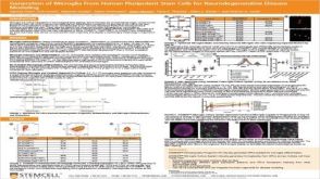 科学海报Generation of Microglia From Human Pluripotent Stem Cells for Neurodegenerative Disease Modeling
 实验方案How to Co-Culture Human Airway Epithelial and Immune Cells for RSV Infection