Chemically defined serum-free conditions for cartilage regeneration from human embryonic stem cells.
AIMS The aim of this study was to improve a method that induce cartilage differentiation of human embryoid stem cells (hESCs) in vitro,and test the effect of in vivo environments on the further maturation of hESCs derived cells. MAIN METHODS Embryoid bodies (EBs) formed from hESCs,with serum-free KSR-based medium and mesodermal specification related factors,CHIR,and Noggin for first 8days. Then cells were digested and cultured as micropellets in serum-free KSR-based chondrogenic medium that was supplemented with PDGF-BB,TGF β3,BMP4 in sequence for 24days. The morphology,FACS,histological staining as well as the expression of chondrogenic specific genes were detected in each stage,and further in vivo experiments,cell injections and tissue transplantations,further verified the formation of chondrocytes. KEY FINDINGS We were able to obtain chondrocyte/cartilage from hESCs using serum-free KSR-based conditioned medium. qPCR analysis showed that expression of the chondroprogenitor genes and the chondrocyte/cartilage matrix genes. Morphology analysis demonstrated we got PG+COL2+COL1-particles. It indicated we obtained hyaline cartilage-like particles. 32-Day differential cells were injected subcutaneous. Staining results showed grafts developed further mature in vivo. But when transplanted in subrenal capsule,their effect was not good as in subcutaneous. Microenvironment might affect the cartilage formation. SIGNIFICANCE The results of this study provide an absolute serum-free and efficient approach for generation of hESC-derived chondrocytes,and cells will become further maturation in vivo. It provides evidence and technology for the hypothesis that hESCs may be a promising therapy for the treatment of cartilage disease.
View Publication
产品类型:
产品号#:
85850
85857
产品名:
mTeSR™1
mTeSR™1
文献
Hannoun Z et al. (APR 2010)
Cellular reprogramming 12 2 133--140
The comparison between conditioned media and serum-free media in human embryonic stem cell culture and differentiation.
Human embryonic stem cells (hESCs) offer an inexhaustible supply of human somatic cell types through their ability to self-renew while retaining pluripotency. As such,hESC-derived cell types are important for applications ranging from in vitro modeling to therapeutic use. However,for their full potential to be realized,both the growth of the undifferentiated cells and their derivatives must be performed in defined culture conditions. Many research groups maintain hESCs using mouse embryonic fibroblasts (MEF) and MEF conditioned medium (CM). The use of murine systems to support hESCs has been imperative in developing hESC technology; however,they suffer from some major limitations including lack of definition,xenobiotic nature,batch-to-batch variation,and labor-intensive production. Therefore,hESC culture definition is essential if hESC lines,and their derivatives are to be quality assured and manufactured to GMP. We have initiated the process of standardizing hESC tissue culture and have employed two serum-free media: mTeSR (MT) and Stem Pro (SP). hESCs were maintained in a pluripotent state,for over 30 passages using MT and SP. Additionally,we present evidence that hESCs maintained in MT and SP generate equivalent levels of human hepatic endoderm as observed with CM. This data suggests that MT and SP are effective replacements for MEF-CM in hESC culture,contributing to the standardization of hESC in vitro models and ultimately their application.
View Publication
产品类型:
产品号#:
85850
85857
产品名:
mTeSR™1
mTeSR™1
文献
R. O. Bak et al. (FEB 2018)
Nature protocols 13 2 358--376
CRISPR/Cas9 genome editing in human hematopoietic stem cells.
Genome editing via homologous recombination (HR) (gene targeting) in human hematopoietic stem cells (HSCs) has the power to reveal gene-function relationships and potentially transform curative hematological gene and cell therapies. However,there are no comprehensive and reproducible protocols for targeting HSCs for HR. Herein,we provide a detailed protocol for the production,enrichment,and in vitro and in vivo analyses of HR-targeted HSCs by combining CRISPR/Cas9 technology with the use of rAAV6 and flow cytometry. Using this protocol,researchers can introduce single-nucleotide changes into the genome or longer gene cassettes with the precision of genome editing. Along with our troubleshooting and optimization guidelines,researchers can use this protocol to streamline HSC genome editing at any locus of interest. The in vitro HSC-targeting protocol and analyses can be completed in 3 weeks,and the long-term in vivo HSC engraftment analyses in immunodeficient mice can be achieved in 16 weeks. This protocol enables manipulation of genes for investigation of gene functions during hematopoiesis,as well as for the correction of genetic mutations in HSC transplantation-based therapies for diseases such as sickle cell disease,$\beta$-thalassemia,and primary immunodeficiencies.
View Publication
Irwin EF et al. (OCT 2011)
Biomaterials 32 29 6912--6919
Engineered polymer-media interfaces for the long-term self-renewal of human embryonic stem cells.
We have developed a synthetic polymer interface for the long-term self-renewal of human embryonic stem cells (hESCs) in defined media. We successfully cultured hESCs on hydrogel interfaces of aminopropylmethacrylamide (APMAAm) for over 20 passages in chemically-defined mTeSR™1 media and demonstrated pluripotency of multiple hESC lines with immunostaining and quantitative RT-PCR studies. Results for hESC proliferation and pluripotency markers were both qualitatively and quantitatively similar to cells cultured on Matrigel™-coated substrates. Mechanistically,it was resolved that bovine serum albumin (BSA) in the mTeSR™1 media was critical for cell adhesion on APMAAm hydrogel interfaces. This study uniquely identified a robust long-term culture surface for the self-renewal of hESCs without the use of biologic coatings (e.g.,peptides,proteins,or Matrigel™) in completely chemically-defined media that employed practical culturing techniques amenable to clinical-scale cell expansion.
View Publication
产品类型:
产品号#:
85850
85857
产品名:
mTeSR™1
mTeSR™1
文献
Renz PF and Beyer TA (FEB 2016)
Methods in molecular biology (Clifton,N.J.) 1341 369--376
A Concise Protocol for siRNA-Mediated Gene Suppression in Human Embryonic Stem Cells.
Human embryonic stem cells hold great promise for future biomedical applications such as disease modeling and regenerative medicine. However,these cells are notoriously difficult to culture and are refractory to common means of genetic manipulation,thereby limiting their range of applications. In this protocol,we present an easy and robust method of gene repression in human embryonic stem cells using lipofection of small interfering RNA (siRNA).
View Publication
产品类型:
产品号#:
05850
05857
05870
05875
05872
05873
07909
07920
85850
85857
85870
85875
产品名:
IV型胶原酶(1mg /mL)
ACCUTASE™
mTeSR™1
mTeSR™1
文献
Perez-Campo FM et al. (JUN 2014)
STEM CELLS 32 6 1591--1601
MOZ-Mediated Repression of p16 INK 4 a Is Critical for the Self-Renewal of Neural and Hematopoietic Stem Cells
Although inhibition of p16(INK4a) expression is critical to preserve the proliferative capacity of stem cells,the molecular mechanisms responsible for silencing p16(INK4a) expression remain poorly characterized. Here,we show that the histone acetyltransferase (HAT) monocytic leukemia zinc finger protein (MOZ) controls the proliferation of both hematopoietic and neural stem cells by modulating the transcriptional repression of p16(INK4a) . In the absence of the HAT activity of MOZ,expression of p16(INK4a) is upregulated in progenitor and stem cells,inducing an early entrance into replicative senescence. Genetic deletion of p16(INK4a) reverses the proliferative defect in both Moz(HAT) (-) (/) (-) hematopoietic and neural progenitors. Our results suggest a critical requirement for MOZ HAT activity to silence p16(INK4a) expression and to protect stem cells from early entrance into replicative senescence.
View Publication
产品类型:
产品号#:
05700
05701
05702
05707
产品名:
NeuroCult™ 基础培养基(小鼠和大鼠)
NeuroCult™ 扩增添加物(小鼠和大鼠)
NeuroCult™扩增试剂盒(小鼠和大鼠)
NeuroCult™化学解离试剂盒(小鼠)
文献
Kang M and Han Y-M (APR 2014)
PloS one 9 4 e94888
Differentiation of human pluripotent stem cells into nephron progenitor cells in a serum and feeder free system.
OBJECTIVES Kidney disease is emerging as a critical medical problem worldwide. Because of limited treatment options for the damaged kidney,stem cell treatment is becoming an alternative therapeutic approach. Of many possible human stem cell sources,pluripotent stem cells are most attractive due to their self-renewal and pluripotent capacity. However,little is known about the derivation of renal lineage cells from human pluripotent stem cells (hPSCs). In this study,we developed a novel protocol for differentiation of nephron progenitor cells (NPCs) from hPSCs in a serum- and feeder-free system. MATERIALS AND METHODS We designed step-wise protocols for differentiation of human pluripotent stem cells toward primitive streak,intermediate mesoderm and NPCs by recapitulating normal nephrogenesis. Expression of key marker genes was examined by RT-PCR,real time RT-PCR and immunocytochemistry. Each experiment was independently performed three times to confirm its reproducibility. RESULTS After modification of culture period and concentration of exogenous factors,hPSCs can differentiate into NPCs that markedly express specific marker genes such as SIX2,GDNF,HOXD11,WT1 and CITED1 in addition to OSR1,PAX2,SALL1 and EYA1. Moreover,NPCs possess the potential of bidirectional differentiation into both renal tubular epithelial cells and glomerular podocytes in defined culture conditions. In particular,approximately 70% of SYN-positive cells were obtained from hPSC-derived NPCs after podocytes induction. NPCs can also form in vitro tubule-like structures in three dimensional culture systems. CONCLUSIONS Our novel protocol for hPSCs differentiation into NPCs can be useful for producing alternative sources of cell replacement therapy and disease modeling for human kidney diseases.
View Publication

 EasySep™小鼠TIL(CD45)正选试剂盒


 文献
文献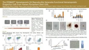 科学海报The STEMdiff™ Hematopoietic Kit Reproducibly Generates Functional Hematopoietic Progenitor Cells from Human Pluripotent Stem Cells
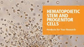 产品手册Hematopoietic Stem and Progenitor Cells - Products for Your Research
 产品手册Products for Human Pluripotent Stem Cells
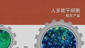 产品手册Products for Human Pluripotent Stem Cells
