Zandstra PW et al. (APR 1997)
Proceedings of the National Academy of Sciences of the United States of America 94 9 4698--703
Cytokine manipulation of primitive human hematopoietic cell self-renewal.
Previous studies have shown that primitive human hematopoietic cells detectable as long-term culture-initiating cells (LTC-ICs) and colony-forming cells (CFCs) can be amplified when CD34(+) CD38(-) marrow cells are cultured for 10 days in serum-free medium containing flt3 ligand (FL),Steel factor (SF),interleukin (IL)-3,IL-6,and granulocyte colony-stimulating factor. We now show that the generation of these two cell types in such cultures is differentially affected at the single cell level by changes in the concentrations of these cytokines. Thus,maximal expansion of LTC-ICs (60-fold) was obtained in the presence of 30 times more FL,SF,IL-3,IL-6,and granulocyte colony-stimulating factor than could concomitantly stimulate the near-maximal (280-fold) amplification of CFCs. Furthermore,the reduced ability of suboptimal cytokine concentrations to support the production of LTC-ICs could be ascribed to a differential response of the stimulated cells since this was not accompanied by a change in the number of input CD34(+) CD38(-) cells that proliferated. Reduced LTC-IC amplification in the absence of a significant effect on CFC generation also occurred when the concentrations of FL and SF were decreased but the concentration of IL-3 was high (as compared with cultures containing high levels of all three cytokines). To our knowledge,these findings provide the first evidence suggesting that extrinsically acting cytokines can alter the self-renewal behavior of primary human hematopoietic stem cells independent of effects on their viability or proliferation.
View Publication
产品类型:
产品号#:
05150
产品名:
MyeloCult™H5100
文献
Sumitomo A et al. (OCT 2010)
Molecular and cellular biology 30 20 4818--27
The transcriptional mediator subunit MED1/TRAP220 in stromal cells is involved in hematopoietic stem/progenitor cell support through osteopontin expression.
MED1/TRAP220,a subunit of the transcriptional Mediator/TRAP complex,is crucial for various biological events through its interaction with distinct activators,such as nuclear receptors and GATA family activators. In hematopoiesis,MED1 plays a pivotal role in optimal nuclear receptor-mediated myelomonopoiesis and GATA-1-induced erythropoiesis. In this study,we present evidence that MED1 in stromal cells is involved in supporting hematopoietic stem and/or progenitor cells (HSPCs) through osteopontin (OPN) expression. We found that the proliferation of bone marrow (BM) cells cocultured with MED1 knockout (Med1(-/-)) mouse embryonic fibroblasts (MEFs) was significantly suppressed compared to the control. Furthermore,the number of long-term culture-initiating cells (LTC-ICs) was attenuated for BM cells cocultured with Med1(-/-) MEFs. The vitamin D receptor (VDR)- and Runx2-mediated expression of OPN,as well as Mediator recruitment to the Opn promoter,was specifically attenuated in the Med1(-/-) MEFs. Addition of OPN to these MEFs restored the growth of cocultured BM cells and the number of LTC-ICs,both of which were attenuated by the addition of the anti-OPN antibody to Med1(+/+) MEFs and to BM stromal cells. Consequently,MED1 in niche appears to play an important role in supporting HSPCs by upregulating VDR- and Runx2-mediated transcription on the Opn promoter.
View Publication
产品类型:
产品号#:
03334
03434
03444
09500
产品名:
MethoCult™M3334
MethoCult™GF M3434
MethoCult™GF M3434
BIT 9500血清替代物
文献
X. Li et al. (jul 2019)
Stem cells (Dayton,Ohio) 37 7 937--947
p53-TP53-Induced Glycolysis Regulator Mediated Glycolytic Suppression Attenuates DNA Damage and Genomic Instability in Fanconi Anemia Hematopoietic Stem Cells.
Emerging evidence has shown that resting quiescent hematopoietic stem cells (HSCs) prefer to utilize anaerobic glycolysis rather than mitochondrial respiration for energy production. Compelling evidence has also revealed that altered metabolic energetics in HSCs underlies the onset of certain blood diseases; however,the mechanisms responsible for energetic reprogramming remain elusive. We recently found that Fanconi anemia (FA) HSCs in their resting state are more dependent on mitochondrial respiration for energy metabolism than on glycolysis. In the present study,we investigated the role of deficient glycolysis in FA HSC maintenance. We observed significantly reduced glucose consumption,lactate production,and ATP production in HSCs but not in the less primitive multipotent progenitors or restricted hematopoietic progenitors of Fanca-/- and Fancc-/- mice compared with that of wild-type mice,which was associated with an overactivated p53 and TP53-induced glycolysis regulator,the TIGAR-mediated metabolic axis. We utilized Fanca-/- HSCs deficient for p53 to show that the p53-TIGAR axis suppressed glycolysis in FA HSCs,leading to enhanced pentose phosphate pathway and cellular antioxidant function and,consequently,reduced DNA damage and attenuated HSC exhaustion. Furthermore,by using Fanca-/- HSCs carrying the separation-of-function mutant p53R172P transgene that selectively impairs the p53 function in apoptosis but not cell-cycle control,we demonstrated that the cell-cycle function of p53 was not required for glycolytic suppression in FA HSCs. Finally,ectopic expression of the glycolytic rate-limiting enzyme PFKFB3 specifically antagonized p53-TIGAR-mediated metabolic reprogramming in FA HSCs. Together,our results suggest that p53-TIGAR metabolic axis-mediated glycolytic suppression may play a compensatory role in attenuating DNA damage and proliferative exhaustion in FA HSCs. Stem Cells 2019;37:937-947.
View Publication
De Kock J et al. (SEP 2011)
Toxicology in vitro : an international journal published in association with BIBRA 25 6 1191--202
Evaluation of the multipotent character of human foreskin-derived precursor cells.
In the present study,the trilineage differentiation capacity of human foreskin-derived precursor cells (hSKP) was evaluated upon exposure to various (non)commercial (i and ii) ectodermal,(iii) mesodermal and (iv) endodermal differentiation media. (i) Upon sequential exposure of the cells to keratinocyte growth (CnT-07® or CnT-057®) and differentiation (CnT-02® or Epilife®) media,keratinocyte-like cells (filaggrin(+)/involucrin(+)) were obtained. The preferred keratinocyte differentiation strategy was exposure to CnT-07®. (ii) When hSKP were subsequently exposed to NeuroCult® media,cells underwent a weak neuro-ectodermal differentiation expressing nestin,myelin binding protein (MBP),vimentin and alpha-foetoprotein (AFP). Sequential exposure to NPMM® and NPDM® generated cells with an inferior neuro-ectodermal phenotype (nestin(+)/vimentin(+)/MBP(-)/AFP(-)). (iii) Upon exposure of hSKP to insulin-transferrin-selenite (ITS) and dexamethasone,small lipid droplets were observed,suggesting their differentiation potential towards adipocyte-like cells. (iv) Finally,after sequential exposure to hepatogenic growth factors and cytokines,an immature hepatic cell population was generated. The presence of pre-albumin suggests that a sequential exposure strategy is here superior to a cocktail approach. In summary,a considerable impact of different (non)commercial media on the lineage-specific differentiation efficiency of hSKP is shown. In addition,we demonstrate here for the first time that,in a suitable keratinocyte stimulating micro-environment,hSKP can generate keratinocyte-like progeny in vitro.
View Publication
产品类型:
产品号#:
05751
产品名:
NeuroCult™ NS-A 扩增试剂盒(人)
文献
Lianguzova MS et al. (APR 2007)
Cell biology international 31 4 330--7
Phosphoinositide 3-kinase inhibitor LY294002 but not serum withdrawal suppresses proliferation of murine embryonic stem cells.
Mouse embryonic stem (mES) cells have short duration of their cell cycle and are capable of proliferating in the absence of growth factors. To find out which signaling pathways contribute to the regulation of the mES cell cycle,we used pharmacological inhibitors of MAP and PI3 kinase cascades. The MAP kinase inhibitors as well as serum withdrawal did not affect mES cell cycle distribution,whereas the inhibitor of PI3K activity,LY294002,induced accumulation of cells in G(1) phase followed by apoptotic cell death. Serum withdrawal also causes apoptosis,but it does not change the content and activity of cell cycle regulators. In contrast,in mES cells treated with LY294002,the activities of Cdk2 and E2F were significantly decreased. Interestingly,LY294002had a much stronger effect on cell cycle distribution in low serum conditions,implying that serum can promote G(1)--textgreaterS transition of mES cells by a LY294002-resistant mechanism. Thus,proliferation of mES cells is maintained by at least two separate mechanisms: a LY294002-sensitive pathway,which is active even in the absence of serum,and LY294002-resistant,but serum-dependent,pathway.
View Publication
Kwok CTD et al. (MAR 2016)
Stem Cell Research 16 3 651--661
The Forkhead box transcription factor FOXM1 is required for the maintenance of cell proliferation and protection against oxidative stress in human embryonic stem cells
Human embryonic stem cells (hESCs) exhibit unique cell cycle structure,self-renewal and pluripotency. The Forkhead box transcription factor M1 (FOXM1) is critically required for the maintenance of pluripotency in mouse embryonic stem cells and mouse embryonal carcinoma cells,but its role in hESCs remains unclear. Here,we show that FOXM1 expression was enriched in undifferentiated hESCs and was regulated in a cell cycle-dependent manner with peak levels detected at the G2/M phase. Expression of FOXM1 did not correlate with OCT4 and NANOG during in vitro differentiation of hESCs. Importantly,knockdown of FOXM1 expression led to aberrant cell cycle distribution with impairment in mitotic progression but showed no profound effect on the undifferentiated state. Interestingly,FOXM1 depletion sensitized hESCs to oxidative stress. Moreover,genome-wide analysis of FOXM1 targets by ChIP-seq identified genes important for M phase including CCNB1 and CDK1,which were subsequently confirmed by ChIP and RNA interference analyses. Further peak set comparison against a differentiating hESC line and a cancer cell line revealed a substantial difference in the genomic binding profile of FOXM1 in hESCs. Taken together,our findings provide the first evidence to support FOXM1 as an important regulator of cell cycle progression and defense against oxidative stress in hESCs.
View Publication
产品类型:
产品号#:
05110
85850
85857
产品名:
STEMdiff™权威内胚层检测试剂盒
mTeSR™1
mTeSR™1
文献
Ortiz-Sá et al. (JAN 2009)
Leukemia 23 1 59--70
Enhanced cytotoxicity of an anti-transferrin receptor IgG3-avidin fusion protein in combination with gambogic acid against human malignant hematopoietic cells: functional relevance of iron, the receptor, and reactive oxygen species.
The human transferrin receptor (hTfR) is a target for cancer immunotherapy due to its overexpression on the surface of cancer cells. We previously developed an antibody-avidin fusion protein that targets hTfR (anti-hTfR IgG3-Av) and exhibits intrinsic cytotoxicity against certain malignant cells. Gambogic acid (GA),a drug that also binds hTfR,induces cytotoxicity in several malignant cell lines. We now report that anti-hTfR IgG3-Av and GA induce cytotoxicity in a new broader panel of hematopoietic malignant cell lines. Our results show that the effect of anti-hTfR IgG3-Av is iron-dependent whereas that of GA is iron-independent in all cells tested. In addition,we observed that GA exerts a TfR-independent cytotoxicity. We also found that GA increases the generation of reactive oxygen species that may play a role in the cytotoxicity induced by this drug. Additive cytotoxicity was observed by simultaneous combination treatment with these drugs and synergy by using anti-hTfR IgG3-Av as a chemosensitizing agent. In addition,we found a concentration of GA that is toxic to malignant hematopoietic cells but not to human hematopoietic progenitor cells. Our results suggest that these two compounds may be effective,alone or in combination,for the treatment of human hematopoietic malignancies.
View Publication
产品类型:
产品号#:
04434
04444
产品名:
MethoCult™H4434经典
MethoCult™H4434经典
文献
Denning-Kendall P et al. (JAN 2003)
Stem cells (Dayton,Ohio) 21 6 694--701
Cobblestone area-forming cells in human cord blood are heterogeneous and differ from long-term culture-initiating cells.
The long-term culture-initiating cell (LTC-IC) assay is a physiological approach to the quantitation of primitive human hematopoietic cells. The readout using identification of cobblestone area-forming cells (CAFC) has gained popularity over the LTC-IC readout where cells are subcultured in a colony-forming cell assay. However,comparing the two assays,cord blood (CB) mononuclear cell (MNC) samples were found to contain a higher frequency of CAFC than LTC-IC (126 +/- 83 versus 40 +/- 31 per 10(5) cells,p = 0.0001). Overall,60% of week-5 cobblestones produced by CB MNC were not functional LTC-IC and were classified as false." Separation of CB MNC using immunomagnetic columns showed that false cobblestones were CD34(-)/lineage(+). Purified CD34(+) cells�
View Publication
Smad4 binds Hoxa9 in the cytoplasm and protects primitive hematopoietic cells against nuclear activation by Hoxa9 and leukemia transformation.
We studied leukemic stem cells (LSCs) in a Smad4(-/-) mouse model of acute myelogenous leukemia (AML) induced either by the HOXA9 gene or by the fusion oncogene NUP98-HOXA9. Although Hoxa9-Smad4 complexes accumulate in the cytoplasm of normal hematopoietic stem cells and progenitor cells (HSPCs) transduced with these oncogenes,there is no cytoplasmic stabilization of HOXA9 in Smad4(-/-) HSPCs,and as a consequence increased levels of Hoxa9 is observed in the nucleus leading to increased immortalization in vitro. Loss of Smad4 accelerates the development of leukemia in vivo because of an increase in transformation of HSPCs. Therefore,the cytoplasmic binding of Hoxa9 by Smad4 is a mechanism to protect Hoxa9-induced transformation of normal HSPCs. Because Smad4 is a potent tumor suppressor involved in growth control,we developed a strategy to modify the subcellular distribution of Smad4. We successfully disrupted the interaction between Hoxa9 and Smad4 to activate the TGF-β pathway and apoptosis,leading to a loss of LSCs. Together,these findings reveal a major role for Smad4 in the negative regulation of leukemia initiation and maintenance induced by HOXA9/NUP98-HOXA9 and provide strong evidence that antagonizing Smad4 stabilization by these oncoproteins might be a promising novel therapeutic approach in leukemia.
View Publication

 EasySep™小鼠TIL(CD45)正选试剂盒


 文献
文献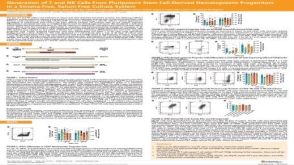 科学海报Generation of T and NK Cells From Pluripotent Stem Cell-Derived Hematopoietic Progenitors in a Stroma-Free, Serum-Free Culture System
 挂图Identification of Colonies Derived from Human Hematopoietic Progenitors Representative colony images and tips for identifying progenitor subtypes in CFU assays
