Liu Y et al. (MAR 2015)
Journal of Biomedical Materials Research - Part A 103 3 1053--1059
Native nucleus pulposus tissue matrix promotes notochordal differentiation of human induced pluripotent stem cells with potential for treating intervertebral disc degeneration
Native porcine nucleus pulposus (NP) tissue harbors a number of notochordal cells (NCs). Whether the native NP matrix supports the homeostasis of notochordal cells is poorly understood. We hypothesized the NP matrix alone may contain sufficient regulatory factors and can serve as stimuli to generate notochordal cells (NCs) from human pluripotent stem cells. NCs are a promising cell sources for cell-based therapy to treat some types of intervertebral disc (IVD) degeneration. One major limitation of this emerging technique is the lack of available NCs as a potential therapeutic cell source. Human pluripotent stem cells derived from reprogramming or somatic cell nuclear transfer technique may yield stable and unlimited source for therapeutic use. We devised a new method to use porcine NP matrix to direct notochordal differentiation of human induced pluripotent stem cells (hiPSCs). The results showed that hiPSCs successfully differentiated into NC-like cells under the influence of devitalized porcine NP matrix. The NC-like cells expressed typical notochordal marker genes including brachyury (T),cytokeratin-8 (CK-8) and cytokeratin-18 (CK-18),and they displayed the ability to generate NP-like tissue in vitro,which was rich in aggrecan and collagen type II. These findings demonstrated the proof of concept for using native NP matrix to direct notochordal differentiation of hiPSCs. It provides a foundation for further understanding the biology of NCs,and eventually towards regenerative therapies for disc degeneration.
View Publication
Huang EH et al. (APR 2009)
Cancer research 69 8 3382--9
Aldehyde dehydrogenase 1 is a marker for normal and malignant human colonic stem cells (SC) and tracks SC overpopulation during colon tumorigenesis.
Although the concept that cancers originate from stem cells (SC) is becoming scientifically accepted,mechanisms by which SC contribute to tumor initiation and progression are largely unknown. For colorectal cancer (CRC),investigation of this problem has been hindered by a paucity of specific markers for identification and isolation of SC from normal and malignant colon. Accordingly,aldehyde dehydrogenase 1 (ALDH1) was investigated as a possible marker for identifying colonic SC and for tracking them during cancer progression. Immunostaining showed that ALDH1(+) cells are sparse and limited to the normal crypt bottom,where SCs reside. During progression from normal epithelium to mutant (APC) epithelium to adenoma,ALDH1(+) cells increased in number and became distributed farther up the crypt. CD133(+) and CD44(+) cells,which are more numerous and broadly distributed in normal crypts,showed similar changes during tumorigenesis. Flow cytometric isolation of cancer cells based on enzymatic activity of ALDH (Aldefluor assay) and implantation of these cells in nonobese diabetic-severe combined immunodeficient mice (a) generated xenograft tumors (Aldefluor(-) cells did not),(b) generated them after implanting as few as 25 cells,and (c) generated them dose dependently. Further isolation of cancer cells using a second marker (CD44(+) or CD133(+) serially) only modestly increased enrichment based on tumor-initiating ability. Thus,ALDH1 seems to be a specific marker for identifying,isolating,and tracking human colonic SC during CRC development. These findings also support our original hypothesis,derived previously from mathematical modeling of crypt dynamics,that progressive colonic SC overpopulation occurs during colon tumorigenesis and drives CRC development.
View Publication
产品类型:
产品号#:
01700
01705
产品名:
ALDEFLUOR™工具
ALDEFLUOR™DEAB试剂
文献
Eaves CJ et al. (JUL 1991)
Blood 78 1 110--7
Mechanisms that regulate the cell cycle status of very primitive hematopoietic cells in long-term human marrow cultures. II. Analysis of positive and negative regulators produced by stromal cells within the adherent layer.
Numerous factors that can influence the proliferation and differentiation in vitro of cells at various stages of hematopoiesis have been identified,but the mechanisms used by stromal cells to regulate the cycling status of the most primitive human hematopoietic cells are still poorly understood. Previous studies of long-term cultures (LTC) of human marrow have suggested that cytokine-induced variations in stromal cell production of one or more stimulators and inhibitors of hematopoiesis may be important. To identify the specific regulators involved,we performed Northern analyses on RNA extracted from human marrow LTC adherent layers,or stromal cell types derived from or related to those present in the adherent layer. These analyses showed marked increases in interleukin-1 beta (IL-1 beta),IL-6,and granulocyte colony-stimulating factor (G-CSF) mRNA levels within 8 hours after treatments that lead to the activation within 2 days of primitive hematopoietic progenitors in such cultures. Increases in granulocyte-macrophage (GM)-CSF and M-CSF mRNA were also sometimes seen. Bioassays using cell lines responsive to G-CSF,GM-CSF,and IL-6 showed significant elevation in growth factor levels 24 hours after IL-1 beta stimulation. Neither IL-3 nor IL-4 mRNA was detectable at any time. In contrast,transforming growth factor-beta (TGF-beta) mRNA and nanogram levels of TGF-beta bioactivity in the medium were detected at all times in established LTC,and these levels were not consistently altered by any of the manipulations that stimulated hematopoietic growth factor production and primitive progenitor cycling. We also found that addition of anti-TGF-beta antibody could prolong or reactivate primitive progenitor proliferation when added to previously stimulated or quiescent cultures,respectively. Together,these results indicate a dominant negative regulatory role of endogenously produced TGF-beta in unperturbed LTC,with activation of primitive hematopoietic cells being achieved by mechanisms that stimulate stromal cells to produce G-CSF,GM-CSF,and IL-6. Given the similarities between the LTC system and the marrow microenvironment,it seems likely that the control of human stem cell activation in vivo may involve similar variations in the production of these factors by stromal cells.
View Publication
产品类型:
产品号#:
05150
产品名:
MyeloCult™H5100
文献
L. Li et al. (OCT 2018)
Cell metabolism
TLR8-Mediated Metabolic Control of Human Treg Function: A Mechanistic Target for Cancer Immunotherapy.
Regulatory T (Treg) cells induce an immunosuppressive microenvironment that is a major obstacle for successful tumor immunotherapy. Dissecting the regulatory mechanisms between energy metabolism and functionality in Treg cells will provide insight toward developing novel immunotherapies against cancer. Here we report that human naturally occurring and tumor-associated Treg cells exhibit distinct metabolic profiles with selectivity for glucose metabolism compared with effector T cells. Treg-mediated accelerated glucose consumption induces cellular senescence and suppression of responder T cells through cross-talk. TLR8 signaling selectively inhibits glucose uptake and glycolysis in human Treg cells,resulting in reversal of Treg suppression. Importantly,TLR8 signaling-mediated reprogramming of glucose metabolism and function in human Treg cells can enhance anti-tumor immunity in vivo in a melanoma adoptive transfer T cell therapy model. Our studies identify mechanistic links between innate signaling and metabolic regulation of human Treg suppression,which may be used as a strategy to advance tumor immunotherapy.
View Publication
产品类型:
产品号#:
产品名:
文献
Allan LL et al. (SEP 2009)
Blood 114 12 2411--6
Apolipoprotein-mediated lipid antigen presentation in B cells provides a pathway for innate help by NKT cells.
Natural killer T (NKT) cells are innate-like lymphocytes that recognize lipid antigens and have been shown to enhance B-cell activation and antibody production. B cells typically recruit T-cell help by presenting internalized antigens recognized by their surface antigen receptor. Here,we demonstrate a highly efficient means whereby human B cells present lipid antigens to NKT cells,capturing the antigen using apolipoprotein E (apoE) and the low-density lipoprotein receptor (LDL-R). ApoE dramatically enhances B-cell presentation of alpha-galactosylceramide (alphaGalCer),an exogenous CD1d presented antigen,inducing activation of NKT cells and the subsequent activation of B cells. B cells express the LDL-R on activation,and the activation of NKT cells by B cells is completely LDL-R dependent,as shown by blocking experiments and the complete lack of presentation when using apoE2,an isoform of apoE incapable of LDL-R binding. The dependence on apoE and the LDL-R is much more pronounced in B cells than we had previously seen in dendritic cells,which can apparently use alternate pathways of lipid antigen uptake. Thus,B cells use an apolipoprotein-mediated pathway of lipid antigen presentation,which constitutes a form of innate help for B cells by NKT cells.
View Publication
产品类型:
产品号#:
19054
19054RF
产品名:
EasySep™人B细胞富集试剂盒
RoboSep™ 人B细胞富集试剂盒含滤芯吸头
文献
Chlon TM et al. (OCT 2014)
Journal of virology 88 19 11315--11326
High-risk human papillomavirus E6 protein promotes reprogramming of Fanconi anemia patient cells through repression of p53 but does not allow for sustained growth of induced pluripotent stem cells.
DNA repair plays a crucial role in embryonic and somatic stem cell biology and cell reprogramming. The Fanconi anemia (FA) pathway,which promotes error-free repair of DNA double-strand breaks,is required for somatic cell reprogramming to induced pluripotent stem cells (iPSC). Thus,cells from Fanconi anemia patients,which lack this critical pathway,fail to be reprogrammed to iPSC under standard conditions unless the defective FA gene is complemented. In this study,we utilized the oncogenes of high-risk human papillomavirus 16 (HPV16) to overcome the resistance of FA patient cells to reprogramming. We found that E6,but not E7,recovers FA iPSC colony formation and,furthermore,that p53 inhibition is necessary and sufficient for this activity. The iPSC colonies resulting from each of these approaches stained positive for alkaline phosphatase,NANOG,and Tra-1-60,indicating that they were fully reprogrammed into pluripotent cells. However,FA iPSC were incapable of outgrowth into stable iPSC lines regardless of p53 suppression,whereas their FA-complemented counterparts grew efficiently. Thus,we conclude that the FA pathway is required for the growth of iPSC beyond reprogramming and that p53-independent mechanisms are involved. IMPORTANCE A novel approach is described whereby HPV oncogenes are used as tools to uncover DNA repair-related molecular mechanisms affecting somatic cell reprogramming. The findings indicate that p53-dependent mechanisms block FA cells from reprogramming but also uncover a previously unrecognized defect in FA iPSC proliferation independent of p53.
View Publication
产品类型:
产品号#:
05850
05857
05870
05875
85850
85857
85870
85875
产品名:
mTeSR™1
mTeSR™1
文献
Fraga AM et al. (MAR 2011)
Cell Transplantation 20 3 431--40
Establishment of a Brazilian line of human embryonic stem cells in defined medium: implications for cell therapy in an ethnically diverse population.
Pluripotent human embryonic stem (hES) cells are an important experimental tool for basic and applied research,and a potential source of different tissues for transplantation. However,one important challenge for the clinical use of these cells is the issue of immunocompatibility,which may be dealt with by the establishment of hES cell banks to attend different populations. Here we describe the derivation and characterization of a line of hES cells from the Brazilian population,named BR-1,in commercial defined medium. In contrast to the other hES cell lines established in defined medium,BR-1 maintained a stable normal karyotype as determined by genomic array analysis after 6 months in continuous culture (passage 29). To our knowledge,this is the first reported line of hES cells derived in South America. We have determined its genomic ancestry and compared the HLA-profile of BR-1 and another 22 hES cell lines established elsewhere with those of the Brazilian population,finding they would match only 0.011% of those individuals. Our results highlight the challenges involved in hES cell banking for populations with a high degree of ethnic admixture.
View Publication

 EasySep™小鼠TIL(CD45)正选试剂盒


 科学海报NeuroFluor™ CDr3: A Novel Tool for the Detection of Live CNS and Human Pluripotent Stem Cell-Derived Neural Stem and Progenitor Cells
 文献
文献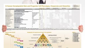 挂图Human Hematopoietic Stem and Progenitor Cell Phenotyping Overview of subset surface markers, frequencies and assays for analysis
 科学海报A Serum-Free Workflow for the Isolation, Expansion and Differentiation of Human Myogenic Progenitors
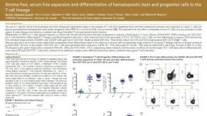 科学海报Stroma-Free, Serum-Free Expansion and Differentiation of Hematopoietic Stem and Progenitor Cells to the T Cell Lineage
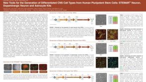 科学海报New Tools for the Generation of Differentiated CNS Cell Types from Human Pluripotent Stem Cells: STEMdiff™ Neuron, Dopaminergic Neuron and Astrocyte Kits