Seiwert TY et al. ( 2009)
Cancer research 69 7 3021--3031
The MET receptor tyrosine kinase is a potential novel therapeutic target for head and neck squamous cell carcinoma.
Recurrent/metastatic head and neck cancer remains a devastating disease with insufficient treatment options. We investigated the MET receptor tyrosine kinase as a novel target for the treatment of head and neck squamous cell carcinoma (HNSCC). MET/phosphorylated MET and HGF expression was analyzed in 121 tissues (HNSCC/normal) by immunohistochemistry,and in 20 HNSCC cell lines by immunoblotting. The effects of MET inhibition using small interfering RNA/two small-molecule inhibitors (SU11274/PF-2341066) on signaling,migration,viability,and angiogenesis were determined. The complete MET gene was sequenced in 66 head and neck cancer tissue samples and eight cell lines. MET gene copy number was determined in 14 cell lines and 23 tumor tissues. Drug combinations of SU11274 with cisplatin or erlotinib were tested in SCC35/HN5 cell lines. Eighty-four percent of the HNSCC samples showed MET overexpression,whereas 18 of 20 HNSCC cell lines (90%) expressed MET. HGF overexpression was present in 45% of HNSCC. MET inhibition with SU11274/PF-2341066 abrogated MET signaling,cell viability,motility/migration in vitro,and tumor angiogenesis in vivo. Mutational analysis of 66 tumor tissues and 8 cell lines identified novel mutations in the semaphorin (T230M/E168D/N375S),juxtamembrane (T1010I/R988C),and tyrosine kinase (T1275I/V1333I) domains (incidence: 13.5%). Increased MET gene copy number was present with textgreater10 copies in 3 of 23 (13%) tumor tissues. A greater-than-additive inhibition of cell growth was observed when combining a MET inhibitor with cisplatin or erlotinib and synergy may be mediated via erbB3/AKT signaling. MET is functionally important in HNSCC with prominent overexpression,increased gene copy number,and mutations. MET inhibition abrogated MET functions,including proliferation,migration/motility,and angiogenesis. MET is a promising,novel target for HNSCC and combination approaches with cisplatin or EGFR inhibitors should be explored.
View Publication
产品类型:
产品号#:
73432
产品名:
SU11274
文献
Palombella VJ et al. (SEP 1994)
Cell 78 5 773--85
The ubiquitin-proteasome pathway is required for processing the NF-kappa B1 precursor protein and the activation of NF-kappa B.
We demonstrate an essential role for the proteasome complex in two proteolytic processes required for activation of the transcription factor NF-kappa B. The p105 precursor of the p50 subunit of NF-kappa B is processed in vitro by an ATP-dependent process that requires proteasomes and ubiquitin conjugation. The C-terminal region of p105 is rapidly degraded,leaving the N-terminal p50 domain. p105 processing can be blocked in intact cells with inhibitors of the proteasome or in yeast with proteasome mutants. These inhibitors also block the activation of NF-kappa B and the rapid degradation of I kappa B alpha induced by tumor necrosis factor alpha. Thus,the ubiquitin-proteasome pathway functions not only in the complete degradation of polypeptides,but also in the regulated processing of precursors into active proteins.
View Publication
产品类型:
产品号#:
73262
73264
产品名:
(S) -MG132
(S) -MG132
文献
Beckerman SR et al. (SEP 2015)
ASSAY and Drug Development Technologies 13 7 377--388
Phenotypic Assays to Identify Agents That Induce Reactive Gliosis: A Counter-Screen to Prioritize Compounds for Preclinical Animal Studies
Astrocyte phenotypes change in a process called reactive gliosis after traumatic central nervous system (CNS) injury. Astrogliosis is characterized by expansion of the glial fibrillary acidic protein (GFAP) cytoskeleton,adoption of stellate morphologies,and differential expression of some extracellular matrix molecules. The astrocytic response immediately after injury is beneficial,but in the chronic injury phase,reactive astrocytes produce inhibitory factors (i.e.,chondroitin sulfate proteoglycans [CSPGs]) that limit the regrowth of injured axons. There are no drugs that promote axon regeneration or functional recovery after CNS trauma in humans. To develop novel therapeutics for the injured CNS,we screened various libraries in a phenotypic assay to identify compounds that promote neurite outgrowth. However,the effects these compounds have on astrocytes are unknown. Specifically,we were interested in whether compounds could alter astrocytes in a manner that mimics the glial reaction to injury. To test this hypothesis,we developed cell-based phenotypic bioassays to measure changes in (1) GFAP morphology/localization and (2) CSPG expression/immunoreactivity from primary astrocyte cultures. These assays were optimized for six-point dose-response experiments in 96-well plates. The GFAP morphology assay is suitable for counter-screening with a Z-factor of 0.44±0.03 (mean±standard error of the mean; N=3 biological replicates). The CSPG assay is reproducible and informative,but does not satisfy common metrics for a screenable" assay. As proof of principle we tested a small set of hit compounds from our neurite outgrowth bioassay and identified one that can enhance axon growth without exacerbating the deleterious characteristics of reactive gliosis.
View Publication
产品类型:
产品号#:
05711
产品名:
NeuroCult™ SM1 神经添加物
文献
Chaumeil MM et al. ( 2016)
NeuroImage. Clinical 12 180--9
Hyperpolarized (13)C MR imaging detects no lactate production in mutant IDH1 gliomas: Implications for diagnosis and response monitoring.
Metabolic imaging of brain tumors using (13)C Magnetic Resonance Spectroscopy (MRS) of hyperpolarized [1-(13)C] pyruvate is a promising neuroimaging strategy which,after a decade of preclinical success in glioblastoma (GBM) models,is now entering clinical trials in multiple centers. Typically,the presence of GBM has been associated with elevated hyperpolarized [1-(13)C] lactate produced from [1-(13)C] pyruvate,and response to therapy has been associated with a drop in hyperpolarized [1-(13)C] lactate. However,to date,lower grade gliomas had not been investigated using this approach. The most prevalent mutation in lower grade gliomas is the isocitrate dehydrogenase 1 (IDH1) mutation,which,in addition to initiating tumor development,also induces metabolic reprogramming. In particular,mutant IDH1 gliomas are associated with low levels of lactate dehydrogenase A (LDHA) and monocarboxylate transporters 1 and 4 (MCT1,MCT4),three proteins involved in pyruvate metabolism to lactate. We therefore investigated the potential of (13)C MRS of hyperpolarized [1-(13)C] pyruvate for detection of mutant IDH1 gliomas and for monitoring of their therapeutic response. We studied patient-derived mutant IDH1 glioma cells that underexpress LDHA,MCT1 and MCT4,and wild-type IDH1 GBM cells that express high levels of these proteins. Mutant IDH1 cells and tumors produced significantly less hyperpolarized [1-(13)C] lactate compared to GBM,consistent with their metabolic reprogramming. Furthermore,hyperpolarized [1-(13)C] lactate production was not affected by chemotherapeutic treatment with temozolomide (TMZ) in mutant IDH1 tumors,in contrast to previous reports in GBM. Our results demonstrate the unusual metabolic imaging profile of mutant IDH1 gliomas,which,when combined with other clinically available imaging methods,could be used to detect the presence of the IDH1 mutation in vivo.
View Publication
产品类型:
产品号#:
05700
05750
05751
产品名:
NeuroCult™ 基础培养基(小鼠和大鼠)
NeuroCult™ NS-A 基础培养基(人)
NeuroCult™ NS-A 扩增试剂盒(人)
文献
Milush JM et al. (NOV 2009)
Blood 114 23 4823--31
Functionally distinct subsets of human NK cells and monocyte/DC-like cells identified by coexpression of CD56, CD7, and CD4.
The lack of natural killer (NK) cell-specific markers,as well as the overlap among several common surface antigens and functional properties,has obscured the delineation between NK cells and dendritic cells. Here,novel subsets of peripheral blood CD3/14/19(neg) NK cells and monocyte/dendritic cell (DC)-like cells were identified on the basis of CD7 and CD4 expression. Coexpression of CD7 and CD56 differentiates NK cells from CD56+ monocyte/DC-like cells,which lack CD7. In contrast to CD7+CD56+ NK cells,CD7(neg)CD56+ cells lack expression of NK cell-associated markers,but share commonalities in their expression of various monocyte/DC-associated markers. Using CD7,we observed approximately 60% of CD4+CD56+ cells were CD7(neg) cells,indicating the actual frequency of activated CD4+ NK cells is much lower in the blood than previously recognized. Functionally,only CD7+ NK cells secrete gamma interferon (IFNgamma) and degranulate after interleukin-12 (IL-12) plus IL-18 or K562 target cell stimulation. Furthermore,using CD7 to separate CD56+ NK cells and CD56+ myeloid cells,we demonstrate that unlike resting CD7+CD56+ NK cells,the CD7(neg)CD56+ myeloid cells stimulate a potent allogeneic response. Our data indicate that CD7 and CD56 coexpression discriminates NK cells from CD7(neg)CD56+ monocyte/DC-like cells,thereby improving our ability to study the intricacies of NK-cell subset phenotypes and functions in vivo.
View Publication
产品类型:
产品号#:
19051
19051RF
产品名:
EasySep™人T细胞富集试剂盒
RoboSep™ 人T细胞富集试剂盒含滤芯吸头
文献
Lian RH et al. (MAY 2002)
Journal of immunology (Baltimore,Md. : 1950) 168 10 4980--7
Orderly and nonstochastic acquisition of CD94/NKG2 receptors by developing NK cells derived from embryonic stem cells in vitro.
In mice there are two families of MHC class I-specific receptors,namely the Ly49 and CD94/NKG2 receptors. The latter receptors recognize the nonclassical MHC class I Qa-1(b) and are thought to be responsible for the recognition of missing-self and the maintenance of self-tolerance of fetal and neonatal NK cells that do not express Ly49. Currently,how NK cells acquire individual CD94/NKG2 receptors during their development is not known. In this study,we have established a multistep culture method to induce differentiation of embryonic stem (ES) cells into the NK cell lineage and examined the acquisition of CD94/NKG2 by NK cells as they differentiate from ES cells in vitro. ES-derived NK (ES-NK) cells express NK cell-associated proteins and they kill certain tumor cell lines as well as MHC class I-deficient lymphoblasts. They express CD94/NKG2 heterodimers,but not Ly49 molecules,and their cytotoxicity is inhibited by Qa-1(b) on target cells. Using RT-PCR analysis,we also report that the acquisition of these individual receptor gene expressions during different stages of differentiation from ES cells to NK cells follows a predetermined order,with their order of acquisition being first CD94; subsequently NKG2D,NKG2A,and NKG2E; and finally,NKG2C. Single-cell RT-PCR showed coexpression of CD94 and NKG2 genes in most ES-NK cells,and flow cytometric analysis also detected CD94/NKG2 on most ES-NK cells,suggesting that the acquisition of these receptors by ES-NK cells in vitro is nonstochastic,orderly,and cumulative.
View Publication
产品类型:
产品号#:
06902
06952
00321
00322
00323
00324
00325
产品名:
文献
Frazer-Abel AA et al. (NOV 2004)
The Journal of pharmacology and experimental therapeutics 311 2 758--69
Nicotine activates nuclear factor of activated T cells c2 (NFATc2) and prevents cell cycle entry in T cells.
We used primary peripheral blood T cells,a population that exists in G(0) and can be stimulated to enter the cell cycle synchronously,to define more precisely the effects of nicotine on pathways that control cell cycle entry and progression. Our data show that nicotine decreased the ability of T cells to transit through the G(0)/G(1) boundary (acquire competence) and respond to progression signals. These effects were due to nuclear factor of activated T cells c2 (NFATc2)-dependent repression of cyclin-dependent kinase 4 (CDK4) expression. Growth arrest at the G(0)/G(1) boundary was further enforced by inhibition of cyclin D2 expression and by increased expression and stabilization of p27Kip1. Intriguingly,T cells from habitual users of tobacco products and from NFATc2-deficient mice constitutively expressed CDK4 and were resistant to the antiproliferative effects of nicotine. These results indicate that nicotine impairs T cell cycle entry through NFATc2-dependent mechanisms and suggest that,in the face of chronic nicotine exposure,selection may favor cells that can evade these effects. We postulate that cross talk between nicotinic acetylcholine receptors and growth factor receptor-activated pathways offers a novel mechanism by which nicotine may directly impinge on cell cycle progression. This offers insight into possible reasons that underlie the unique effects of nicotine on distinct cell types and identifies new targets that may be useful control tobacco-related diseases.
View Publication
产品类型:
产品号#:
产品名:
文献
Ouchi T et al. (MAY 2015)
Differentiation
LNGFR+THY-1+ human pluripotent stem cell-derived neural crest-like cells have the potential to develop into mesenchymal stem cells
Mesenchymal stem cells (MSCs) are defined as non-hematopoietic,plastic-adherent,self-renewing cells that are capable of tri-lineage differentiation into bone,cartilage or fat in vitro. Thus,MSCs are promising candidates for cell-based medicine. However,classifications of MSCs have been defined retrospectively; moreover,this conventional criterion may be inaccurate due to contamination with other hematopoietic lineage cells. Human MSCs can be enriched by selection for LNGFR and THY-1,and this population may be analogous to murine PDGFR??+Sca-1+ cells,which are developmentally derived from neural crest cells (NCCs). Murine NCCs were labeled by fluorescence,which provided definitive proof of neural crest lineage,however,technical considerations prevent the use of a similar approach to determine the origin of human LNGFR+THY-1+ MSCs. To further clarify the origin of human MSCs,human embryonic stem cells (ESCs) and human induced pluripotent stem cells (iPSCs) were used in this study. Under culture conditions required for the induction of neural crest cells,human ESCs and iPSCs-derived cells highly expressed LNGFR and THY-1. These LNGFR+THY-1+ neural crest-like cells,designated as LT-NCLCs,showed a strong potential to differentiate into both mesenchymal and neural crest lineages. LT-NCLCs proliferated to form colonies and actively migrated in response to serum concentration. Furthermore,we transplanted LT-NCLCs into chick embryos,and traced their potential for survival,migration and differentiation in the host environment. These results suggest that LNGFR+THY-1+ cells identified following NCLC induction from ESCs/iPSCs shared similar potentials with multipotent MSCs.
View Publication
产品类型:
产品号#:
85850
85857
产品名:
mTeSR™1
mTeSR™1
文献
Xi J et al. (JAN 2010)
PLoS ONE 5 12 e14457
Human fetal liver stromal cells that overexpress bFGF support growth and maintenance of human embryonic stem cells
In guiding hES cell technology toward the clinic,one key issue to be addressed is to culture and maintain hES cells much more safely and economically in large scale. In order to avoid using mouse embryonic fibroblasts (MEFs) we isolated human fetal liver stromal cells (hFLSCs) from 14 weeks human fetal liver as new human feeder cells. hFLSCs feeders could maintain hES cells for 15 passages (about 100 days). Basic fibroblast growth factor (bFGF) is known to play an important role in promoting self-renewal of human embryonic stem (hES) cells. So,we established transgenic hFLSCs that stably express bFGF by lentiviral vectors. These transgenic human feeder cells--bFGF-hFLSCs maintained the properties of H9 hES cells without supplementing with any exogenous growth factors. H9 hES cells culturing under these conditions maintained all hES cell features after prolonged culture,including the developmental potential to differentiate into representative tissues of all three embryonic germ layers,unlimited and undifferentiated proliferative ability,and maintenance of normal karyotype. Our results demonstrated that bFGF-hFLSCs feeder cells were central to establishing the signaling network among bFGF,insulin-like growth factor 2 (IGF-2),and transforming growth factor β (TGF-β),thereby providing the framework in which hES cells were instructed to self-renew or to differentiate. We also found that the conditioned medium of bFGF-hFLSCs could maintain the H9 hES cells under feeder-free conditions without supplementing with bFGF. Taken together,bFGF-hFLSCs had great potential as feeders for maintaining pluripotent hES cell lines more safely and economically.
View Publication
Kandasamy M et al. (MAR 2017)
Cell and Tissue Research 368 3 531--549
Glycoconjugates reveal diversity of human neural stem cells (hNSCs) derived from human induced pluripotent stem cells (hiPSCs)
Neural stem cells (NSCs) have the ability to self-renew and to differentiate into various cell types of the central nervous system. This potential can be recapitulated by human induced pluripotent stem cells (hiPSCs) in vitro. The differentiation capacity of hiPSCs is characterized by several stages with distinct morphologies and the expression of various marker molecules. We used the monoclonal antibodies (mAbs) 487(LeX),5750(LeX) and 473HD to analyze the expression pattern of particular carbohydrate motifs as potential markers at six differentiation stages of hiPSCs. Mouse ESCs were used as a comparison. At the pluripotent stage,487(LeX)-,5750(LeX)- and 473HD-related glycans were differently expressed. Later,cells of the three germ layers in embryoid bodies (hEBs) and,even after neuralization of hEBs,subpopulations of cells were labeled with these surface antibodies. At the human rosette-stage of NSCs (hR-NSC),LeX- and 473HD-related epitopes showed antibody-specific expression patterns. We also found evidence that these surface antibodies could be used to distinguish the hR-NSCs from the hSR-NSCs stages. Characterization of hNSCs(FGF-2/EGF) derived from hSR-NSCs revealed that both LeX antibodies and the 473HD antibody labeled subpopulations of hNSCs(FGF-2/EGF). Finally,we identified potential LeX carrier molecules that were spatiotemporally regulated in early and late stages of differentiation. Our study provides new insights into the regulation of glycoconjugates during early human stem cell development. The mAbs 487(LeX),5750(LeX) and 473HD are promising tools for identifying distinct stages during neural differentiation.
View Publication
产品类型:
产品号#:
05832
85850
85857
产品名:
STEMdiff™ 神经花环选择试剂
mTeSR™1
mTeSR™1
文献
Huat T et al. (JUL 2014)
BMC Neuroscience 15 1 91
IGF-1 enhances cell proliferation and survival during early differentiation of mesenchymal stem cells to neural progenitor-like cells
BACKGROUND There has been increasing interest recently in the plasticity of mesenchymal stem cells (MSCs) and their potential to differentiate into neural lineages. To unravel the roles and effects of different growth factors in the differentiation of MSCs into neural lineages,we have differentiated MSCs into neural lineages using different combinations of growth factors. Based on previous studies of the roles of insulin-like growth factor 1 (IGF-1) in neural stem cell isolation in the laboratory,we hypothesized that IGF-1 can enhance proliferation and reduce apoptosis in neural progenitor-like cells (NPCs) during differentiation of MSCs into NCPs.We induced MSCs differentiation under four different combinations of growth factors: (A) EGF%+%bFGF,(B) EGF%+%bFGF%+%IGF-1,(C) EGF%+%bFGF%+%LIF,(D) EGF%+%bFGF%+%BDNF,and (E) without growth factors,as a negative control. The neurospheres formed were characterized by immunofluorescence staining against nestin,and the expression was measured by flow cytometry. Cell proliferation and apoptosis were also studied by MTS and Annexin V assay,respectively,at three different time intervals (24 hr,3 days,and 5 days). The neurospheres formed in the four groups were then terminally differentiated into neuron and glial cells. RESULTS The four derived NPCs showed a significantly higher expression of nestin than was shown by the negative control. Among the groups treated with growth factors,NPCs treated with IGF-1 showed the highest expression of nestin. Furthermore,NPCs derived using IGF-1 exhibited the highest cell proliferation and cell survival among the treated groups. The NPCs derived from IGF-1 treatment also resulted in a better yield after the terminal differentiation into neurons and glial cells than that of the other treated groups. CONCLUSIONS Our results suggested that IGF-1 has a crucial role in the differentiation of MSCs into neuronal lineage by enhancing the proliferation and reducing the apoptosis in the NPCs. This information will be beneficial in the long run for improving both cell-based and cell-free therapy for neurodegenerative diseases.
View Publication

 EasySep™小鼠TIL(CD45)正选试剂盒


 文献
文献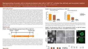 科学海报Reprogramming of Somatic Cells to Induced Pluripotent Stem Cells in TeSR™-E7™, a Feeder-Free, Defined, and Low-Protein Medium