Wang YI et al. (JUL 2016)
Biotechnology and Bioengineering
Microfluidic blood-brain barrier model provides in vivo-like barrier properties for drug permeability screening
Efficient delivery of therapeutics across the neuroprotective blood-brain barrier (BBB) remains a formidable challenge for central nervous system drug development. High-fidelity in vitro models of the BBB could facilitate effective early screening of drug candidates targeting the brain. In this study,we developed a microfluidic BBB model that is capable of mimicking in vivo BBB characteristics for a prolonged period and allows for reliable in vitro drug permeability studies under recirculating perfusion. We derived brain microvascular endothelial cells (BMECs) from human induced pluripotent stem cells (hiPSCs) and cocultured them with rat primary astrocytes on the two sides of a porous membrane on a pumpless microfluidic platform for up to 10 days. The microfluidic system was designed based on the blood residence time in human brain tissues,allowing for medium recirculation at physiologically relevant perfusion rates with no pumps or external tubing,meanwhile minimizing wall shear stress to test whether shear stress is required for in vivo-like barrier properties in a microfluidic BBB model. This BBB-on-a-chip model achieved significant barrier integrity as evident by continuous tight junction formation and in vivo-like values of trans-endothelial electrical resistance (TEER). The TEER levels peaked above 4000 $$ textperiodcentered cm(2) on day 3 on chip and were sustained above 2000 $$ textperiodcentered cm(2) up to 10 days,which are the highest sustained TEER values reported in a microfluidic model. We evaluated the capacity of our microfluidic BBB model to be used for drug permeability studies using large molecules (FITC-dextrans) and model drugs (caffeine,cimetidine,and doxorubicin). Our analyses demonstrated that the permeability coefficients measured using our model were comparable to in vivo values. Our BBB-on-a-chip model closely mimics physiological BBB barrier functions and will be a valuable tool for screening of drug candidates. The residence time-based design of a microfluidic platform will enable integration with other organ modules to simulate multi-organ interactions on drug response. Biotechnol. Bioeng. 2016;9999: 1-11. textcopyright 2016 Wiley Periodicals,Inc.
View Publication
产品类型:
产品号#:
85850
85857
产品名:
mTeSR™1
mTeSR™1
文献
Wang E et al. (AUG 1991)
The Journal of biological chemistry 266 22 14486--90
Inhibition of sphingolipid biosynthesis by fumonisins. Implications for diseases associated with Fusarium moniliforme.
Culture materials and grains contaminated with certain isolates of Fusarium moniliforme cause equine leucoencephalomalacia,porcine pulmonary edema syndrome,and liver cancer in rats. The causative agents are thought to be a family of compounds called fumonisins,which bear considerable structural similarity to the long-chain (sphingoid) base backbones of sphingolipids. Incubation of rat hepatocytes with fumonisins inhibited incorporation of [14C]serine into the sphingosine moiety of cellular sphingolipids with an IC50 of 0.1 microM for fumonisin B1. In contrast,fumonisin B1 increased the amount of the biosynthetic intermediate sphinganine,which suggests that fumonisins inhibit the conversion of [14C]sphinganine to N-acyl-[14C]sphinganines,a step that is thought to precede introduction of the 4,5-trans double bond of sphingosine (Merrill,A.H.,Jr. and Wang,E. (1986) J. Biol. Chem. 261,3764-3769). In agreement with this mechanism,fumonisin B1 inhibited the activity of sphingosine N-acyltransferase (ceramide synthase) in rat liver microsomes with 50% inhibition at approximately 0.1 microM and reduced the conversion of [3H]sphingosine to [3H]ceramide by intact hepatocytes. As far as we are aware,this is the first discovery of a naturally occurring inhibitor of this step of sphingolipid metabolism. These findings suggest that disruption of the de novo pathway of sphingolipid biosynthesis may be a critical event in the diseases that have been associated with consumption of fumonisins.
View Publication
产品类型:
产品号#:
73682
73684
产品名:
Fumonisin B1
Fumonisin B1
文献
Bouscary D et al. (MAY 2003)
Blood 101 9 3436--43
Critical role for PI 3-kinase in the control of erythropoietin-induced erythroid progenitor proliferation.
The production of red blood cells is tightly regulated by erythropoietin (Epo). The phosphoinositide 3-kinase (PI 3-kinase) pathway was previously shown to be activated in response to Epo. We studied the role of this pathway in the control of Epo-induced survival and proliferation of primary human erythroid progenitors. We show that phosphoinositide 3 (PI 3)-kinase associates with 4 tyrosine-phosphorylated proteins in primary human erythroid progenitors,namely insulin receptor substrate-2 (IRS2),Src homology 2 domain-containing inositol 5'-phosphatase (SHIP),Grb2-associated binder-1 (Gab1),and the Epo receptor (EpoR). Using different in vitro systems,we demonstrate that 3 alternative pathways independently lead to Epo-induced activation of PI 3-kinase and phosphorylation of its downstream effectors,Akt,FKHRL1,and P70S6 kinase: through direct association of PI 3-kinase with the last tyrosine residue (Tyr479) of the Epo receptor (EpoR),through recruitment and phosphorylation of Gab proteins via either Tyr343 or Tyr401 of the EpoR,or through phosphorylation of IRS2 adaptor protein. The mitogen-activated protein (MAP) kinase pathway was also activated by Epo in erythroid progenitors,but we found that this process is independent of PI 3-kinase activation. In erythroid progenitors,the functional role of PI 3-kinase was both to prevent apoptosis and to stimulate cell proliferation in response to Epo stimulation. Finally,our results show that PI 3-kinase-mediated proliferation of erythroid progenitors in response to Epo occurs mainly through modulation of the E3 ligase SCF(SKP2),which,in turn,down-regulates p27(Kip1) cyclin-dependent kinase (CDK) inhibitor via proteasome degradation.
View Publication
Girardot T et al. (OCT 2016)
Journal of immunological methods
An optimized protocol for adenosine triphosphate quantification in T lymphocytes of lymphopenic patients.
In several clinical contexts,the measurement of ATP concentration in T lymphocytes has been proposed as a biomarker of immune status,predictive of secondary infections. However,the use of such biomarker in lymphopenic patients requires some adaptations in the ATP dosage protocol. We used blood from healthy volunteers to determine the optimal experimental settings. We investigated technical aspects such as the type of anticoagulant for blood sampling,the effect of freeze and thaw cycles,the reagent and sample mixing sequence,and the optimal dilution buffer. We also shortened the incubation time to 8h,and even showed that a 30min incubation may be sufficient. To evaluate the ATP rise upon lymphocyte activation,the optimal dose of stimulant was defined to be 4μg/mL of phytohaemagglutinin. Lastly,we determined that the number of T cells needed for this measurement was as low as 50,000,which is compatible with the existing lymphopenia in clinical settings. This optimized protocol appears ready to be assessed in lymphopenic patients to further investigate the interconnection between T lymphocyte metabolism and impaired phenotype and functions.
View Publication
产品类型:
产品号#:
17851
17851RF
15021
15061
85415
85420
85450
85460
86415
86420
86450
86460
产品名:
EasySep™人CD3正选试剂盒II
RoboSep™ 人CD3正选试剂盒II
RosetteSep™人T细胞富集抗体混合物
RosetteSep™人T细胞富集抗体混合物
SepMate™-15 (IVD)
SepMate™-15 (IVD)
SepMate™-50 (IVD)
SepMate™-50 (IVD)
SepMate™-15 (RUO)
SepMate™-15 (RUO)
SepMate™-50 (RUO)
SepMate™-50 (RUO)
文献
E. E. Ford et al. (may 2023)
Journal of immunology (Baltimore,Md. : 1950) 210 10 1607--1619
FLAIRR-Seq: A Method for Single-Molecule Resolution of Near Full-Length Antibody H Chain Repertoires.
Current Adaptive Immune Receptor Repertoire sequencing (AIRR-seq) using short-read sequencing strategies resolve expressed Ab transcripts with limited resolution of the C region. In this article,we present the near-full-length AIRR-seq (FLAIRR-seq) method that uses targeted amplification by 5' RACE,combined with single-molecule,real-time sequencing to generate highly accurate (99.99%) human Ab H chain transcripts. FLAIRR-seq was benchmarked by comparing H chain V (IGHV),D (IGHD),and J (IGHJ) gene usage,complementarity-determining region 3 length,and somatic hypermutation to matched datasets generated with standard 5' RACE AIRR-seq using short-read sequencing and full-length isoform sequencing. Together,these data demonstrate robust FLAIRR-seq performance using RNA samples derived from PBMCs,purified B cells,and whole blood,which recapitulated results generated by commonly used methods,while additionally resolving H chain gene features not documented in IMGT at the time of submission. FLAIRR-seq data provide,for the first time,to our knowledge,simultaneous single-molecule characterization of IGHV,IGHD,IGHJ,and IGHC region genes and alleles,allele-resolved subisotype definition,and high-resolution identification of class switch recombination within a clonal lineage. In conjunction with genomic sequencing and genotyping of IGHC genes,FLAIRR-seq of the IgM and IgG repertoires from 10 individuals resulted in the identification of 32 unique IGHC alleles,28 (87%) of which were previously uncharacterized. Together,these data demonstrate the capabilities of FLAIRR-seq to characterize IGHV,IGHD,IGHJ,and IGHC gene diversity for the most comprehensive view of bulk-expressed Ab repertoires to date.
View Publication
产品类型:
产品号#:
19554
18000
产品名:
EasySep™人Pan-B细胞富集试剂盒
EasySep™磁极
文献
Nolte SM et al. (APR 2013)
Journal of the National Cancer Institute 105 8 551--562
A cancer stem cell model for studying brain metastases from primary lung cancer.
BACKGROUND Brain metastases are most common in adults with lung cancer,predicting uniformly poor patient outcome,with a median survival of only months. Despite their frequency and severity,very little is known about tumorigenesis in brain metastases. METHODS We applied previously developed primary solid tumor-initiating cell models to the study of brain metastases from the lung to evaluate the presence of a cancer stem cell population. Patient-derived brain metastases (n = 20) and the NCI-H1915 cell line were cultured as stem-enriching tumorspheres. We used in vitro limiting-dilution and sphere-forming assays,as well as intracranial human-mouse xenograft models. To determine genes overexpressed in brain metastasis tumorspheres,we performed comparative transcriptome analysis. All statistical analyses were two-sided. RESULTS Patient-derived brain metastasis tumorspheres had a mean sphere-forming capacity of 33 spheres/2000 cells (SD = 33.40) and median stem-cell frequency of 1/60 (range = 0-1/141),comparable to that of primary brain tumorspheres (P = .53 and P = .20,respectively). Brain metastases also expressed CD15 and CD133,markers suggestive of a stemlike population. Through intracranial xenotransplantation,brain metastasis tumorspheres were found to recapitulate the original patient tumor heterogeneity. We also identified several genes overexpressed in brain metastasis tumorspheres as statistically significant predictors of poor survival in primary lung cancer. CONCLUSIONS For the first time,we demonstrate the presence of a stemlike population in brain metastases from the lung. We also show that NCI-H1915 tumorspheres could be useful in studying self-renewal and tumor initiation in brain metastases. Our candidate genes may be essential to metastatic stem cell populations,where pathway interference may be able to transform a uniformly fatal disease into a more localized and treatable one.
View Publication
产品类型:
产品号#:
01700
01705
产品名:
ALDEFLUOR™工具
ALDEFLUOR™DEAB试剂
文献
Prowse A et al. (JUL 2009)
BioTechniques 47 1 599--606
A rapid, cost-effective method for counting human embryonic stem cell numbers as clumps.
Enumeration of human embryonic stem cell (hESC) numbers through single cell digestion can be time consuming especially in high-throughput or multi-factorial analysis containing 50+ samples. We have developed a reproducible,cost-effective method of counting hESCs in clumps circumventing the need to manually dissociate each sample to single cells. The method is based on the DNA binding capacity of propidium iodide (PI) and subsequent fluorescent signal detection. Standard curves generated for cell numbers versus PI fluorescence as single cells or clumps showed an almost identical relationship in the lines of best fit. The reproducibility of the assay was first demonstrated by seeding hESC clumps at specific cell densities ranging 0.05[x02013]2x10⁵ cells/well and then secondly by using the assay to count cell numbers after different growth conditions. Validation tests showed that consistent seeding densities are important in maintaining undifferentiated hESC culture and that the assay can be used to estimate relative cell numbers and growth curves with high accuracy.
View Publication
产品类型:
产品号#:
05850
05857
05870
05875
85850
85857
85870
85875
产品名:
mTeSR™1
mTeSR™1
文献
Berndt A et al. ( 2010)
Nature chemical biology 6 2 117--124
The p110 delta structure: mechanisms for selectivity and potency of new PI(3)K inhibitors.
Deregulation of the phosphoinositide-3-OH kinase (PI(3)K) pathway has been implicated in numerous pathologies including cancer,diabetes,thrombosis,rheumatoid arthritis and asthma. Recently,small-molecule and ATP-competitive PI(3)K inhibitors with a wide range of selectivities have entered clinical development. In order to understand the mechanisms underlying the isoform selectivity of these inhibitors,we developed a new expression strategy that enabled us to determine to our knowledge the first crystal structure of the catalytic subunit of the class IA PI(3)K p110 delta. Structures of this enzyme in complex with a broad panel of isoform- and pan-selective class I PI(3)K inhibitors reveal that selectivity toward p110 delta can be achieved by exploiting its conformational flexibility and the sequence diversity of active site residues that do not contact ATP. We have used these observations to rationalize and synthesize highly selective inhibitors for p110 delta with greatly improved potencies.
View Publication
产品类型:
产品号#:
73152
产品名:
GDC- 0941
文献
Volpe DA and Warren MK (JUN 2003)
Toxicology in vitro : an international journal published in association with BIBRA 17 3 271--7
Myeloid clonogenic assays for comparison of the in vitro toxicity of alkylating agents.
A battery of clonal assays for myeloid progenitor cells (HPP-CFC,CFU-gemm,CFU-gm,CFU-g) was utilized to evaluate the myelotoxicity of a series of alkylating agents representing the spectrum of clinical times to nadir. Bone marrow aspirates from normal volunteers were incubated with mechlorethamine,busulfan,melphalan,carmustine or lomustine for 1 h and then cultured in methylcellulose with 30% serum and cytokines. There was a concentration-dependent inhibition of colony formation and often a differential toxicity to the myeloid progenitors with the alkylators tested. On a molar basis,mechlorethamine and melphalan were the most toxic of the alkylator drugs to the myeloid precursors. The most sensitive progenitor was CFU-gemm with the lowest inhibitory concentration IC(70) concentrations for mechlorethamine,melphalan,carmustine and lomustine. Generally,there was great similarity for drug effects between CFU-g and CFU-gm with overlapping inhibition curves. HPP-CFC proved to be the least sensitive of the progenitors to the toxic actions of the drugs. While there was no correlation between the time to clinical neutropenic nadir and the most sensitive progenitor in the clonal assays,the CFU-gm assay remains a suitable method for determining the myelotoxic potential of cytotoxic agents.
View Publication
Rahman M et al. (MAR 2015)
Anatomy & cell biology 48 1 25--35
Neurosphere and adherent culture conditions are equivalent for malignant glioma stem cell lines.
Certain limitations of the neurosphere assay (NSA) have resulted in a search for alternative culture techniques for brain tumor-initiating cells (TICs). Recently,reports have described growing glioblastoma (GBM) TICs as a monolayer using laminin. We performed a side-by-side analysis of the NSA and laminin (adherent) culture conditions to compare the growth and expansion of GBM TICs. GBM cells were grown using the NSA and adherent culture conditions. Comparisons were made using growth in culture,apoptosis assays,protein expression,limiting dilution clonal frequency assay,genetic affymetrix analysis,and tumorigenicity in vivo. In vitro expansion curves for the NSA and adherent culture conditions were virtually identical (P=0.24) and the clonogenic frequencies (5.2% for NSA vs. 5.0% for laminin,P=0.9) were similar as well. Likewise,markers of differentiation (glial fibrillary acidic protein and beta tubulin III) and proliferation (Ki67 and MCM2) revealed no statistical difference between the sphere and attachment methods. Several different methods were used to determine the numbers of dead or dying cells (trypan blue,DiIC,caspase-3,and annexin V) with none of the assays noting a meaningful variance between the two methods. In addition,genetic expression analysis with microarrays revealed no significant differences between the two groups. Finally,glioma cells derived from both methods of expansion formed large invasive tumors exhibiting GBM features when implanted in immune-compromised animals. A detailed functional,protein and genetic characterization of human GBM cells cultured in serum-free defined conditions demonstrated no statistically meaningful differences when grown using sphere (NSA) or adherent conditions. Hence,both methods are functionally equivalent and remain suitable options for expanding primary high-grade gliomas in tissue culture.
View Publication

 EasySep™小鼠TIL(CD45)正选试剂盒


 文献
文献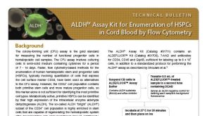 技术公告ALDHbr Assay Kit for Enumeration of HSPCs in Cord Blood by Flow Cytometry