Chromosome 13 abnormalities identified by FISH analysis and serum beta2-microglobulin produce a powerful myeloma staging system for patients receiving high-dose therapy.
A careful prognostic evaluation of patients referred for high-dose therapy (HDT) is warranted to identify those who maximally benefit from HDT as well as those who clearly fail current HDT and are candidates for more innovative treatments. In a series of 110 patients with myeloma who received HDT as first-line therapy,times to event (disease progression and death) were studied through proportional hazard models,in relation to different prognostic factors,including a chromosome 13 fluorescence in situ hybridization (FISH) analysis using a D13S319 probe. Delta13 was detected in 42 patients (38%). Follow-up time among surviving patients and survival time were 48 +/- 3 and 51 +/- 7 months,respectively (median +/- SE). In the univariate analysis,Delta13 was the most powerful adverse prognostic factor for all times to event,especially for the survival time (P textless.0001) and was followed by beta2-microglobulin (beta2m) levels 2.5 mg/L or higher (P =.0001). The comparison of survival prognostic models including beta2m 2.5 mg/L or greater and another factor favored the Delta13/beta2m combination. In 22 patients (20%) with no unfavorable factor,the median survival time was not reached at 111 months. In contrast,among 55 patients (50%) with one unfavorable factor and 33 patients (30%) with 2 unfavorable factors,median survival times were 47.3 +/- 4.6 months and 25.3 +/- 3.2 months,respectively (P textless.0001). We conclude that delta13,adequately detected by FISH analysis,is a very strong factor related to poor survival,especially when associated with a beta2m level of 2.5 mg/L or higher. Routine FISH Delta13 assessment is strongly recommended for patients considered for HDT.
View Publication
产品类型:
产品号#:
产品名:
文献
Gerrits A et al. (APR 2010)
Blood 115 13 2610--8
Cellular barcoding tool for clonal analysis in the hematopoietic system.
Clonal analysis is important for many areas of hematopoietic stem cell research,including in vitro cell expansion,gene therapy,and cancer progression and treatment. A common approach to measure clonality of retrovirally transduced cells is to perform integration site analysis using Southern blotting or polymerase chain reaction-based methods. Although these methods are useful in principle,they generally provide a low-resolution,biased,and incomplete assessment of clonality. To overcome those limitations,we labeled retroviral vectors with random sequence tags or barcodes." On integration�
View Publication
Sumitomo A et al. (OCT 2010)
Molecular and cellular biology 30 20 4818--27
The transcriptional mediator subunit MED1/TRAP220 in stromal cells is involved in hematopoietic stem/progenitor cell support through osteopontin expression.
MED1/TRAP220,a subunit of the transcriptional Mediator/TRAP complex,is crucial for various biological events through its interaction with distinct activators,such as nuclear receptors and GATA family activators. In hematopoiesis,MED1 plays a pivotal role in optimal nuclear receptor-mediated myelomonopoiesis and GATA-1-induced erythropoiesis. In this study,we present evidence that MED1 in stromal cells is involved in supporting hematopoietic stem and/or progenitor cells (HSPCs) through osteopontin (OPN) expression. We found that the proliferation of bone marrow (BM) cells cocultured with MED1 knockout (Med1(-/-)) mouse embryonic fibroblasts (MEFs) was significantly suppressed compared to the control. Furthermore,the number of long-term culture-initiating cells (LTC-ICs) was attenuated for BM cells cocultured with Med1(-/-) MEFs. The vitamin D receptor (VDR)- and Runx2-mediated expression of OPN,as well as Mediator recruitment to the Opn promoter,was specifically attenuated in the Med1(-/-) MEFs. Addition of OPN to these MEFs restored the growth of cocultured BM cells and the number of LTC-ICs,both of which were attenuated by the addition of the anti-OPN antibody to Med1(+/+) MEFs and to BM stromal cells. Consequently,MED1 in niche appears to play an important role in supporting HSPCs by upregulating VDR- and Runx2-mediated transcription on the Opn promoter.
View Publication
产品类型:
产品号#:
03334
03434
03444
09500
产品名:
MethoCult™M3334
MethoCult™GF M3434
MethoCult™GF M3434
BIT 9500血清替代物
文献
X. Li et al. (jul 2019)
Stem cells (Dayton,Ohio) 37 7 937--947
p53-TP53-Induced Glycolysis Regulator Mediated Glycolytic Suppression Attenuates DNA Damage and Genomic Instability in Fanconi Anemia Hematopoietic Stem Cells.
Emerging evidence has shown that resting quiescent hematopoietic stem cells (HSCs) prefer to utilize anaerobic glycolysis rather than mitochondrial respiration for energy production. Compelling evidence has also revealed that altered metabolic energetics in HSCs underlies the onset of certain blood diseases; however,the mechanisms responsible for energetic reprogramming remain elusive. We recently found that Fanconi anemia (FA) HSCs in their resting state are more dependent on mitochondrial respiration for energy metabolism than on glycolysis. In the present study,we investigated the role of deficient glycolysis in FA HSC maintenance. We observed significantly reduced glucose consumption,lactate production,and ATP production in HSCs but not in the less primitive multipotent progenitors or restricted hematopoietic progenitors of Fanca-/- and Fancc-/- mice compared with that of wild-type mice,which was associated with an overactivated p53 and TP53-induced glycolysis regulator,the TIGAR-mediated metabolic axis. We utilized Fanca-/- HSCs deficient for p53 to show that the p53-TIGAR axis suppressed glycolysis in FA HSCs,leading to enhanced pentose phosphate pathway and cellular antioxidant function and,consequently,reduced DNA damage and attenuated HSC exhaustion. Furthermore,by using Fanca-/- HSCs carrying the separation-of-function mutant p53R172P transgene that selectively impairs the p53 function in apoptosis but not cell-cycle control,we demonstrated that the cell-cycle function of p53 was not required for glycolytic suppression in FA HSCs. Finally,ectopic expression of the glycolytic rate-limiting enzyme PFKFB3 specifically antagonized p53-TIGAR-mediated metabolic reprogramming in FA HSCs. Together,our results suggest that p53-TIGAR metabolic axis-mediated glycolytic suppression may play a compensatory role in attenuating DNA damage and proliferative exhaustion in FA HSCs. Stem Cells 2019;37:937-947.
View Publication
Lianguzova MS et al. (APR 2007)
Cell biology international 31 4 330--7
Phosphoinositide 3-kinase inhibitor LY294002 but not serum withdrawal suppresses proliferation of murine embryonic stem cells.
Mouse embryonic stem (mES) cells have short duration of their cell cycle and are capable of proliferating in the absence of growth factors. To find out which signaling pathways contribute to the regulation of the mES cell cycle,we used pharmacological inhibitors of MAP and PI3 kinase cascades. The MAP kinase inhibitors as well as serum withdrawal did not affect mES cell cycle distribution,whereas the inhibitor of PI3K activity,LY294002,induced accumulation of cells in G(1) phase followed by apoptotic cell death. Serum withdrawal also causes apoptosis,but it does not change the content and activity of cell cycle regulators. In contrast,in mES cells treated with LY294002,the activities of Cdk2 and E2F were significantly decreased. Interestingly,LY294002had a much stronger effect on cell cycle distribution in low serum conditions,implying that serum can promote G(1)--textgreaterS transition of mES cells by a LY294002-resistant mechanism. Thus,proliferation of mES cells is maintained by at least two separate mechanisms: a LY294002-sensitive pathway,which is active even in the absence of serum,and LY294002-resistant,but serum-dependent,pathway.
View Publication
产品类型:
产品号#:
72152
72154
产品名:
LY294002
LY294002
文献
Qué et al. (JUN 2011)
Blood 117 22 5918--30
Smad4 binds Hoxa9 in the cytoplasm and protects primitive hematopoietic cells against nuclear activation by Hoxa9 and leukemia transformation.
We studied leukemic stem cells (LSCs) in a Smad4(-/-) mouse model of acute myelogenous leukemia (AML) induced either by the HOXA9 gene or by the fusion oncogene NUP98-HOXA9. Although Hoxa9-Smad4 complexes accumulate in the cytoplasm of normal hematopoietic stem cells and progenitor cells (HSPCs) transduced with these oncogenes,there is no cytoplasmic stabilization of HOXA9 in Smad4(-/-) HSPCs,and as a consequence increased levels of Hoxa9 is observed in the nucleus leading to increased immortalization in vitro. Loss of Smad4 accelerates the development of leukemia in vivo because of an increase in transformation of HSPCs. Therefore,the cytoplasmic binding of Hoxa9 by Smad4 is a mechanism to protect Hoxa9-induced transformation of normal HSPCs. Because Smad4 is a potent tumor suppressor involved in growth control,we developed a strategy to modify the subcellular distribution of Smad4. We successfully disrupted the interaction between Hoxa9 and Smad4 to activate the TGF-β pathway and apoptosis,leading to a loss of LSCs. Together,these findings reveal a major role for Smad4 in the negative regulation of leukemia initiation and maintenance induced by HOXA9/NUP98-HOXA9 and provide strong evidence that antagonizing Smad4 stabilization by these oncoproteins might be a promising novel therapeutic approach in leukemia.
View Publication
产品类型:
产品号#:
03434
03444
03236
产品名:
MethoCult™GF M3434
MethoCult™GF M3434
MethoCult™SF M3236
文献
Conneally E et al. (JAN 1996)
Blood 87 2 456--64
Rapid and efficient selection of human hematopoietic cells expressing murine heat-stable antigen as an indicator of retroviral-mediated gene transfer.
Recombinant retroviruses offer many advantages for the genetic modification of human hematopoietic cells,although their use in clinical protocols has thus far given disappointing results. There is therefore an important need to develop new strategies that will allow effectively transduced primitive hematopoietic target populations to be both rapidly characterized and isolated free of residual nontransduced but biologically equivalent cells. To address this need,we constructed a murine stem cell virus (MSCV)-based retroviral vector containing the 228-bp coding sequence of the murine heat-stable antigen (HSA) and generated helper virus-free amphotropic MSCV-HSA producer cells by transfection of GP-env AM12 packaging cells. Light density and,in some cases,lineage marker-negative (lin-) normal human marrow or mobilized peripheral blood cells preactivated by exposure to interleukin-3 (IL-3),IL-6,and Steel factor in vitro for 48 hours were then infected by cocultivation with these MSCV-HSA producer cells for a further 48 hours in the presence of the same cytokines. Fluorescence-activated cell sorting (FACS) analysis of the cells 24 hours later showed 21% to 41% (mean,27%) of those that were still CD34+ to have acquired the ability to express HSA. The extent of gene transfer to erythroid and granulopoietic progenitors (burst-forming unit-erythroid and colony-forming unit-granulocyte-macrophage),as assessed by the ability of these cells to form colonies of mature progeny in the presence of normally toxic concentrations of G418,averaged 11% and 12%,respectively,in 6 experiments. These values could be increased to 100% and 77%,respectively,by prior isolation of the CD34+HSA+ cell fraction and were correspondingly decreased to an average of 2% and 5%,respectively,in the CD34+HSA- cells. In addition,the extent of gene transfer to long-term culture-initiating cells (LTC-IC) was assessed by G418 resistance. The average gene transfer to LTC-IC-derived colony-forming cells in the unsorted population was textless or = 7% in 4 experiments. FACS selection of the initially CD34+HSA+ cells increased this value to 86% and decreased it to 3% for the LTC-IC plated from the CD34+HSA- cells. Transfer of HSA gene expression to a phenotypically defined more primitive subpopulation of CD34+ cells,ie,those expressing little or no CD38,could also be shown by FACS analysis of infected populations 24 hours after infection. These findings underscore the potential use of retroviral vectors encoding HSA for the specific identification and non-toxic selection immediately after infection of retrovirally transduced populations of primitive human hematopoietic cells. In addition,such vectors should facilitate the subsequent tracking of their marked progeny using multiparameter flow cytometry.
View Publication
Haenebalcke L et al. (FEB 2013)
Cell reports 3 2 335--41
The ROSA26-iPSC mouse: a conditional, inducible, and exchangeable resource for studying cellular (De)differentiation.
Control of cellular (de)differentiation in a temporal,cell-specific,and exchangeable manner is of paramount importance in the field of reprogramming. Here,we have generated and characterized a mouse strain that allows iPSC generation through the Cre/loxP conditional and doxycycline/rtTA-controlled inducible expression of the OSKM reprogramming factors entirely from within the ROSA26 locus. After reprogramming,these factors can be replaced by genes of interest-for example,to enhance lineage-directed differentiation-with the use of a trap-coupled RMCE reaction. We show that,similar to ESCs,Dox-controlled expression of the cardiac transcriptional regulator Mesp1 together with Wnt inhibition enhances the generation of functional cardiomyocytes upon in vitro differentiation of such RMCE-retargeted iPSCs. This ROSA26-iPSC mouse model is therefore an excellent tool for studying both cellular reprogramming and lineage-directed differentiation factors from the same locus and will greatly facilitate the identification and ease of functional characterization of the genetic/epigenetic determinants involved in these complex processes.
View Publication

 EasySep™小鼠TIL(CD45)正选试剂盒


 文献
文献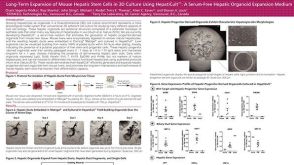 科学海报Long-Term Expansion of Mouse Hepatic Stem Cells in 3D Culture Using HepatiCult™: A Serum-Free Hepatic Organoid Expansion Medium
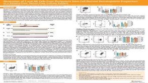 科学海报Generation of T and NK Cells From Pluripotent Stem Cell-Derived Hematopoietic Progenitors in a Stroma-Free, Serum-Free Culture System