Pierre-Louis O et al. (OCT 2009)
Stem cells (Dayton,Ohio) 27 10 2552--62
Dual SP/ALDH functionalities refine the human hematopoietic Lin-CD34+CD38- stem/progenitor cell compartment.
Identification of prevalent specific markers is crucial to stem/progenitor cell purification. Determinants such as the surface antigens CD34 and CD38 are traditionally used to analyze and purify hematopoietic stem/progenitor cells (HSCs/HPCs). However,the variable expression of these membrane antigens poses some limitations to their use in HSC/HPC purification. Techniques based on drug/stain efflux through the ATP-binding cassette (ABC)G2 pump (side population [SP] phenotype) or on detection of aldehyde dehydrogenase (ALDH) activity have been independently developed and distinguish the SP and ALDH(Bright) (ALDH(Br)) cell subsets for their phenotype and proliferative capability. In this study,we developed a multiparametric flow cytometric method associating both SP and ALDH activities on human lineage negative (Lin(-)) bone marrow cells and sorted different cell fractions according to their SP/ALDH activity level. We find that Lin(-)CD34(+)CD38(Low/-) cells are found throughout the spectrum of ALDH expression and are enriched especially in ALDH(Br) cells when associated with SP functionality (SP/ALDH(Br) fraction). Furthermore,the SP marker identified G(0) cells in all ALDH fractions,allowing us to sort quiescent cells regardless of ALDH activity. Moreover,we show that,within the Lin(-)CD34(+)CD38(-)ALDH(Br) population,the SP marker identifies cells with higher primitive characteristics,in terms of stemness-related gene expression and in vitro and in vivo proliferative potential,than the Lin(-)CD34(+) CD38(-)ALDH(Br) main population cells. In conclusion,our study shows that the coexpression of SP and ALDH markers refines the Lin(-)CD34(+)CD38(-) hematopoietic compartment and identifies an SP/ALDH(Br) cell subset enriched in quiescent primitive HSCs/HPCs.
View Publication
产品类型:
产品号#:
01700
01705
01701
01702
产品名:
ALDEFLUOR™工具
ALDEFLUOR™DEAB试剂
ALDEFLUOR™测定缓冲液
文献
Ryan MA et al. (OCT 2010)
Nature medicine 16 10 1141--6
Mobilization of hematopoietic stem and progenitor cells (HSPCs) from bone marrow into peripheral blood by the cytokine granulocyte colony-stimulating factor (G-CSF) has become the preferred source of HSPCs for stem cell transplants. However,G-CSF fails to mobilize sufficient numbers of stem cells in up to 10% of donors,precluding autologous transplantation in those donors or substantially delaying transplant recovery time. Consequently,new regimens are needed to increase the number of stem cells in peripheral blood upon mobilization. Using a forward genetic approach in mice,we mapped the gene encoding the epidermal growth factor receptor (Egfr) to a genetic region modifying G-CSF-mediated HSPC mobilization. Amounts of EGFR in HSPCs inversely correlated with the cells' ability to be mobilized by G-CSF,implying a negative role for EGFR signaling in mobilization. In combination with G-CSF treatment,genetic reduction of EGFR activity in HSPCs (in waved-2 mutant mice) or treatment with the EGFR inhibitor erlotinib increased mobilization. Increased mobilization due to suppression of EGFR activity correlated with reduced activity of cell division control protein-42 (Cdc42),and genetic Cdc42 deficiency in vivo also enhanced G-CSF-induced mobilization. Our findings reveal a previously unknown signaling pathway regulating stem cell mobilization and provide a new pharmacological approach for improving HSPC mobilization and thereby transplantation outcomes.
View Publication
产品类型:
产品号#:
03234
产品名:
MethoCult™M3234
文献
Morrow M et al. (MAY 2004)
Blood 103 10 3890--6
TEL-AML1 promotes development of specific hematopoietic lineages consistent with preleukemic activity.
The t(12;21)(p13;q22) translocation is the most common chromosomal abnormality yet identified in any pediatric leukemia and gives rise to the TEL-AML1 fusion product. To investigate the effects of TEL-AML1 on hematopoiesis,fetal liver hematopoietic progenitor cells (HPCs) were transduced with retroviral vectors expressing this fusion protein. We show that TEL-AML1 dramatically alters differentiation of HPCs in vitro,preferentially promoting B-lymphocyte development,enhancing self-renewal of B-cell precursors,and leading to the establishment of long-term growth factor-dependent pre-B-cell lines. However,it had no effect on myeloid development in vitro. Further experiments were performed to determine whether TEL-AML1 also demonstrates lineage-specific activity in vivo. TEL-AML1-expressing HPCs displayed a competitive advantage in reconstituting both B-cell and myeloid lineages in vivo but had no effect on reconstitution of the T-cell lineage. Despite promoting these alterations in hematopoiesis,TEL-AML1 did not induce leukemia in transplanted mice. Our study provides a unique insight into the role of TEL-AML1 in leukemia predisposition and a potential model to study the mechanism of leukemogenesis associated with this fusion.
View Publication
产品类型:
产品号#:
03534
03231
产品名:
MethoCult™GF M3534
MethoCult™M3231
文献
Liu B et al. (MAR 2014)
PLoS ONE 9 3 e90615
Nanog1 in NTERA-2 and recombinant NanogP8 from somatic cancer cells adopt multiple protein conformations and migrate at multiple M.W species
Human Nanog1 is a 305-amino acid (aa) homeodomain-containing transcription factor critical for the pluripotency of embryonic stem (ES) and embryonal carcinoma (EC) cells. Somatic cancer cells predominantly express a retrogene homolog of Nanog1 called NanogP8,which is ˜99% similar to Nanog at the aa level. Although the predicted M.W of Nanog1/NanogP8 is ∼35 kD,both have been reported to migrate,on Western blotting (WB),at apparent molecular masses of 29-80 kD. Whether all these reported protein bands represent authentic Nanog proteins is unclear. Furthermore,detailed biochemical studies on Nanog1/NanogpP8 have been lacking. By combining WB using 8 anti-Nanog1 antibodies,immunoprecipitation,mass spectrometry,and studies using recombinant proteins,here we provide direct evidence that the Nanog1 protein in NTERA-2 EC cells exists as multiple M.W species from ˜22 kD to 100 kD with a major 42 kD band detectable on WB. We then demonstrate that recombinant NanogP8 (rNanogP8) proteins made in bacteria using cDNAs from multiple cancer cells also migrate,on denaturing SDS-PAGE,at ˜28 kD to 180 kD. Interestingly,different anti-Nanog1 antibodies exhibit differential reactivity towards rNanogP8 proteins,which can spontaneously form high M.W protein species. Finally,we show that most long-term cultured cancer cell lines seem to express very low levels of or different endogenous NanogP8 protein that cannot be readily detected by immunoprecipitation. Altogether,the current study reveals unique biochemical properties of Nanog1 in EC cells and NanogP8 in somatic cancer cells.
View Publication
Fenouille N et al. (DEC 2010)
Cancer research 70 23 9659--70
Persistent activation of the Fyn/ERK kinase signaling axis mediates imatinib resistance in chronic myelogenous leukemia cells through upregulation of intracellular SPARC.
SPARC is an extracellular matrix protein that exerts pleiotropic effects on extracellular matrix organization,growth factor availability,cell adhesion,differentiation,and immunity in cancer. Chronic myelogenous leukemia (CML) cells resistant to the BCR-ABL inhibitor imatinib (IM-R cells) were found to overexpress SPARC mRNA. In this study,we show that imatinib triggers SPARC accumulation in a variety of tyrosine kinase inhibitor (TKI)-resistant CML cell lines. SPARC silencing in IM-R cells restored imatinib sensitivity,whereas enforced SPARC expression in imatinib-sensitive cells promoted viability as well as protection against imatinib-mediated apoptosis. Notably,we found that the protective effect of SPARC required intracellular retention inside cells. Accordingly,SPARC was not secreted into the culture medium of IM-R cells. Increased SPARC expression was intimately linked to persistent activation of the Fyn/ERK kinase signaling axis. Pharmacologic inhibition of this pathway or siRNA-mediated knockdown of Fyn kinase resensitized IM-R cells to imatinib. In support of our findings,increased levels of SPARC mRNA were documented in blood cells from CML patients after 1 year of imatinib therapy compared with initial diagnosis. Taken together,our results highlight an important role for the Fyn/ERK signaling pathway in imatinib-resistant cells that is driven by accumulation of intracellular SPARC.
View Publication
产品类型:
产品号#:
04100
产品名:
MethoCult™H4100
文献
Miller TW et al. (APR 2011)
Clinical cancer research : an official journal of the American Association for Cancer Research 17 7 2024--34
A gene expression signature from human breast cancer cells with acquired hormone independence identifies MYC as a mediator of antiestrogen resistance.
PURPOSE: Although most patients with estrogen receptor α (ER)-positive breast cancer initially respond to endocrine therapy,many ultimately develop resistance to antiestrogens. However,mechanisms of antiestrogen resistance and biomarkers predictive of such resistance are underdeveloped. EXPERIMENTAL DESIGN: We adapted four ER(+) human breast cancer cell lines to grow in an estrogen-depleted medium. A gene signature of estrogen independence was developed by comparing expression profiles of long-term estrogen-deprived (LTED) cells to their parental counterparts. We evaluated the ability of the LTED signature to predict tumor response to neoadjuvant therapy with an aromatase inhibitor and disease outcome following adjuvant tamoxifen. We utilized Gene Set Analysis (GSA) of LTED cell gene expression profiles and a loss-of-function approach to identify pathways causally associated with resistance to endocrine therapy. RESULTS: The LTED gene expression signature was predictive of high tumor cell proliferation following neoadjuvant therapy with anastrozole and letrozole,each in different patient cohorts. This signature was also predictive of poor recurrence-free survival in two studies of patients treated with adjuvant tamoxifen. Bioinformatic interrogation of expression profiles in LTED cells revealed a signature of MYC activation. The MYC activation signature and high MYC protein levels were both predictive of poor outcome following tamoxifen therapy. Finally,knockdown of MYC inhibited LTED cell growth. CONCLUSIONS: A gene expression signature derived from ER(+) breast cancer cells with acquired hormone independence predicted tumor response to aromatase inhibitors and associated with clinical markers of resistance to tamoxifen. Activation of the MYC pathway was associated with this resistance.
View Publication
产品类型:
产品号#:
05620
产品名:
MammoCult™ 人源培养基套装
文献
Wang P et al. ( 2017)
Molecular autism 8 11
CRISPR/Cas9-mediated heterozygous knockout of the autism gene CHD8 and characterization of its transcriptional networks in cerebral organoids derived from iPS cells.
BACKGROUND CHD8 (chromodomain helicase DNA-binding protein 8),which codes for a member of the CHD family of ATP-dependent chromatin-remodeling factors,is one of the most commonly mutated genes in autism spectrum disorders (ASD) identified in exome-sequencing studies. Loss of function mutations in the gene have also been found in schizophrenia (SZ) and intellectual disabilities and influence cancer cell proliferation. We previously reported an RNA-seq analysis carried out on neural progenitor cells (NPCs) and monolayer neurons derived from induced pluripotent stem (iPS) cells that were heterozygous for CHD8 knockout (KO) alleles generated using CRISPR-Cas9 gene editing. A significant number of ASD and SZ candidate genes were among those that were differentially expressed in a comparison of heterozygous KO lines (CHD8(+/-)) vs isogenic controls (CHD8(+/-)),including the SZ and bipolar disorder (BD) candidate gene TCF4,which was markedly upregulated in CHD8(+/-) neuronal cells. METHODS In the current study,RNA-seq was carried out on CHD8(+/-) and isogenic control (CHD8(+/+)) cerebral organoids,which are 3-dimensional structures derived from iPS cells that model the developing human telencephalon. RESULTS TCF4 expression was,again,significantly upregulated. Pathway analysis carried out on differentially expressed genes (DEGs) revealed an enrichment of genes involved in neurogenesis,neuronal differentiation,forebrain development,Wnt/β-catenin signaling,and axonal guidance,similar to our previous study on NPCs and monolayer neurons. There was also significant overlap in our CHD8(+/-) DEGs with those found in a transcriptome analysis carried out by another group using cerebral organoids derived from a family with idiopathic ASD. Remarkably,the top DEG in our respective studies was the non-coding RNA DLX6-AS1,which was markedly upregulated in both studies; DLX6-AS1 regulates the expression of members of the DLX (distal-less homeobox) gene family. DLX1 was also upregulated in both studies. DLX genes code for transcription factors that play a key role in GABAergic interneuron differentiation. Significant overlap was also found in a transcriptome study carried out by another group using iPS cell-derived neurons from patients with BD,a condition characterized by dysregulated WNT/β-catenin signaling in a subgroup of affected individuals. CONCLUSIONS Overall,the findings show that distinct ASD,SZ,and BD candidate genes converge on common molecular targets-an important consideration for developing novel therapeutics in genetically heterogeneous complex traits.
View Publication
Prostaglandin E2 enhances hematopoietic stem cell homing, survival, and proliferation.
Adult hematopoietic stem cells (HSCs) are routinely used to reconstitute hematopoiesis after myeloablation; however,transplantation efficacy and multilineage reconstitution can be limited by inadequate HSC number,or poor homing,engraftment,or self-renewal. Here we report that mouse and human HSCs express prostaglandin E2 (PGE2) receptors,and that short-term ex vivo exposure of HSCs to PGE2 enhances their homing,survival,and proliferation,resulting in increased long-term repopulating cell (LTRC) and competitive repopulating unit (CRU) frequency. HSCs pulsed with PGE2 are more competitive,as determined by head-to-head comparison in a competitive transplantation model. Enhanced HSC frequency and competitive advantage is stable and maintained upon serial transplantation,with full multilineage reconstitution. PGE2 increases HSC CXCR4 mRNA and surface expression,enhances their migration to SDF-1 in vitro and homing to bone marrow in vivo,and stimulates HSC entry into and progression through cell cycle. In addition,PGE2 enhances HSC survival,associated with an increase in Survivin mRNA and protein expression and reduction in intracellular active caspase-3. Our results define novel mechanisms of action whereby PGE2 enhances HSC function and supports a strategy to use PGE2 to facilitate hematopoietic transplantation.
View Publication

 EasySep™小鼠TIL(CD45)正选试剂盒


 文献
文献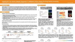 科学海报HepatiCult™ for Human Hepatic Organoids: A Validated Culture System for Drug Toxicity Screening
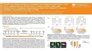 科学海报CRISPR-Cas9 Gene Editing Of CDK5RAP2 In Human Pluripotent Stem Cells, Derivation Of Genetically Stable Clonal Lines And Formation Of Cerebral Organoids
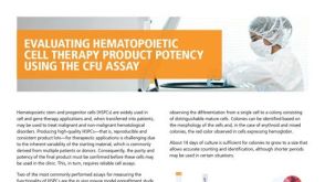 产品手册Evaluating Hematopoietic Cell Therapy Product Potency Using the CFU Assay
 产品手册RoboSep™: Solutions for HIV Research