Lee S-HH et al. (JUN 2000)
Nature biotechnology 18 6 675--9
Efficient generation of midbrain and hindbrain neurons from mouse embryonic stem cells.
Embryonic stem (ES) cells are clonal cell lines derived from the inner cell mass of the developing blastocyst that can proliferate extensively in vitro and are capable of adopting all the cell fates in a developing embryo. Clinical interest in the use of ES cells has been stimulated by studies showing that isolated human cells with ES properties from the inner cell mass or developing germ cells can provide a source of somatic precursors. Previous studies have defined in vitro conditions for promoting the development of specific somatic fates,specifically,hematopoietic,mesodermal,and neurectodermal. In this study,we present a method for obtaining dopaminergic (DA) and serotonergic neurons in high yield from mouse ES cells in vitro. Furthermore,we demonstrate that the ES cells can be obtained in unlimited numbers and that these neuron types are generated efficiently. We generated CNS progenitor populations from ES cells,expanded these cells and promoted their differentiation into dopaminergic and serotonergic neurons in the presence of mitogen and specific signaling molecules. The differentiation and maturation of neuronal cells was completed after mitogen withdrawal from the growth medium. This experimental system provides a powerful tool for analyzing the molecular mechanisms controlling the functions of these neurons in vitro and in vivo,and potentially for understanding and treating neurodegenerative and psychiatric diseases.
View Publication
Chesnokova V et al. (AUG 2013)
Proceedings of the National Academy of Sciences 110 35 E3331--E3339
Growth hormone is a cellular senescence target in pituitary and nonpituitary cells
Premature proliferative arrest in benign or early-stage tumors induced by oncoproteins,chromosomal instability,or DNA damage is associated with p53/p21 activation,culminating in either senescence or apoptosis,depending on cell context. Growth hormone (GH) elicits direct peripheral metabolic actions as well as growth effects mediated by insulin-like growth factor 1 (IGF1). Locally produced peripheral tissue GH,in contrast to circulating pituitary-derived endocrine GH,has been proposed to be both proapoptotic and prooncogenic. Pituitary adenomas expressing and secreting GH are invariably benign and exhibit DNA damage and a senescent phenotype. We therefore tested effects of nutlin-induced p53-mediated senescence in rat and human pituitary cells. We show that DNA damage senescence induced by nutlin triggers the p53/p21 senescent pathway,with subsequent marked induction of intracellular pituitary GH in vitro. In contrast,GH is not induced in cells devoid of p53. Furthermore we show that p53 binds specific GH promoter motifs and enhances GH transcription and secretion in senescent pituitary adenoma cells and also in nonpituitary (human breast and colon) cells. In vivo,treatment with nutlin results in up-regulation of both p53 and GH in the pituitary gland,as well as increased GH expression in nonpituitary tissues (lung and liver). Intracrine GH acts in pituitary cells as an apoptosis switch for p53-mediated senescence,likely protecting the pituitary adenoma from progression to malignancy. Unlike in the pituitary,in nonpituitary cells GH exerts antiapoptotic properties. Thus,the results show that GH is a direct p53 transcriptional target and fulfills criteria as a p53 target gene. Induced GH is a readily measurable cell marker for p53-mediated cellular senescence.
View Publication
产品类型:
产品号#:
产品名:
文献
Daynac M et al. (DEC 2014)
STEM CELLS 32 12 3257--3265
TGFβ Lengthens the G1 Phase of Stem Cells in Aged Mouse Brain
Neurogenesis decreases during aging causing a progressive cognitive decline but it is still controversial whether proliferation defects in neurogenic niches result from a loss of neural stem cells or from an impairment of their progression through the cell cycle. Using an accurate fluorescence-activated cell sorting technique,we show that the pool of neural stem cells is maintained in the subventricular zone of middle-aged mice while they have a reduced proliferative potential eventually leading to the subsequent decrease of their progeny. In addition,we demonstrate that the G1 phase is lengthened during aging specifically in activated stem cells,but not in transit-amplifying cells,and directly impacts on neurogenesis. Finally,we report that inhibition of TGFβ signaling restores cell cycle progression defects in stem cells. Our data highlight the significance of cell cycle dysregulation in stem cells in the aged brain and provide an attractive foundation for the development of anti-TGFβ regenerative therapies based on stimulating endogenous neural stem cells.
View Publication
产品类型:
产品号#:
05700
05701
05702
产品名:
NeuroCult™ 基础培养基(小鼠&大鼠)
NeuroCult™ 扩增添加物 (小鼠&大鼠)
NeuroCult™ 扩增试剂盒 (小鼠&大鼠)
文献
A. H. Nile et al. (JUN 2018)
Nature chemical biology 14 6 582--590
A selective peptide inhibitor of Frizzled 7 receptors disrupts intestinal stem cells.
Regeneration of the adult intestinal epithelium is mediated by a pool of cycling stem cells,which are located at the base of the crypt,that express leucine-rich-repeat-containing G-protein-coupled receptor 5 (LGR5). The Frizzled (FZD) 7 receptor (FZD7) is enriched in LGR5+ intestinal stem cells and plays a critical role in their self-renewal. Yet,drug discovery approaches and structural bases for targeting specific FZD isoforms remain poorly defined. FZD proteins interact with Wnt signaling proteins via,in part,a lipid-binding groove on the extracellular cysteine-rich domain (CRD) of the FZD receptor. Here we report the identification of a potent peptide that selectively binds to the FZD7 CRD at a previously uncharacterized site and alters the conformation of the CRD and the architecture of its lipid-binding groove. Treatment with the FZD7-binding peptide impaired Wnt signaling in cultured cells and stem cell function in intestinal organoids. Together,our data illustrate that targeting the lipid-binding groove holds promise as an approach for achieving isoform-selective FZD receptor inhibition.
View Publication
Ovchinnikov DA et al. (JUL 2012)
World journal of stem cells 4 7 71--9
Generation of a human embryonic stem cell line stably expressing high levels of the fluorescent protein mCherry.
AIM: The generation and characterization of a human embryonic stem cell (hESC) line stably expressing red fluorescent mCherry protein.backslashnbackslashnMETHODS: Lentiviral transduction of a ubiquitously-expressed human EF-1α promoter driven mCherry transgene was performed in MEL2 hESC. Red fluore-scence was assessed by immunofluorescence and flow cytometry. Pluripotency of stably transduced hESC was determined by immunofluorescent pluripotency marker expression,flow cytometry,teratoma assays and embryoid body-based differentiation followed by reverse transcriptase-polymerase chain reaction. Quantification of cell motility and survival was performed with time lapse microscopy.backslashnbackslashnRESULTS: Constitutively fluorescently-labeled hESCs are useful tools for facile in vitro and in vivo tracking of survival,motility and cell spreading on various surfaces before and after differentiation. Here we describe the generation and characterization of a hESC line (MEL2) stably expressing red fluorescent protein,mCherry. This line was generated by random integration of a fluorescent protein-expressing cassette,driven by the ubiquitously-expressed human EF-1α promoter. Stably transfected MEL2-mCherry hESC were shown to express pluripotency markers in the nucleus (POU5F1/OCT4,NANOG and SOX2) and on the cell surface (SSEA4,TRA1-60 and TG30/CD9) and were shown to maintain a normal karyotype in long-term (for at least 35 passages) culture. MEL2-mCherry hESC further readily differentiated into representative cell types of the three germ layers in embryoid body and teratoma based assays and,importantly,maintained robust mCherry expression throughout differentiation. The cell line was next adapted to single-cell passaging,rendering it compatible with numerous bioengineering applications such as measurement of cell motility and cell spreading on various protein modified surfaces,quantification of cell attachment to nanoparticles and rapid estimation of cell survival.backslashnbackslashnCONCLUSION: The MEL2-mCherry hESC line conforms to the criteria of bona fide pluripotent stem cells and maintains red fluorescence throughout differentiation,making it a useful tool for bioengineering and in vivo tracking experiments.
View Publication
产品类型:
产品号#:
85850
85857
产品名:
mTeSR™1
mTeSR™1
文献
Jitprasertwong P et al. (FEB 2014)
Cytokine 65 2 222--30
Leptin enhances the secretion of interleukin (IL)-18, but not IL-1β, from human monocytes via activation of caspase-1.
Circulating levels of leptin are elevated in type-2 diabetes mellitus (T2DM) and leptin plays a role in immune responses. Elevated circulating IL-18 levels are associated with clinical complications of T2DM. IL-18 regulates cytokine secretion and the function of a number of immune cells including T-cells,neutrophils and macrophages and as such has a key role in immunity and inflammation. Pro-inflammatory monocytes exhibiting elevated cytokine secretion are closely associated with inflammation in T2DM,however,little is known about the role of leptin in modifying monocyte IL-18 secretion. We therefore aimed to investigate the effect of leptin on IL-18 secretion by monocytes. We report herein that leptin increases IL-18 secretion in THP-1 and primary human monocytes but has no effect on IL-18mRNA. Leptin and LPS signalling in monocytes occurs by overlapping but distinct pathways. Thus,in contrast to a strong stimulation by LPS,leptin has no effect on IL-1βmRNA levels or IL-1β secretion. In addition,LPS stimulates the secretion of IL-6 but leptin did not whereas both treatments up regulate IL-8 secretion from the same cells. Although leptin (and LPS) has a synergistic effect with exogenous ATP on IL-18 secretion in both THP-1 and primary monocytes,experiments involving ATP assays and pharmacological inhibition of ATP signalling failed to provide any evidence that endogenous ATP secreted by leptin-stimulated monocytes was responsible for enhancement of monocyte IL-18 secretion by leptin. Analysis of the action of caspase-1 revealed that leptin up regulates caspase-1 activity and the effect of leptin on IL-18 release is prevented by caspase-1 inhibitor (Ac-YVAD-cmk). These data suggest that leptin activates IL-18 processing rather than IL-18 transcription. In conclusion,leptin enhances IL-18 secretion via modulation of the caspase-1 inflammasome function and acts synergistically with ATP in this regard. This process may contribute to aberrant immune responses in T2DM and other conditions of hyperleptinemia.
View Publication
产品类型:
产品号#:
产品名:
文献
Kitamura T et al. (AUG 1989)
Journal of cellular physiology 140 2 323--34
Establishment and characterization of a unique human cell line that proliferates dependently on GM-CSF, IL-3, or erythropoietin.
We have established a novel cell line,designated as TF-1,from a patient with erythroleukemia,which showed complete growth dependency on granulocyte-macrophage colony-stimulating factor (GM-CSF) or on interleukin-3 (IL-3) and carried a homogeneous chromosomal abnormality (54X). Erythropoietin (EPO) also sustained the short-term growth of TF-1,but did not induce erythroid differentiation. These three hematopoietic growth factors acted on TF-1 synergistically. Transforming growth factor-beta and interferons inhibited the factor-dependent growth of TF-1 cells in a dose-dependent fashion,and monocyte-colony stimulating factor and interkeukin-1 enhanced the GM-CSF-dependent growth of TF-1. Ultrastructural studies revealed some very immature features in this cell line. Although TF-1 cells do not express glycophorin A or carbonyl anhydrase I,the morphological and cytochemical features,and the constitutive expression of globin genes,indicate the commitment of TF-1 to erythroid lineage. When induced to differentiate,TF-1 entered two different pathways. Specifically,hemin and delta-aminolevulinic acid induced hemoglobin synthesis,whereas TPA induced dramatic differentiation of TF-1 into macrophage-like cells. In summary,TF-1 is a cell line of immature erythroid origin that requires GM-CSF,IL-3,or EPO for its growth and that has the ability to undergo differentiation into either more mature erythroid cells or into macrophage-like cells. TF-1 is a useful tool for analyzing the human receptors for IL-3,GM-CSF,and EPO or the signal transduction of these hemopoietic growth factors.
View Publication
产品类型:
产品号#:
产品名:
文献
Dye BR et al. (SEP 2016)
eLife 5
A bioengineered niche promotes in vivo engraftment and maturation of pluripotent stem cell derived human lung organoids.
Human pluripotent stem cell (hPSC) derived tissues often remain developmentally immature in vitro,and become more adult-like in their structure,cellular diversity and function following transplantation into immunocompromised mice. Previously we have demonstrated that hPSC-derived human lung organoids (HLOs) resembled human fetal lung tissue in vitro (Dye et al.,2015). Here we show that HLOs required a bioartificial microporous poly(lactide-co-glycolide) (PLG) scaffold niche for successful engraftment,long-term survival,and maturation of lung epithelium in vivo. Analysis of scaffold-grown transplanted tissue showed airway-like tissue with enhanced epithelial structure and organization compared to HLOs grown in vitro. By further comparing in vitro and in vivo grown HLOs with fetal and adult human lung tissue,we found that in vivo transplanted HLOs had improved cellular differentiation of secretory lineages that is reflective of differences between fetal and adult tissue,resulting in airway-like structures that were remarkably similar to the native adult human lung.
View Publication
产品类型:
产品号#:
85850
85857
产品名:
mTeSR™1
mTeSR™1
文献
Cao X et al. (MAR 2017)
Toxicological sciences : an official journal of the Society of Toxicology 156 1 14--24
Evaluating the Toxicity of Cigarette Whole Smoke Solutions in an Air-Liquid-Interface Human In Vitro Airway Tissue Model.
Exposure to cigarette smoke causes a multitude of pathological changes leading to tissue damage and disease. Quantifying such changes in highly differentiated in vitro human tissue models may assist in evaluating the toxicity of tobacco products. In this methods development study,well-differentiated human air-liquid-interface (ALI) in vitro airway tissue models were used to assess toxicological endpoints relevant to tobacco smoke exposure. Whole mainstream smoke solutions (WSSs) were prepared from 2 commercial cigarettes (R60 and S60) that differ in smoke constituents when machine-smoked under International Organization for Standardization conditions. The airway tissue models were exposed apically to WSSs 4-h per day for 1-5 days. Cytotoxicity,tissue barrier integrity,oxidative stress,mucin secretion,and matrix metalloproteinase (MMP) excretion were measured. The treatments were not cytotoxic and had marginal effects on tissue barrier properties; however,other endpoints responded in time- and dose-dependent manners,with the R60 resulting in higher levels of response than the S60 for many endpoints. Based on the lowest effect dose,differences in response to the WSSs were observed for mucin induction and MMP secretion. Mitigation of mucin induction by cotreatment of cultures with N-acetylcysteine suggests that oxidative stress contributes to mucus hypersecretion. Overall,these preliminary results suggest that quantifying disease-relevant endpoints using ALI airway models is a potential tool for tobacco product toxicity evaluation. Additional research using tobacco samples generated under smoking machine conditions that more closely approximate human smoking patterns will inform further methods development.
View Publication

 EasySep™小鼠TIL(CD45)正选试剂盒


 文献
文献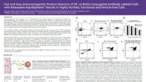 科学海报Positive Selection of PE- or Biotin-Conjugated Antibody Labeled Cells with Releasable Rapidspheres™
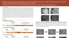 科学海报Defined, Feeder-Free Human Induced Pluripotent Stem Cell (hiPSC) Generation, Selection and Expansion from Multiple Somatic Cell Types

 沪公网安备31010102008431号