Koga C et al. (DEC 2014)
Annals of surgical oncology 21 Suppl 4 4 591--600
Reprogramming Using microRNA-302 Improves Drug Sensitivity in Hepatocellular Carcinoma Cells.
BACKGROUND Although studies have shown that Oct4,Sox2,Klf4,and c-Myc (OKSM)-mediated induced pluripotent stem cell (iPSC) technology sensitizes cancer cells to drugs,the potential risk of inserting c-Myc and random insertions of exogenous sequences into the genome persists. Several authors,including us,have presented microRNA (miRNA)-mediated reprogramming as an alternative approach. Herein,we evaluated the efficacy of miRNA-mediated reprogramming on hepatocellular carcinoma (HCC) cells. METHODS Among three miRNAs (miR-200c,miR-302s,and miR-369s) that were previously presented for miRNA-mediated reprogramming,miR-302 was expressed at low levels in HCC cells. After transfecting three times with miR-302,the cells were incubated in ES medium for 3 weeks and then characterized. RESULTS iPSC-like spheres were obtained after the 3-week incubation. Spheres presented high NANOG and OCT4 expression,low proliferation,high apoptosis,low epithelial-mesenchymal transition marker expression (N-cadherin,TGFBR2),and sensitization to drugs. Several miRNAs were changed (e.g.,low oncomiR miR-21,high miR-29b). cMyc was decreased,and methylation was elevated on histone 3 at lysine 4 (H3K4). Differentiated cells expressed markers of each germ layer (GFAP,FABP4,and ALB). AOF2 (also known as LSD1 or KDM1),one of the targets for miR-302,was repressed in iPSC-like-spheres. Silencing of AOF2 resulted in similar features of iPSC-like-spheres,including cMyc down-regulation and H3K4 methylation. In drug-resistant cells,sensitization was achieved through miR-302-mediated reprogramming. CONCLUSIONS miR-302-mediated iPSC technology reprogrammed HCC cells and improved drug sensitivity through AOF2 down-regulation,which caused H3K4 methylation and c-Myc repression.
View Publication
产品类型:
产品号#:
85850
85857
产品名:
mTeSR™1
mTeSR™1
文献
Flores-Figueroa E et al. (FEB 2005)
Leukemia research 29 2 215--24
Mesenchymal stem cells in myelodysplastic syndromes: phenotypic and cytogenetic characterization.
Bone marrow-derived mesenchymal stem cells (MSC) have been defined as primitive,undifferentiated cells,capable of self-renewal and with the ability to give rise to different cell lineages,including adipocytes,osteocytes,fibroblasts,chondrocytes,and myoblasts. MSC are key components of the hematopoietic microenvironment. Several studies,including some from our own group,suggest that important quantitative and functional alterations are present in the stroma of patients with myelodysplasia (MDS). However,in most of such studies the stroma has been analyzed as a complex network of different cell types and molecules,thus it has been difficult to identify and characterize the cell(s) type(s) that is (are) altered in MDS. In the present study,we have focused on the biological characterization of MSC from MDS. As a first approach,we have quantified their numbers in bone marrow,and have worked on their phenotypic (morphology and immunophenotype) and cytogenetic properties. MSC were obtained by a negative selection procedure and cultured in a MSC liquid culture medium. In terms of morphology,as well as the expression of certain cell markers,no differences were observed between MSC from MDS patients and those derived from normal marrow. In both cases,MSC expressed CD29,CD90,CD105 and Prolyl-4-hydroxylase; in contrast,they did not express CD14,CD34,CD68,or alkaline phosphatase. Interestingly,in five out of nine MDS patients,MSC developed in culture showed cytogenetic abnormalities,usually involving the loss of chromosomal material. All those five cases also showed cytogenetic abnormalities in their hematopoietic cells. Interestingly,in some cases there was a complete lack of overlap between the karyotypes of hematopoietic cells and MSC. To the best of our knowledge,the present study is the first in which a pure population of MSC from MDS patients is analyzed in terms of their whole karyotype and demonstrates that in a significant proportion of patients,MSC are cytogenetically abnormal. Although the reason of this is still unclear,such alterations may have an impact on the physiology of these cells. Further studies are needed to assess the functional integrity of MDS-derived MSC.
View Publication
产品类型:
产品号#:
产品名:
文献
Foresta C et al. (NOV 2006)
The Journal of clinical endocrinology and metabolism 91 11 4599--602
Reduced number of circulating endothelial progenitor cells in hypogonadal men.
CONTEXT: Endothelial dysfunction seems to be the first step of the atherosclerotic process. In the past few years,it has been demonstrated that injured endothelial monolayer is restored by a premature pool of circulating progenitor cells (PCs) and a more mature one of circulating endothelial PCs (EPCs). Even though there is increasing evidence that estrogens play a beneficial role on EPCs and,even if debated,on the cardiovascular system,less is known about androgens. OBJECTIVE: Our objective was to evaluate the levels of circulating PCs and EPCs in men with hypogonadotropic hypogonadism (HH) and the effect of prolonged testosterone (T) replacement therapy on these cells. DESIGN AND SETTING: We conducted a prospective study on males with HH at a university andrological center. PATIENTS: The study included 10 young HH patients (28.6 +/- 3.1 yr) and 25 age-matched controls. INTERVENTIONS: Idiopathic HH patients were treated with T gel therapy,50 mg/d for 6 months. MAIN OUTCOME MEASURES: We assessed circulating PC and EPC concentrations and immunocytochemistry for androgen receptor expression on cultured EPCs. RESULTS: At baseline,HH patients showed a significant reduction of both PCs and EPCs with respect to controls. T replacement therapy induced a significant increase of these cells with respect to baseline. Immunocytochemistry on cultured EPCs showed strong expression of the androgen receptor. CONCLUSIONS: Hypotestosteronemia is associated with a low number of circulating PCs and EPCs in young HH subjects. T treatment is able to induce an increase in these cells through a possible direct effect on the bone marrow.
View Publication
产品类型:
产品号#:
产品名:
文献
Tomihara K et al. (JUN 2010)
Journal of immunology (Baltimore,Md. : 1950) 184 11 6151--60
Antigen-specific immunity and cross-priming by epithelial ovarian carcinoma-induced CD11b(+)Gr-1(+) cells.
Both innate and adaptive immune systems are considered important for cancer prevention,immunosurveillance,and control of cancer progression. It is known that,although both systems initially eliminate emerging tumor cells efficiently,tumors eventually escape immune attack by a variety of mechanisms,including differentiation and recruitment of immunosuppressive CD11b(+)Gr-1(+) myeloid suppressor cells into the tumor microenvironment. However,we show that CD11b(+)Gr-1(+) cells found in ascites of epithelial ovarian cancer-bearing mice at advanced stages of disease are immunostimulatory rather than being immunosuppressive. These cells consist of a homogenous population of cells that morphologically resemble neutrophils. Moreover,like dendritic cells,immunostimulatory CD11b(+)Gr-1(+) cells can strongly cross-prime,augmenting the proliferation of functional CTLs via signaling through the expression of costimulatory molecule CD80. Adoptive transfer of these immunostimulatory CD11b(+)Gr-1(+) cells from ascites of ovarian cancer-bearing mice results in the significant regression of s.c. tumors even without being pulsed with exogenous tumor Ag prior to adoptive transfer. We now show for the first time that adaptive immune responses against cancer can be augmented by these cancer-induced granulocyte-like immunostimulatory myeloid (CD11b(+)Gr-1(+)) cells,thereby mediating highly effective antitumor immunity in an adoptive transfer model of immunity.
View Publication
产品类型:
产品号#:
21000
20119
20155
产品名:
RoboSep™- S
RoboSep™ 吸头组件抛光剂
RoboSep™分选试管套装(9个塑料管+吸头保护器)
文献
Zhang W et al. (JUL 2010)
PloS one 5 7 e11917
Retinoic acids potentiate BMP9-induced osteogenic differentiation of mesenchymal progenitor cells.
BACKGROUND As one of the least studied bone morphogenetic proteins (BMPs),BMP9 is one of the most osteogenic BMPs. Retinoic acid (RA) signaling is known to play an important role in development,differentiation and bone metabolism. In this study,we investigate the effect of RA signaling on BMP9-induced osteogenic differentiation of mesenchymal progenitor cells (MPCs). METHODOLOGY/PRINCIPAL FINDINGS Both primary MPCs and MPC line are used for BMP9 and RA stimulation. Recombinant adenoviruses are used to deliver BMP9,RARalpha and RXRalpha into MPCs. The in vitro osteogenic differentiation is monitored by determining the early and late osteogenic markers and matrix mineralization. Mouse perinatal limb explants and in vivo MPC implantation experiments are carried out to assess bone formation. We find that both 9CRA and ATRA effectively induce early osteogenic marker,such as alkaline phosphatase (ALP),and late osteogenic markers,such as osteopontin (OPN) and osteocalcin (OC). BMP9-induced osteogenic differentiation and mineralization is synergistically enhanced by 9CRA and ATRA in vitro. 9CRA and ATRA are shown to induce BMP9 expression and activate BMPR Smad-mediated transcription activity. Using mouse perinatal limb explants,we find that BMP9 and RAs act together to promote the expansion of hypertrophic chondrocyte zone at growth plate. Progenitor cell implantation studies reveal that co-expression of BMP9 and RXRalpha or RARalpha significantly increases trabecular bone and osteoid matrix formation. CONCLUSION/SIGNIFICANCE Our results strongly suggest that retinoid signaling may synergize with BMP9 activity in promoting osteogenic differentiation of MPCs. This knowledge should expand our understanding about how BMP9 cross-talks with other signaling pathways. Furthermore,a combination of BMP9 and retinoic acid (or its agonists) may be explored as effective bone regeneration therapeutics to treat large segmental bony defects,non-union fracture,and/or osteoporotic fracture.
View Publication

 EasySep™小鼠TIL(CD45)正选试剂盒


 人重组IL-6, ACF 白细胞介素6,不含动物成分
 重组人IL-8 (CXCL8) 白介素8
 重组人PDGF-AB, ACF 血小板衍生生长因子,不含动物成分
 重组人RANTES (CCL5) 激活后调节正常T细胞表达和分泌因子
 人TNF-αELISA试剂盒 用于人体TNF - α的检测和测定
 MesenCult™ 增殖试剂盒(人) 检测CFU-F和人间充质干细胞扩增的培养基
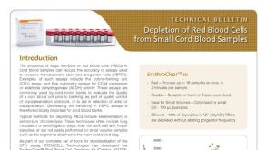 技术公告Depletion of Red Blood Cells from Small Cord Blood Samples