Lassailly F et al. (JUL 2010)
Blood 115 26 5347--54
Microenvironmental contaminations" induced by fluorescent lipophilic dyes used for noninvasive in vitro and in vivo cell tracking."
Determining how normal and leukemic stem cells behave in vivo,in a dynamic and noninvasive way,remains a major challenge. Most optical tracking technologies rely on the use of fluorescent or bioluminescent reporter genes,which need to be stably expressed in the cells of interest. Because gene transfer in primary leukemia samples represents a major risk to impair their capability to engraft in a xenogenic context,we evaluated the possibility to use gene transfer-free labeling technologies. The lipophilic dye 3,3,3',3' tetramethylindotricarbocyanine iodide (DiR) was selected among 4 near-infrared (NIR) staining technologies. Unfortunately we report here a massive transfer of the dye occurring toward the neighbor cells both in vivo and in vitro. We further demonstrate that all lipophilic dyes tested in this study (1,1'-dioctadecyl-3,3,3',3'-tetramethylindotricarbocyanine perchlorate [DiI],DiD,DiR,and PKH26) can give rise to microenvironmental contamination,including when used in suboptimal concentration,after extensive washing procedures and in the absence of phagocytosis or marked cell death. This was observed from all cell types tested. Eventually,we show that this microenvironmental contamination is mediated by both direct cell-cell contacts and diffusible microparticles. We conclude that tracking of labeled cells using non-genetically encoded markers should always be accompanied by drastic cross validation using multimodality approaches.
View Publication
Grimaldi JC et al. (JUN 1999)
Journal of Leukocyte Biology 65 6 846--53
Depletion of eosinophils in mice through the use of antibodies specific for C-C chemokine receptor 3 (CCR3).
We have generated rat monoclonal antibodies specific for the mouse eotaxin receptor,C-C chemokine receptor 3 (CCR3). Several anti-CCR3 mAbs proved to be useful for in vivo depletion of CCR3-expressing cells and immunofluorescent staining. In vivo CCR3 mAbs of the IgG2b isotype substantially depleted blood eosinophil levels in Nippostrongyus brasiliensis-infected mice. Repeated anti-CCR3 mAb treatment in these mice significantly reduced tissue eosinophilia in the lung tissue and bronchoalveolar lavage fluid. Flow cytometry revealed that mCCR3 was expressed on eosinophils but not on stem cells,dendritic cells,or cells from the thymus,lymph node,or spleen of normal mice. Unlike human Th2 cells,mouse Th2 cells did not express detectable levels of CCR3 nor did they give a measurable response to eotaxin. None of the mAbs were antagonists or agonists of CCR3 calcium mobilization. To our knowledge,the antibodies described here are the first mAbs reported to be specific for mouse eosinophils and to be readily applicable for the detection,isolation,and in vivo depletion of eosinophils.
View Publication
产品类型:
产品号#:
03800
03801
03802
03803
03804
03805
03806
产品名:
ClonaCell™-HY杂交瘤试剂盒
ClonaCell™-HY Medium
ClonaCell™-HY Medium
ClonaCell™-HY Medium
ClonaCell™-HY Medium
ClonaCell™-HY Medium
ClonaCell™衔接挂钩
文献
Bareiss PM et al. (SEP 2013)
Cancer research 73 17 5544--5555
SOX2 expression associates with stem cell state in human ovarian carcinoma.
The SRY-related HMG-box family of transcription factors member SOX2 regulates stemness and pluripotency in embryonic stem cells and plays important roles during early embryogenesis. More recently,SOX2 expression was documented in several tumor types including ovarian carcinoma,suggesting an involvement of SOX2 in regulation of cancer stem cells (CSC). Intriguingly,however,studies exploring the predictive value of SOX2 protein expression with respect to histopathologic and clinical parameters report contradictory results in individual tumors,indicating that SOX2 may play tumor-specific roles. In this report,we analyze the functional relevance of SOX2 expression in human ovarian carcinoma. We report that in human serous ovarian carcinoma (SOC) cells,SOX2 expression increases the expression of CSC markers,the potential to form tumor spheres,and the in vivo tumor-initiating capacity,while leaving cellular proliferation unaltered. Moreover,SOX2-expressing cells display enhanced apoptosis resistance in response to conventional chemotherapies and TRAIL. Hence,our data show that SOX2 associates with stem cell state in ovarian carcinoma and induction of SOX2 imposes CSC properties on SOC cells. We propose the existence of SOX2-expressing ovarian CSCs as a mechanism of tumor aggressiveness and therapy resistance in human SOC.
View Publication
产品类型:
产品号#:
01700
01705
产品名:
ALDEFLUOR™ 试剂盒
ALDEFLUOR™DEAB试剂
文献
Zhong X et al. (JUN 2014)
Nature communications 5 May 4047
Generation of three-dimensional retinal tissue with functional photoreceptors from human iPSCs.
Many forms of blindness result from the dysfunction or loss of retinal photoreceptors. Induced pluripotent stem cells (iPSCs) hold great potential for the modelling of these diseases or as potential therapeutic agents. However,to fulfill this promise,a remaining challenge is to induce human iPSC to recreate in vitro key structural and functional features of the native retina,in particular the presence of photoreceptors with outer-segment discs and light sensitivity. Here we report that hiPSC can,in a highly autonomous manner,recapitulate spatiotemporally each of the main steps of retinal development observed in vivo and form three-dimensional retinal cups that contain all major retinal cell types arranged in their proper layers. Moreover,the photoreceptors in our hiPSC-derived retinal tissue achieve advanced maturation,showing the beginning of outer-segment disc formation and photosensitivity. This success brings us one step closer to the anticipated use of hiPSC for disease modelling and open possibilities for future therapies.
View Publication
产品类型:
产品号#:
85850
85857
产品名:
mTeSR™1
mTeSR™1
文献
Wen Z et al. (NOV 2014)
Nature 515 7527 414--418
Synaptic dysregulation in a human iPS cell model of mental disorders
Dysregulated neurodevelopment with altered structural and functional connectivity is believed to underlie many neuropsychiatric disorders,and /`a disease of synapses/' is the major hypothesis for the biological basis of schizophrenia. Although this hypothesis has gained indirect support from human post-mortem brain analyses and genetic studies,little is known about the pathophysiology of synapses in patient neurons and how susceptibility genes for mental disorders could lead to synaptic deficits in humans. Genetics of most psychiatric disorders are extremely complex due to multiple susceptibility variants with low penetrance and variable phenotypes. Rare,multiply affected,large families in which a single genetic locus is probably responsible for conferring susceptibility have proven invaluable for the study of complex disorders. Here we generated induced pluripotent stem (iPS) cells from four members of a family in which a frameshift mutation of disrupted in schizophrenia 1 (DISC1) co-segregated with major psychiatric disorders and we further produced different isogenic iPS cell lines via gene editing. We showed that mutant DISC1 causes synaptic vesicle release deficits in iPS-cell-derived forebrain neurons. Mutant DISC1 depletes wild-type DISC1 protein and,furthermore,dysregulates expression of many genes related to synapses and psychiatric disorders in human forebrain neurons. Our study reveals that a psychiatric disorder relevant mutation causes synapse deficits and transcriptional dysregulation in human neurons and our findings provide new insight into the molecular and synaptic etiopathology of psychiatric disorders.
View Publication
产品类型:
产品号#:
85850
85857
产品名:
mTeSR™1
mTeSR™1
文献
Dye BR et al. (MAR 2015)
eLife 4 e05098
In vitro generation of human pluripotent stem cell derived lung organoids.
Recent breakthroughs in 3-dimensional (3D) organoid cultures for many organ systems have led to new physiologically complex in vitro models to study human development and disease. Here,we report the step-wise differentiation of human pluripotent stem cells (hPSCs) (embryonic and induced) into lung organoids. By manipulating developmental signaling pathways hPSCs generate ventral-anterior foregut spheroids,which are then expanded into human lung organoids (HLOs). HLOs consist of epithelial and mesenchymal compartments of the lung,organized with structural features similar to the native lung. HLOs possess upper airway-like epithelium with basal cells and immature ciliated cells surrounded by smooth muscle and myofibroblasts as well as an alveolar-like domain with appropriate cell types. Using RNA-sequencing,we show that HLOs are remarkably similar to human fetal lung based on global transcriptional profiles,suggesting that HLOs are an excellent model to study human lung development,maturation and disease.
View Publication
产品类型:
产品号#:
85850
85857
产品名:
mTeSR™1
mTeSR™1
文献
Nageshappa S et al. (FEB 2016)
Molecular psychiatry 21 2 178--188
Altered neuronal network and rescue in a human MECP2 duplication model.
Increased dosage of methyl-CpG-binding protein-2 (MeCP2) results in a dramatic neurodevelopmental phenotype with onset at birth. We generated induced pluripotent stem cells (iPSCs) from patients with the MECP2 duplication syndrome (MECP2dup),carrying different duplication sizes,to study the impact of increased MeCP2 dosage in human neurons. We show that cortical neurons derived from these different MECP2dup iPSC lines have increased synaptogenesis and dendritic complexity. In addition,using multi-electrodes arrays,we show that neuronal network synchronization was altered in MECP2dup-derived neurons. Given MeCP2 functions at the epigenetic level,we tested whether these alterations were reversible using a library of compounds with defined activity on epigenetic pathways. One histone deacetylase inhibitor,NCH-51,was validated as a potential clinical candidate. Interestingly,this compound has never been considered before as a therapeutic alternative for neurological disorders. Our model recapitulates early stages of the human MECP2 duplication syndrome and represents a promising cellular tool to facilitate therapeutic drug screening for severe neurodevelopmental disorders.
View Publication
产品类型:
产品号#:
85850
85857
产品名:
mTeSR™1
mTeSR™1
文献
Shetty DK et al. (MAR 2016)
Stem Cell Research 16 2 246--248
Generation of OCIAD1 inducible overexpression human embryonic stem cell line: BJNhem20-OCIAD1-Tet-On
Human embryonic stem cell line BJNhem20-OCIAD1-Tet-On was generated using non-viral method. The constructs pCAG-Tet-On and pTRE-Tight vector driving OCIAD1 expression were transfected using microporation procedure. pCAG-Tet-On cells can be used for inducible expression of any coding sequence cloned into pTRE-Tight vector. For example,in human embryonic stem cells,Tet-On system has been used to generate SOX2 overexpression cell line (Adachi et al.,2010).
View Publication
产品类型:
产品号#:
85850
85857
产品名:
mTeSR™1
mTeSR™1
文献
Aranha M et al. (JAN 2010)
BMC genomics 11 514
Apoptosis-associated microRNAs are modulated in mouse, rat and human neural differentiation.
BACKGROUND MicroRNAs (miRs or miRNAs) regulate several biological processes in the cell. However,evidence for miRNAs that control the differentiation program of specific neural cell types has been elusive. Recently,we have shown that apoptosis-associated factors,such as p53 and caspases participate in the differentiation process of mouse neural stem (NS) cells. To identify apoptosis-associated miRNAs that might play a role in neuronal development,we performed global miRNA expression profiling experiments in NS cells. Next,we characterized the expression of proapoptotic miRNAs,including miR-16,let-7a and miR-34a in distinct models of neural differentiation,including mouse embryonic stem cells,PC12 and NT2N cells. In addition,the expression of antiapoptotic miR-19a and 20a was also evaluated. RESULTS The expression of miR-16,let-7a and miR-34a was consistently upregulated in neural differentiation models. In contrast,expression of miR-19a and miR-20a was downregulated in mouse NS cell differentiation. Importantly,differential expression of specific apoptosis-related miRNAs was not associated with increased cell death. Overexpression of miR-34a increased the proportion of postmitotic neurons of mouse NS cells. CONCLUSIONS In conclusion,the identification of miR-16,let-7a and miR-34a,whose expression patterns are conserved in mouse,rat and human neural differentiation,implicates these specific miRNAs in mammalian neuronal development. The results provide new insights into the regulation of neuronal differentiation by apoptosis-associated miRNAs.
View Publication
产品类型:
产品号#:
72792
72794
产品名:
LY411575
LY411575
文献
Perez SA et al. (MAY 2003)
Blood 101 9 3444--50
A novel myeloid-like NK cell progenitor in human umbilical cord blood.
Natural killer (NK) cell differentiation from pluripotent CD34(+) human hematopoietic stem cells or oligopotent lymphoid progenitors has already been reported. In the present study,long-term cultures of the CD56(-)/CD34(-) myeloid-like adherent cell fraction (ACF) from umbilical cord blood (UCB),characterized by the expression of CD14(+) as well as other myeloid markers,were set up with flt3 ligand (FL) and interleukin-15 (IL-15). The UCB/ACF gradually expressed the CD56 marker,which reached fairly high levels (approximately 90% of the cells were CD56(+)) by day 15. FL plus IL-15-driven ACF/CD56(+) cells progressively expressed a mature NK functional program lysing both NK- and lymphokine-activate killer (LAK)-sensitive tumor targets and producing high levels of interferon-gamma (IFN-gamma),granulocyte-macrophage colony-stimulating factor,tumor necrosis factor alpha,and IL-10 upon stimulation with IL-12 and IL-18. Similar results were obtained when highly purified CD14(+) cells from UCB were cultured with FL and IL-15. In contrast,UCB/CD34(+) cells cultured under the same conditions showed a delayed expression of CD56 and behaved functionally differently in that they exhibited NK but not LAK cytotoxicity and produced significantly fewer cytokines. Kinetic studies on the phenotype of UCB/ACF or UCB/CD14(+) cells cultured in the presence of FL and IL-15 showed a rapid decrease in CD14 expression after day 5,which reached levels of zero by day 20. Approximately 60% of the CD56(+) derived from the UCB/ACF or the UCB/CD14(+) cells coexpressed CD14 by day 5. Taken together,our data support the role of CD14(+) myeloid-like cells within UCB as a novel progenitor for lymphoid NK cells.
View Publication

 EasySep™小鼠TIL(CD45)正选试剂盒


 文献
文献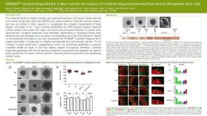 科学海报STEMdiff™ Cerebral Organoid Kit: A New Tool for the Culture of 3D Brain Organoids Derived from hPSCs
