mDYRK3 kinase is expressed selectively in late erythroid progenitor cells and attenuates colony-forming unit-erythroid development.
DYRKs are a new subfamily of dual-specificity kinases that was originally discovered on the basis of homology to Yak1,an inhibitor of cell cycle progression in yeast. At present,mDYRK-3 and mDYRK-2 have been cloned,and mDYRK-3 has been characterized with respect to kinase activity,expression among tissues and hematopoietic cells,and possible function during erythropoiesis. In sequence,mDYRK-3 diverges markedly in noncatalytic domains from mDYRK-2 and mDYRK-1a,but is 91.3% identical overall to hDYRK-3. Catalytically,mDYRK-3 readily phosphorylated myelin basic protein (but not histone 2B) and also appeared to autophosphorylate in vitro. Expression of mDYRK-1a,mDYRK-2,and mDYRK-3 was high in testes,but unlike mDYRK1a and mDYRK 2,mDYRK-3 was not expressed at appreciable levels in other tissues examined. Among hematopoietic cells,however,mDYRK-3 expression was selectively elevated in erythroid cell lines and primary pro-erythroid cells. In developmentally synchronized erythroid progenitor cells,expression peaked sharply following exposure to erythropoietin plus stem cell factor (SCF) (but not SCF alone),and in situ hybridizations of sectioned embryos revealed selective expression of mDYRK-3 in fetal liver. Interestingly,antisense oligonucleotides to mDYRK-3 were shown to significantly and specifically enhance colony-forming unit-erythroid colony formation. Thus,it is proposed that mDYRK-3 kinase functions as a lineage-restricted,stage-specific suppressor of red cell development. (Blood. 2001;97:901-910)
View Publication
产品类型:
产品号#:
04971
04902
04901
04963
04962
产品名:
MegaCult™-C细胞因子完整试剂盒
胶原蛋白溶液
MegaCult™-C细胞因子培养基
双室载玻片试剂盒
MegaCult™-C cfu染色试剂盒
文献
N. Allende-Vega et al. (jan 2022)
Scientific reports 12 1 1341
Metformin sensitizes leukemic cells to cytotoxic lymphocytes by increasing expression of intercellular adhesion molecule-1 (ICAM-1).
Solid tumor cells have an altered metabolism that can protect them from cytotoxic lymphocytes. The anti-diabetic drug metformin modifies tumor cell metabolism and several clinical trials are testing its effectiveness for the treatment of solid cancers. The use of metformin in hematologic cancers has received much less attention,although allogeneic cytotoxic lymphocytes are very effective against these tumors. We show here that metformin induces expression of Natural Killer G2-D (NKG2D) ligands (NKG2DL) and intercellular adhesion molecule-1 (ICAM-1),a ligand of the lymphocyte function-associated antigen 1 (LFA-1). This leads to enhance sensitivity to cytotoxic lymphocytes. Overexpression of anti-apoptotic Bcl-2 family members decrease both metformin effects. The sensitization to activated cytotoxic lymphocytes is mainly mediated by the increase on ICAM-1 levels,which favors cytotoxic lymphocytes binding to tumor cells. Finally,metformin decreases the growth of human hematological tumor cells in xenograft models,mainly in presence of monoclonal antibodies that recognize tumor antigens. Our results suggest that metformin could improve cytotoxic lymphocyte-mediated therapy.
View Publication
产品类型:
产品号#:
17751
产品名:
EasySep™ Release人CD3正选试剂盒
文献
Oeda S et al. (JAN 2013)
The International journal of developmental biology 57 5 383--9
Induction of intermediate mesoderm by retinoic acid receptor signaling from differentiating mouse embryonic stem cells.
Renal lineages including kidney are derived from intermediate mesoderm,which are differentiated from a subset of caudal undifferentiated mesoderm. The inductive mechanisms of mammalian intermediate mesoderm and renal lineages are still poorly understood. Mouse embryonic stem cells (mESCs) can be a good in vitro model to reconstitute the developmental pathway of renal lineages and to analyze the mechanisms of the sequential differentiation. We examined the effects of Activin A and retinoic acid (RA) on the induction of intermediate mesoderm from mESCs under defined,serum-free,adherent,monolayer culture conditions. We measured the expression level of intermediate mesodermal marker genes and examined the developmental potential of the differentiated cells into kidney using an ex vivo transplantation assay. Adding Activin A followed by RA to mESC cultures induced the expression of marker genes and proteins for intermediate mesoderm,odd-skipped related 1 (Osr1) and Wilms Tumor 1 (Wt1). These differentiated cells integrated into laminin-positive tubular cells and Pax2-positive renal cells in cultured embryonic kidney explants. We demonstrated that intermediate mesodermal marker expression was also induced by RA receptor (RAR) agonist,but not by retinoid X receptor (RXR) agonists. Furthermore,the expression of these markers was decreased by RAR antagonists. We directed the differentiation of mESCs into intermediate mesoderm using Activin A and RA and revealed the role of RAR signaling in this differentiation. These methods and findings will improve our understanding of renal lineage development and could contribute to the regenerative medicine of kidney.
View Publication
产品类型:
产品号#:
72892
产品名:
TTNPB
文献
Golay J et al. (MAR 2006)
Haematologica 91 3 322--30
The sensitivity of acute lymphoblastic leukemia cells carrying the t(12;21) translocation to campath-1H-mediated cell lysis.
BACKGROUND AND OBJECTIVES: Campath-1H is used in conditioning regimens and more recently as an anti-leukemic therapy in acute lymphoblastic leukemias (ALL). We therefore investigated CD52 expression and campath-1H-mediated lysis of ALL cells in vitro. DESIGN AND METHODS: Complement-mediated cytotoxicity assays were performed on freshly isolated neoplastic cells and cell lines using human serum. Antibody-dependent cellular cytotoxicity (ADCC) was performed by calcein-AM release assays. RESULTS: CD52 was expressed in four out of eight ALL cell lines studied. Among 61 freshly isolated ALL samples CD52 was expressed at varying levels in 87% of cases. Whereas ADCC was equivalent in different CD52+ lines,complement-dependent cytotoxicity (CDC) was variable. The REH cell line bearing the t(12;21) translocation showed 47-60% lysis when treated with 10 microg/mL campath-1H compared to 0-6% for the other cell lines expressing equivalent amounts of CD52. Furthermore all nine ALL samples with t(12;21) showed very high CDC (mean 97%) compared to the other 24 CD52+cases (mean 24%)(ptextless0.0001). In t(12;21) samples,efficient CDC was obtained with as little as 1 microg/mL campath-1H. CDC correlated in part with CD52 levels,suggesting that CD52 expression and other yet undefined factors contribute to the particular sensitivity of t(12;21) cells. The resistance of non t(12;21) ALL cases could be overcome to a limited extent by increasing the concentration of campath-1H,blocking the CD55 and CD59 complement inhibitors,and more effectively by combining campath-1H with fludarabine. INTERPRETATION AND CONCLUSIONS: We conclude that most ALL samples express CD52 to a variable level and that campath-1H has cytotoxic activity against CD52+ALL,alone or in combination with cytotoxic drugs.
View Publication
产品类型:
产品号#:
09600
09650
产品名:
StemSpan™ SFEM
StemSpan™ SFEM
文献
Moussaieff A et al. (MAR 2015)
Cell Metabolism 21 3 392--402
Glycolysis-mediated changes in acetyl-CoA and histone acetylation control the early differentiation of embryonic stem cells
Loss of pluripotency is a gradual event whose initiating factors are largely unknown. Here we report the earliest metabolic changes induced during the first hours of differentiation. High-resolution NMR identified 44 metabolites and a distinct metabolic transition occurring during early differentiation. Metabolic and transcriptional analyses showed that pluripotent cells produced acetyl-CoA through glycolysis and rapidly lost this function during differentiation. Importantly,modulation of glycolysis blocked histone deacetylation and differentiation in human and mouse embryonic stem cells. Acetate,a precursor of acetyl-CoA,delayed differentiation and blocked early histone deacetylation in a dose-dependent manner. Inhibitors upstream of acetyl-CoA caused differentiation of pluripotent cells,while those downstream delayed differentiation. Our results show a metabolic switch causing a loss of histone acetylation and pluripotent state during the first hours of differentiation. Our data highlight the important role metabolism plays in pluripotency and suggest that a glycolytic switch controlling histone acetylation can release stem cells from pluripotency.
View Publication
产品类型:
产品号#:
85850
85857
产品名:
mTeSR™1
mTeSR™1
文献
Ramakrishnan VM et al. (AUG 2015)
Scientific reports 5 13231
Restoration of Physiologically Responsive Low-Density Lipoprotein Receptor-Mediated Endocytosis in Genetically Deficient Induced Pluripotent Stem Cells.
Acquiring sufficient amounts of high-quality cells remains an impediment to cell-based therapies. Induced pluripotent stem cells (iPSC) may be an unparalleled source,but autologous iPSC likely retain deficiencies requiring correction. We present a strategy for restoring physiological function in genetically deficient iPSC utilizing the low-density lipoprotein receptor (LDLR) deficiency Familial Hypercholesterolemia (FH) as our model. FH fibroblasts were reprogrammed into iPSC using synthetic modified mRNA. FH-iPSC exhibited pluripotency and differentiated toward a hepatic lineage. To restore LDLR endocytosis,FH-iPSC were transfected with a 31 kb plasmid (pEHZ-LDLR-LDLR) containing a wild-type LDLR (FH-iPSC-LDLR) controlled by 10 kb of upstream genomic DNA as well as Epstein-Barr sequences (EBNA1 and oriP) for episomal retention and replication. After six months of selective culture,pEHZ-LDLR-LDLR was recovered from FH-iPSC-LDLR and transfected into Ldlr-deficient CHO-a7 cells,which then exhibited feedback-controlled LDLR-mediated endocytosis. To quantify endocytosis,FH-iPSC ± LDLR were differentiated into mesenchymal cells (MC),pretreated with excess free sterols,Lovastatin,or ethanol (control),and exposed to DiI-LDL. FH-MC-LDLR demonstrated a physiological response,with virtually no DiI-LDL internalization with excess sterols and an ˜2-fold increase in DiI-LDL internalization by Lovastatin compared to FH-MC. These findings demonstrate the feasibility of functionalizing genetically deficient iPSC using episomal plasmids to deliver physiologically responsive transgenes.
View Publication
Xu X et al. (MAR 2017)
Stem Cell Reports 8 3 619--633
Reversal of Phenotypic Abnormalities by CRISPR/Cas9-Mediated Gene Correction in Huntington Disease Patient-Derived Induced Pluripotent Stem Cells
Huntington disease (HD) is a dominant neurodegenerative disorder caused by a CAG repeat expansion in HTT. Here we report correction of HD human induced pluripotent stem cells (hiPSCs) using a CRISPR-Cas9 and piggyBac transposon-based approach. We show that both HD and corrected isogenic hiPSCs can be differentiated into excitable,synaptically active forebrain neurons. We further demonstrate that phenotypic abnormalities in HD hiPSC-derived neural cells,including impaired neural rosette formation,increased susceptibility to growth factor withdrawal,and deficits in mitochondrial respiration,are rescued in isogenic controls. Importantly,using genome-wide expression analysis,we show that a number of apparent gene expression differences detected between HD and non-related healthy control lines are absent between HD and corrected lines,suggesting that these differences are likely related to genetic background rather than HD-specific effects. Our study demonstrates correction of HD hiPSCs and associated phenotypic abnormalities,and the importance of isogenic controls for disease modeling using hiPSCs.
View Publication

 EasySep™小鼠TIL(CD45)正选试剂盒


 文献
文献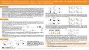 科学海报A Feeder-Free and Serum-Free Culture System Supports Expansion of CD34+ AML and CML Stem/Progenitor Cells
 产品手册Tools for Cancer Research